Digital Poster
Relaxometry
Joint Annual Meeting ISMRM-ESMRMB & ISMRT 31st Annual Meeting • 07-12 May 2022 • London, UK

| Computer # | ||||
|---|---|---|---|---|
2851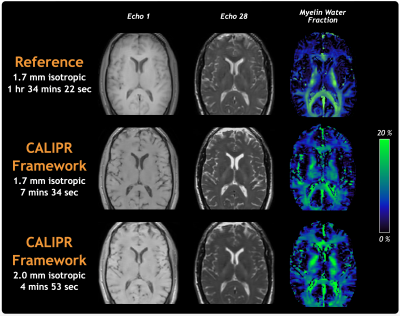 |
92 | The CALIPR framework comprehensively improves acquisition, reconstruction & analysis of multi-component relaxation imaging
Adam V. Dvorak1,2, Dushyant Kumar3, Guillaume Gilbert4, Cornelia Laule1,2,5,6, G.R. Wayne Moore2,6, Alex L. MacKay1,5,7, and Shannon H. Kolind1,2,5,8
1Physics and Astronomy, University of British Columbia, Vancouver, BC, Canada, 2International Collaboration on Repair Discoveries, University of British Columbia, Vancouver, BC, Canada, 3Radiology, University of Pennsylvania, Philadelphia, PA, United States, 4MR Clinical Science, Philips Canada, Mississauga, ON, Canada, 5Radiology, University of British Columbia, Vancouver, BC, Canada, 6Pathology and Laboratory Medicine, University of British Columbia, Vancouver, BC, Canada, 7UBC MRI Research Centre, University of British Columbia, Vancouver, BC, Canada, 8Medicine (Neurology), University of British Columbia, Vancouver, BC, Canada We introduce the CALIPR framework, applied here to multi-component T2 relaxation myelin water imaging (MWI) to achieve a simultaneous increase in data quality and decrease in acquisition time. CALIPR showed robust results for data acquired with aggressive under sampling acceleration, strong agreement with a newly developed “gold-standard” reference MWI acquisition, excellent scan-rescan repeatability, and the ability to acquire whole-brain MWI in under 5 minutes. While this work demonstrates dramatic improvements the CALIPR framework brings to MWI, its utility can be extended to a wide range of both quantitative and qualitative MRI applications across different scanner vendors and field strengths. |
||
2852 |
93 | MP2UTE: T1 mapping of short-T2 components
Stefan Sommer1,2,3, Tom Hilbert3,4,5, Tobias Kober3,4,5, Natalie Hinterholzer2, Daniel Nanz2,6, and Constantin von Deuster1,2,3
1Siemens Healthcare AG, Zurich, Switzerland, 2Swiss Center for Musculoskeletal Imaging (SCMI), Balgrist Campus, Zurich, Switzerland, 3Advanced Clinical Imaging Technology (ACIT), Siemens Healthcare AG, Lausanne, Switzerland, 4Department of Radiology, Lausanne University Hospital (CHUV), Lausanne, Switzerland, 5LTS5, École Polytechnique Fédérale de Lausanne, Lausanne, Switzerland, 6University of Zurich, Zurich, Switzerland
T1 mapping of species with very fast T2/T2* relaxation is impossible with conventional methods measuring echoes refocused by gradients or radiofrequency pulses. We therefore present a new approach to estimate T1 relaxation times of short-T2 tissue such as bone, tendon, or ligament by sampling transversal magnetization at different inversion times with a short-T2 sensitive ultra-short-TE readout. The presented T1 mapping method is efficient and B1-insensitive. By acquiring multiple echoes, it is possible to obtain T1 relaxation times at short and long echo-time and therefore assess the influence of short-T2 components.
|
||
2853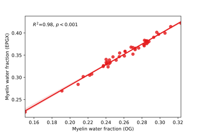 |
94 | Application of a two-pool model of water exchange to myelin water fraction in multiple sclerosis spinal cord
Sharada Balaji1, Irene M. Vavasour2,3, Adam Dvorak1, Poljanka Johnson4, Alex MacKay1,2, and Shannon H. Kolind1,2,3,4
1Physics and Astronomy, University of British Columbia, Vancouver, BC, Canada, 2Radiology, University of British Columbia, Vancouver, BC, Canada, 3International Collaboration on Repair Discoveries, Vancouver, BC, Canada, 4Medicine, University of British Columbia, Vancouver, BC, Canada
Myelin water fraction (MWF) quantifies myelin content in the central nervous system. MWF analysis typically assumes there is no water exchange between different water pools in tissue during the measurement. Here we investigate the effect of incorporating a two-pool model of exchange in a study of MWF in multiple sclerosis (MS) spinal cord and compare with results from the original algorithm. Including exchange resulted in higher MWF values and myelin water residence times that were correlated with MWF, but maintained the expected relationship in MWF between MS subtypes and controls.
|
||
2854 |
95 | Fat-water separated T1 mapping with multi-echo MP2RAGE
Marc-Antoine Fortin1,2, Véronique Fortier1,3, Jorge Campos Pazmino1, Andre van der Kouwe4, and Ives Roger Levesque1,5
1Medical Physics Unit, McGill University, Montreal, QC, Canada, 2Physics, NTNU, Trondheim, Norway, 3Medical Imaging, McGill University Health Centre, Montreal, QC, Canada, 4Athinoula A. Martinos Center for Biomedical Imaging, Havard Medical School, Boston, MA, United States, 5Research Institute of the McGill University Health Centre, Montreal, QC, Canada To design a novel fat-water separated T1 mapping technique from a multi echo (ME) version of the Magnetization Prepared Two Rapid Acquisition of Gradient Echoes (MP2RAGE) sequence by adapting the MP2RAGE sequence to shorter T1 values resembling values observed for fat (between 200 and 800 ms) and introducing a 3-point Dixon fat-water separation step in the analysis. |
||
2855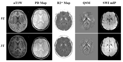 |
96 | An initial multi-parametric imaging experience of MULTIPLEX at 5T whole body system
Yongquan Ye1, Xiaodi Liu2, Ying Wu2, Zhongqi Zhang1, and Jian Xu1
1UIH America, Inc., Houston, TX, United States, 2United Imaging Healthcare, Shanghai, China
In this work, we evaluated the performance of a recently proposed high resolution multi-parametric method, i.e. MULTIPLEX, on a state-of-the-art 5T whole body scanner. Comparing to the 3T counterpart, MULTIPLEX on 5T generally offers improved SNR, image quality and imaging efficiency, while maintaining satisfactory performance on B1/T1 mapping.
|
||
2856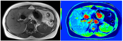 |
97 | Native T1 Mapping Combined With Liver Stiffness Measurement in Identifying Significant Liver Fibrosis in Chronic Hepatitis B Patients Video Not Available
Zhi-Yuan Chen1, Yu-Pin Liu1, and Yunzhu Wu2
1Department of Radiology, The Second Affiliated Hospital of Guangzhou University of Chinese Medicine, Guangdong Provincial Hospital of Traditional Chinese Medicine, Guangzhou, China, 2MR Scientific Marketing, SIEMENS Healthineers, Shanghai, China
Noninvasive identification of significant liver fibrosis in chronic hepatitis B patients is of great value because it can be an important indication for anti-viral therapy according to EASL guideline. This abstract was to evaluate the diagnostic capability of variable flip angle native T1 mapping combined with liver stiffness measurement (LT1 model) in identifying significant liver fibrosis in chronic hepatitis B patients. The LT1 score had good sensitivity and specificity for identifying significant liver fibrosis in chronic hepatitis B patients, and could provide a reliable basis for clinical diagnosis and treatment.
|
||
2857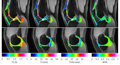 |
98 | Quantitative characterization of fibrous tissues in the knee joint using magnetization prepared 3D spiral UTE sequence at 3 T
Petros Martirosian1, Cecilia Zhang2, Martin Schwartz1,3, Thomas Benkert4, and Fritz Schick1
1Section on Experimental Radiology, University Hospital of Tübingen, Tübingen, Germany, 2Department of Diagnostic and Interventional Radiology, University Hospital of Tübingen, Tübingen, Germany, 3Institute of Signal Processing and System Theory, University of Stuttgart, Stuttgart, Germany, 4MR Application Predevelopment, Siemens Healthcare GmbH, Erlangen, Germany
Magnetization prepared 3D spiral UTE sequences were developed for T1, T2, T1rho and MTR measurements of fibrous structures. The robustness of the parameter mapping for tendons, ligaments, menisci, cartilage and muscle of the human knee joint was evaluated in a test-retest repeatabilty study. Multiparametric UTE imaging was successfully performed for both test and retest examination. The results of the study demonstrate good repeatability of the applied multiparametric protocol. Parameter maps such as T2 and T1rho show less robustness than T1 and MTR measurements. Especially with small structures as tendons and ligaments, there is the least agreement between scan and rescan.
|
||
2858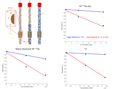 |
99 | SPION relaxivity at low field strengths with hyperpolarized 129Xe
Nicholas Bryden1, Christian T McHugh1, Michele Kelley1, and Rosa T Branca1
1University of North Carolina Chapel Hill, Chapel Hill, NC, United States We provide superparamagnetic iron oxide longitudinal relaxivity measurements at both high (11.7 T) and low (0.6 – 2.1 mT) magnetic field strengths, for both 1H and hyperpolarized 129Xe nuclei. These measurements show significant increase in relaxivity at low field for both nuclei. We also provide some preliminary in vivo results for hyperpolarized 129Xe that suggest a depolarization mechanism similar to that of SPIONs from the hemoglobin found in whole blood. |
||
2859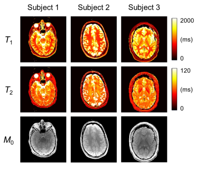 |
100 | Practical and readily-available relaxometry method
Bruno Madore1, Michael Jerosch-Herold1, Jr-Yuan George Chiou1, Cheng-Chieh Cheng2, Srinivasan Mukundan1, Jeffrey Guenette1, and Georgeta Mihai1
1Radiology, Brigham and Women's Hospital, Harvard Medical School, Boston, MA, United States, 2Computer Science and Engineering, National Sun Yat-sen University, Kaohsiung, Taiwan
Although many methods already exist to map T1, T2 and M0, these often involve special sequences not readily available on clinical scanners and/or may require long scan times. In contrast, the proposed method can run on most scanners, it offers flexible tradeoffs between scan time and image quality, and it generates spatially-aligned parameter maps. Validation was performed in gel phantoms with varying concentrations of contrast agents, and in vivo examples are presented from three neuroradiology patients. Compared to other quantitative mapping methods, the present method is meant to stand out in terms of its practicality and availability.
|
||
2860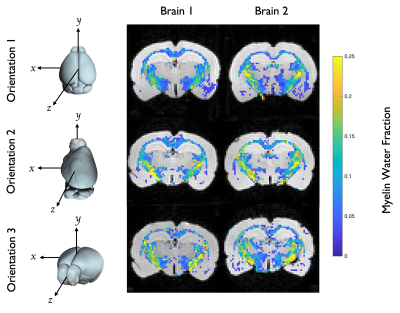 |
101 | Orientation Dependence of Myelin Water Fraction as Measured by MET2
Hannah E. Alderson1,2 and Mark D. Does1,2
1Biomedical Engineering, Vanderbilt University, Nashville, TN, United States, 2Vanderbilt University Institute of Imaging Science, Nashville, TN, United States This study investigated the orientation dependence of myelin water fraction (MWF) as evaluated with a 3D multiple-spin echo sequence (MSE). MSE and diffusion tensor scans were acquired at 7-T from two excised rat brains at three different orientations with respect to the main magnetic field. Region of interest analysis of the corpus callosum and the internal capsule revealed that there is no clear orientation dependency for MWF in these white matter tracts. This result is also evidenced by the minimal orientational modulation of the T2 spectra for both ROIs across both brains. |
||
2861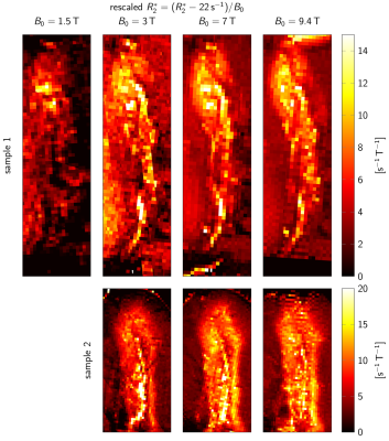 |
102 | Field dependence of T2* contrast in human substantia nigra
Malte Brammerloh1,2, Evgeniya Kirilina1,3, Renat Sibgatulin4, Karl-Heinz Herrmann4, Tilo Reinert1,5, Carsten Jäger1,6, Primož Pelicon7, Kerrin J. Pine1, Primož Vavpetič7, Andreas Deistung8, Markus Morawski6, Jürgen R. Reichenbach4, and Nikolaus Weiskopf1,5
1Department of Neurophysics, Max Planck Institute for Human Cognitive and Brain Sciences, Leipzig, Germany, 2Leipzig University, Leipzig, Germany, 3Center for Cognitive Neuroscience Berlin, Freie Universität Berlin, Berlin, Germany, 4Medical Physics Group, Institute of Diagnostic and Interventional Radiology, University Hospital Jena, Jena, Germany, 5Felix Bloch Institute for Solid State Physics, Leipzig University, Leipzig, Germany, 6Paul Flechsig Institute of Brain Research, Leipzig University, Leipzig, Germany, 7Department for Low and Medium Energy Physics, Jožef Stefan Institute, Ljubljana, Slovenia, 86University Clinic and Outpatient Clinic for Radiology, University Hospital Halle (Saale), Halle (Saale), Germany MRI holds high promise to diagnose Parkinson’s disease (PD) at clinical field strength B0. However, it remains unclear which B0 optimizes T2* contrast in substantia nigra, which provides high diagnostic accuracy. We used quantitative MRI at B0=1.5T-9.4T, MR microscopy, and histochemistry to characterize the field dependence of the major contributors to R2* (1/T2*): dopaminergic neurons, ferritin, and myelin. R2* maps were similar at B0=3T-9.4T, and all contributions scaled approximately linearly with B0. Hence, the contrast mechanisms are similar across currently available MRI field strengths in vivo, which informs the design of novel PD biomarkers. |
||
2862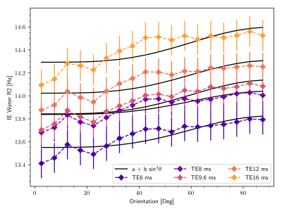 |
103 | T2 measured with multiple spin-echo depends on echo spacing as well as orientation
Jonathan Doucette1,2, Manuel Bauer1,3, Lara Bartels1,2, Alexander Jaffray1,2, and Alexander Rauscher1,2,4,5
1UBC MRI Research Centre, Vancouver, BC, Canada, 2Physics & Astronomy, University of British Columbia, Vancouver, BC, Canada, 3Institute for Physical Chemistry, Albert-Ludwigs-University, Freiburg, Germany, 4Radiology, University of British Columbia, Vancouver, BC, Canada, 5Pediatrics, University of British Columbia, Vancouver, BC, Canada
The MRI signal in white matter tissue is composed of constituent signals from two different water pools: intra/extracellular water and myelin water. When using a multi spin-echo acquisition, the relaxation rate measured in each pool has been shown to depend on the orientation of the containing white matter fibre with respect to the main magnetic field. Here, we show an additional influence of the acquisition echo spacing on the apparent intra/extracellular rate relaxation rate, and using numerical simulation, we provide evidence that decoherence mediated by diffusion through local field inhomogeneities created by blood vessels cannot explain the observed orientation dependence.
|
||
2863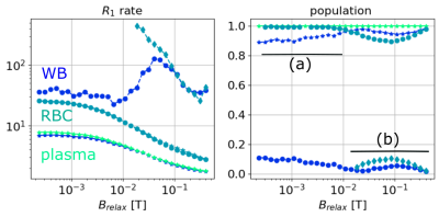 |
104 | Mechanisms of relaxation in blood - Nuclear magnetic resonance dispersion profile of rat’s blood, plasma, and red cells at different temperatures
Mohsine Mekhfi 1, Aimé Labbé 1, Dimitri Kereselidze1, Erwan Selingue 2, Lionel Broche3, Anne-Laure Rollet 4, and Marie Poirier-quinot1
1Université Paris Saclay, CEA, CNRS, Inserm, Laboratoire d’Imagerie biomédicale multimodale Paris Saclay (BioMaps), Orsay, France, 2Université Paris-Saclay, CEA, CNRS, Baobab, NeuroSpin, Gif-sur-Yvette, France, 3Aberdeen Biomedical Imaging Centre, University of Aberdeen, Foresterhill, AB25 2ZD, Aberdeen, UK, Aberdeen, Scotland, 4Laboratoire PHysico-chimie des Electrolytes et Nanosystèmes InterfaciauX, PHENIX, Sorbonne Université, CNRS, Paris, France
Measuring whole blood relaxation time is as a reliable diagnosis tool. Usually, one compartmental model is used to fit data, which seems a very simplistic approach considering the complexity of blood. The aim of this work is to test the validity of this assumption by studying the relaxation rate of whole blood, plasma and red blood cells (rbc) of rats at different temperatures. The whole blood is well described by a two-compartment approach, one corresponding to plasma and another for red blood cells. Once frozen, there is only one relaxation rate population, which is considered to be the rbc population.
|
||
 Back to Meeting Home
 Back to the Program-at-a-Glance
The International Society for Magnetic Resonance in Medicine is accredited by the Accreditation Council for Continuing Medical Education to provide continuing medical education for physicians.
 Back to Meeting Home
 Back to the Program-at-a-Glance
 View the Presentation