Digital Poster
Electromagnetic Properties & Oximetry
Joint Annual Meeting ISMRM-ESMRMB & ISMRT 31st Annual Meeting • 07-12 May 2022 • London, UK

Digital Poster
Electromagnetic Properties & Oximetry
| Computer # | ||||
|---|---|---|---|---|
2910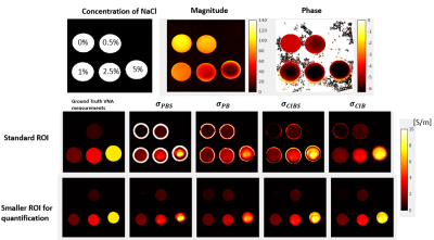 |
105 | Comparison and validation of multiple MR-EPT methods with ground truth vector network analyzer measurements
Zhongzheng He1, Martin Doguet1, Paul Soullié1, Paulo Loureiro de Sousa2, Pauline Lefebvre1, and Freddy Odille1,3
1IADI U1254, INSERM, Université de Lorraine, Nancy, France, 2ICube, Université de Strasbourg, CNRS, Strasbourg, France, 3CIC-IT 1433, CHRU Nancy, INSERM, Université de Lorraine, Nancy, France
Many MR-EPT reconstruction methods have been published, however, there is a lack of comparative experiments to verify the reconstructed electrical properties (EP) maps. In this work, we compared and validated the phase-based (PB), complex-image-based (CIB), and their simplified methods with ground truth vector network analyzer measurements on different conductivity phantoms. After serval comparative experiment repetitions, the complex-image-based method by Soullié et al.[1] was the most consistent to the VNA ground truth measurements. The proposed setup can be used to compare other MR-EPT reconstruction methods.
|
||
2911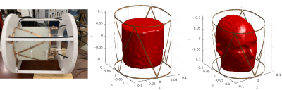 |
106 | A Novel Volume-Surface Integral Equation Formulation of Global Maxwell Tomography: Simulations and Experiments.
Ilias Giannakopoulos1,2, Jose Seralles3, Jan Paska1,2, Georgy Guryev3, Carlotta Ianniello1,2, Luca Daniel3, Jacob White3, Ryan Brown1,2,4, Daniel Sodickson1,2,4, and Riccardo Lattanzi1,2,4
1Center for Advanced Imaging Innovation and Research (CAI2R), Department of Radiology, New York University Grossman School of Medicine, New York, NY, United States, 2The Bernard and Irene Schwartz Center for Biomedical Imaging (CBI), Department of Radiology, New York University Grossman School of Medicine, New York, NY, United States, 3Electrical Engineering and Computer Science, Massachusetts Institute of Technology, Cambridge, MA, United States, 4Vilcek Institute of Graduate Biomedical Sciences, New York University Grossman School of Medicine, New York, NY, United States
Global Maxwell Tomography (GMT) is an inverse scattering technique that estimates electrical properties (EP) from magnetic resonance measurements. The original GMT relies on an approximation of the incident fields, which is not accurate for experiments, especially in-vivo. We propose a new GMT formulation, based on the volume-surface integral equation (VSIE) method, where we implicitly re-estimate the incident fields accounting for the updated EP in every GMT iteration. We show average EP reconstruction error below 8% for a uniform phantom experiment and below 10% for a simulation experiment with a heterogeneous head model and realistic SNR.
|
||
2912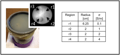 |
107 | Automatic selection of the optimal kernel size for Helmholtz-based EPT
Alessandro Arduino1, Stefano Mandija2,3, Francesca Pennecchi1, Cornelis A. T. Van Den Berg2,3, and Luca Zilberti1
1Istituto Nazionale di Ricerca Metrologica (INRiM), Torino, Italy, 2Department of Radiotherapy, University Medical Center Utrecht, Utrecht, Netherlands, 3Computational Imaging Group for MR Diagnostics & Therapy, Center for Image Sciences, University Medical Center Utrecht, Utrecht, Netherlands
In this work, a procedure for the automatic selection of the optimal kernel size in Helmholtz-based electric properties tomography (MR-EPT) with phase-based approximation is presented and tested on experimental data acquired on a phantom with a 3 T MRI scanner. The procedure is exclusively based on the map of the transceive phase, making no assumptions on the data in addition to those used in the derivation of the Helmholtz-based MR-EPT technique. As a by-product, the approach provides a map indicating the reliability of the reconstructed electric conductivity pixel-by-pixel.
|
||
2913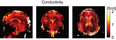 |
108 | Conductivity mapping at 0.55 T with balanced steady state free precession
Santhosh Iyyakkunnel1,2 and Oliver Bieri1,2
1Department of Radiology, University Hospital Basel, Basel, Switzerland, 2Department of Biomedical Engineering, University of Basel, Basel, Switzerland
Conductivity mapping depends sensitively on the signal-to-noise ratio (SNR) of the transmit phase estimation. It is thus questionable, whether conductivity mapping can be performed at low field. Due to reduced off-resonances, however, a possible solution for the reduced SNR might be offered by balanced steady state free precession (bSSFP). Brain conductivity mapping with bSSFP was investigated at 0.55 T and appears to be feasible but besides SNR also the reduced curvature of the transmit field becomes challenging.
|
||
2914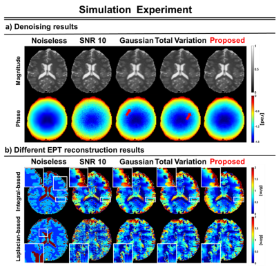 |
109 | Deep prior for suppressing noise amplification and edge preservation in Phase-based EPT with Low-SNR image
Chuanjiang Cui1, Jun-Hyeong Kim1, Kyu-Jin Jung1, Jaeuk Yi1, and Dong-Hyun Kim1
1Department of Electrical and Electronic Engineering, Yonsei University, Seoul, Korea, Republic of
Phase-based EPT algorithm is extremely sensitive to noise. Although many studies have investigated such as linear(Gaussian filter) or non-linear filter(TV norm) to cope with amplification, textured noise and staircasing effect still remain in phase image, which lead to conductivity error such as broadening boundary artifact or high std value in reconstructed conductivity maps. In this study, we propose a deep prior based denoising method, which achieve to not only suppress instability brought by noise amplification but reduce boundary error.
|
||
2915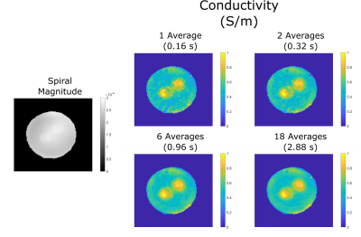 |
110 | Investigating Spiral Trajectories for Sub-second Conductivity Imaging in MREPT
Safa Özdemir1, Efe Ilicak1, Lothar R. Schad1, and Frank G. Zöllner1,2
1Computer Assisted Clinical Medicine, Medical Faculty Mannheim, Heidelberg University, Mannheim, Germany, 2Mannheim Institute for Intelligent Systems in Medicine, Medical Faculty Mannheim, Heidelberg University, Mannheim, Germany
Magnetic Resonance Electrical Properties Tomography (MREPT) technique is used to obtain conductivity (σ) and permittivity (ε), using phase of the B1+ information. While spin echo and bSSFP based pulse sequences were shown to successfully obtain phase images, they are either very slow or suffer from artifacts and require multi-acquisitions. To overcome these limitations, in this work we investigate the use of spiral trajectory-based sequences in MREPT. Our results indicate that even for sub-second acquisition times, conductivity maps can be successfully acquired via spiral trajectory-based sequence, and therefore can improve the utility of MREPT techniques by shortening the acquisition time drastically.
|
||
2916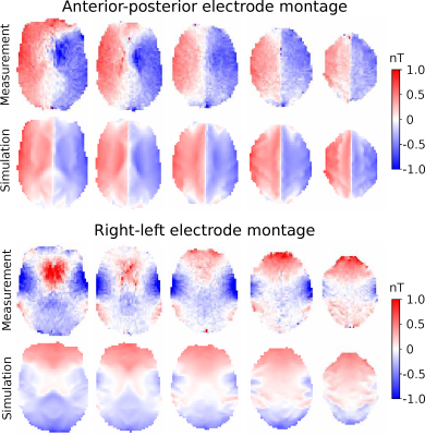 |
111 | Robust measurements of current-induced magnetic fields in the human brain by EPI
Fróði Gregersen1,2,3, Cihan Göksu2,4, Hasan Eroğlu1,2, Zhentao Zuo3,5,6, Axel Thielscher1,2, and Lars Hanson1,2
1Section for Magnetic Resonance, DTU Health Tech, Technical University of Denmark, Kgs. Lyngby, Denmark, 2Danish Research Centre for Magnetic Resonance, Centre for Functional and Diagnostic Imaging and Research, Copenhagen University Hospital - Amager and Hvidovre, Copenhagen, Denmark, 3Sino-Danish College, University of Chinese Academy of Sciences, Beijing, China, 4High-Field Magnetic Resonance Center, Max-Planck-Institute for Biological Cybernetics, Tübingen, Germany, 5State Key Laboratory of Brain and Cognitive Science, Beijing MRI Center for Brain Research, Institute of Biophysics, Chinese Academy of Sciences, Beijing, China, 6Center for Excellence in Brain and Science and Intelligence Technology, Chinese Academy of Sciences, Beijing, China
Magnetic resonance current density imaging (MRCDI) can measure the magnetic fields created in the human brain from currents injected via surface electrodes. Previous methods have demonstrated high sensitivity sufficient for low current strengths (~1 mA). However, they have also proven susceptible to physiological noise. Here we increase the temporal resolution of the method and thereby the robustness to physiological noise by using echo-planar imaging (EPI) for the acquisition. We show that the method produces reliable magnetic field measurements with an average sensitivity of 52 pT for a 2 minutes scan with 3 mm isotropic resolution.
|
||
2917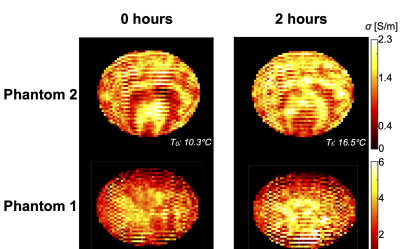 |
112 | In vitro MR-based Electric Properties Tomography during Temperature Increase
Jessica A. Martinez1, Alessandro Arduino1, Kevin Moulin2,3, Adriano Troia1, Oriano Bottauscio1, and Luca Zilberti1
1Advanced Materials Metrology and Life Science, Istituto Nazionale di Ricerca Metrologica, Turin, Italy, 2CREATIS, Lyon, France, 3University Hospital of Saint-Etienne, Saint-Etienne, France
The feasibility of EPT to obtain electrical conductivity data during temperature increase was analyzed. Experiments were performed in two homogeneous phantoms with different saline concentrations during temperature increase.
|
||
2918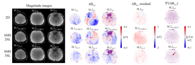 |
113 | Mapping of weak current-induced magnetic fields in a 3D volume of the human brain at high resolution: 2D vs. Simultaneous multi slice
Cihan Göksu1,2, Klaus Scheffler2,3, Fróði Gregersen1,4,5, Hasan Hüseyin Eroğlu1,4, Rahel Heule2,3, Hartwig R. Siebner1,6,7, Lars G. Hanson1,4, and Axel Thielscher1,4
1Danish Research Centre for Magnetic Resonance, Centre for Functional and Diagnostic Imaging and Research, Copenhagen University Hospital, Amager and Hvidovre, Denmark, 2High-Field Magnetic Resonance Center, Max-Planck-Institute for Biological Cybernetics, Tübingen, Germany, 3Department of Biomedical Magnetic Resonance, University of Tübingen, Tübingen, Germany, 4Section for Magnetic Resonance, DTU Health Tech, Technical University of Denmark, Kgs Lyngby, Denmark, 5Sino-Danish Center for Education and Research, Aarhus, Denmark, 6Department of Neurology, Copenhagen University Hospital, Bispebjerg, Denmark, 7Institute for Clinical Medicine, Faculty of Medical and Health Sciences, University of Copenhagen, Copenhagen, Denmark
Exact knowledge of current distributions induced by transcranial electrical stimulation (TES) in the brain is important for effective clinical use of TES. MRCDI uses MRI to measure the TES-induced magnetic fields for estimating the underlying current flow distributions. The estimation methods can benefit from highly sensitive volume MRCDI measurements at a high spatial resolution. Here, we advanced our 2D spoiled gradient-echo-based MRCDI method for a sparse volume acquisition by using simultaneous-multi-slice (SMS) acquisition. Our SMS strategy demonstrated 25% improvement in noise floors against 2D. We test the performance of our methods by phantom and human in-vivo experiments using cable-loop currents.
|
||
2919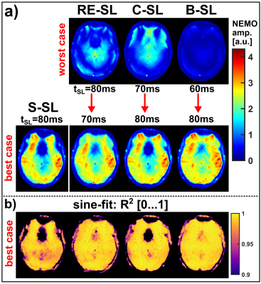 |
114 | Toward static-inhomogeneity-insensitive detection of neuro-electro-magnetic fields via compensated rotary saturation
Maximilian Gram1,2, Petra Albertova1,2, Verena Schirmer2, Martin Blaimer3, Matthias Gamer4, Martin J. Herrmann5, Peter Michael Jakob2, and Peter Nordbeck1,6
1Department of Internal Medicine I, University Hospital Würzburg, Würzburg, Germany, 2Experimental Physics 5, University of Würzburg, Würzburg, Germany, 3Magnetic Resonance and X-ray Imaging Department Fraunhofer IIS, Fraunhofer Institute for Integrated Circuits IIS, Würzburg, Germany, 4Department of Psychology, University of Würzburg, Würzburg, Germany, 5Center of Mental Health, Dept. of Psychiatry, Psychosomatics, and Psychotherapy, University Hospital Würzburg, Würzburg, Germany, 6Comprehensive Heart Failure Center (CHFC), University Hospital Würzburg, Würzburg, Germany
Resonant absorption during spin-lock preparation can be used to measure tiny oscillating magnetic fields acting as direct evidence of electrical neuronal activity. Different spin-locking techniques were compared with respect to their sensitivity in magnetic field detection. As a specialty, the oscillating magnetic fields were generated by the built-in gradient system in an offcenter slice. The spin-lock time was identified as the crucial parameter for the performance of NEMO (neuro-electro-magnetic-oscillations) detection, since minima and maxima in the signal amplitude emerged in phantom and in vivo experiments. Affirmative, the experimental results show an excellent agreement with simulation results.
|
||
2920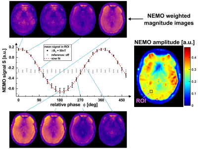 |
115 | Gradient based emulation of neuro-electro-magnetic oscillations: A validation technique for MRI-based detection of Biomagnetism
Petra Albertova1,2, Maximilian Gram1,2, Verena Schirmer1, Martin Blaimer3, Martin J. Herrmann4, Matthias Gamer5, Peter Nordbeck2,6, and Peter Michael Jakob1
1Experimental Physics 5, University of Würzburg, Würzburg, Germany, 2Department of Internal Medicine I, University Hospital Würzburg, Würzburg, Germany, 3Magnetic Resonance and X-ray Imaging Department, Fraunhofer IIS, Fraunhofer Institute for Integrated Circuits IIS, Würzburg, Germany, 4Center of Mental Health, Dept. of Psychiatry, Psychosomatics, and Psychotherapy, University Hospital of Würzburg, Würzburg, Germany, 5Department of Psychology, University of Würzburg, Würzburg, Germany, 6Comprehensive Heart Failure Center (CHFC), University Hospital Würzburg, Würzburg, Germany
Spin-lock based absorption of magnetic oscillations offers potential for direct detection of electrical neuronal activity. We propose a novel versatile validation and calibration technique which paves the way for emulation and quantification of biomagnetic fields. Using ultra-weak gradient waveforms, the method mimics brain activity and thus projects artificial fields onto the tissue under investigation. The method applicable for sequence validation or signal calibration was tested in phantom and in vivo experiments with the built-in gradient system providing sinusoidal field modulations down to 1 nT. It proved to be reliable and reproducible and hence can potentially enable quantification of biomagnetic fields.
|
||
2921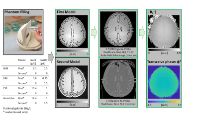 |
116 | A semi-realistic and reusable 3D printed brain phantom for MR-based Electrical Properties Tomography
Thierry G. Meerbothe1,2, Sammy Florczak3, Peter R. S. Stijnman1,2, Cornelis A. T. van den Berg1,2, Riccardo Levato3,4, and Stefano Mandija1,2
1Department of Radiotherapy, Division of Imaging and Oncology, University Medical Center Utrecht, Utrecht, Netherlands, 2Computational Imaging Group for MR Diagnostics and Therapy, Center for Image Sciences, University Medical Center Utrecht, Utrecht, Netherlands, 3Department of Orthopaedics, University Medical Center Utrecht, Utrecht, Netherlands, 4Department of Clinical Sciences, Faculty of Veterinary Medicine, Utrecht University, Utrecht, Netherlands
This work presents a semi-realistic and reusable 3D printed brain phantom to benchmark MR-Electrical-Properties-Tomography reconstruction methods. We show that the hollow compartments of this phantom can be refilled multiple times with different water-based solutions of known electrical properties, which are otherwise not available from in-vivo measurements. Additionally, these brain phantoms can be used inside electromagnetic simulation software, allowing for MR-EPT reconstructions in controlled, simulation settings. In this way, a database comprising of simulated and measured data using these brain models and corresponding 3D printed phantoms will be generated and shared for the first MR-EPT reconstruction challenge.
|
||
2922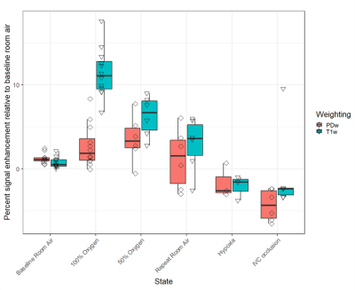 |
117 | Understanding the vasodilatory effects of oxygen in oxygen enhanced functional lung MRI at 0.55T
Björn Wieslander1, Felicia Seemann2, Ahsan Javed2, Christopher G Bruce2, Rajiv Ramasawmy2, Andi Jaimes2, Katherine Lucas2, Victoria Frasier2, Amanda Potersnak2, Robert J Lederman2, and Adrienne E Campbell-Washburn2
1Pulmonary Branch, Division of Intramural Research, National Heart, Lung and Blood Institute, National Institutes of Health, Bethesda, MD, United States, 2Cardiovascular Branch, Division of Intramural Research, National Heart, Lung, and Blood Institute, National Institutes of Health, Bethesda, MD, United States
Oxygen-enhanced lung MRI is promising for assessing regional lung function (ventilation and perfusion) but the physiological contributions to signal enhancement are poorly characterized. We sought to assess how oxygen-induced pulmonary vasodilation may contribute to signal enhancement by altering pulmonary blood volume and therefore lung proton density. We acquired repeated PDw and T1w images using a 0.55 T prototype MRI system in anesthetized pigs exposed to room air, 100% O2, 50% O2, hypoxia and experimental pulmonary hypovolemia. Our findings suggest that T1w signal intensity is affected by oxygen through both T1 shortening and vascular activity.
|
||
2923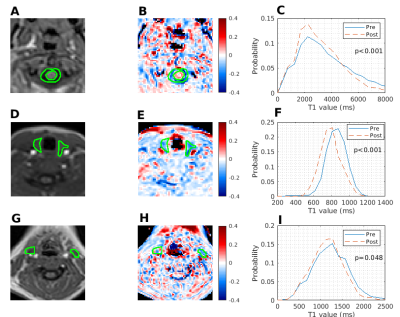 |
118 | Oxygen induced T1 changes in head and neck anatomical structures
Alastair McCabe1,2, Damian Borys3, Judith Christian1, Solange Pereira4, Selene Rowe4, Paul S Morgan5, Jagrit Shah4, Elaine Blackshaw6, Stewart Martin7, and Rafal Panek2,6
1Department of Clinical Oncology, Nottingham University Hospitals NHS Trust, Nottingham, United Kingdom, 2School of Medicine, University of Nottingham, Nottingham, United Kingdom, 3Department of Systems Biology and Engineering, Silesian University of Technology, Gliwice, Poland, 4Department of Radiology, Nottingham University Hospitals NHS Trust, Nottingham, United Kingdom, 5Radiological Sciences, University of Nottingham, Nottingham, United Kingdom, 6Department of Medical Physics & Clinical Engineering, Nottingham University Hospitals NHS Trust, Nottingham, United Kingdom, 7University of Nottingham Biodiscovery Institute, Nottingham, United Kingdom Oxygen enhanced MRI (OE-MRI) is a proposed tumour hypoxia imaging technique that is yet to be widely applied in the head and neck. 7 participants, 3 with suspected head and neck squamous cell carcinoma (HNSCC) were scanned using 3D spoiled GRE-DIXON. After on air scans, oxygen was delivered via a non-rebreather mask. T1 times were determined on in-phase images. Oxygenation led to statistically significant T1 shortening in CSF, thyroid and neck nodes. OE-MRI shows promise in identifying hypoxic regions in the head and neck. Further work investigating the feasibility of OE-MRI to detect hypoxic regions in HNSCC is warranted. |
||
 Back to Meeting Home
 Back to the Program-at-a-Glance
The International Society for Magnetic Resonance in Medicine is accredited by the Accreditation Council for Continuing Medical Education to provide continuing medical education for physicians.
 Back to Meeting Home
 Back to the Program-at-a-Glance
 View the Presentation