Digital Poster
Elastography
Joint Annual Meeting ISMRM-ESMRMB & ISMRT 31st Annual Meeting • 07-12 May 2022 • London, UK

| Computer # | ||||
|---|---|---|---|---|
2236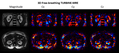 |
49 | Free-Breathing MR Elastography of the Kidneys Using TURBINE-MRE
Yi Sui1, Jiahui Li1, Zheng Zhu1, Kevin J Glaser1, Nana K Owusu1, Joshua D Trzasko1, Arvin Arani1, Ziying Yin1, Phillip J Rossman1, Meng Yin1, and Richard L Ehman1
1Department of Radiology, Mayo Clinic, Rochester, MN, United States
A free-breathing hybrid radial-cartesian 3D MR elastography technique (TURBINE-MRE) was extended for kidneys with a dual driver system. This technique provides wave images and stiffness maps in a single 3.5-minute free-breathing acquisition. The feasibility of the new technique was demonstrated in a healthy volunteer.
|
||
2237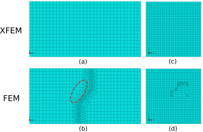 |
50 | Numerical simulation of wave propagation through interfaces using the eXtended FEM for MR elastography
Quanshangze DU1, Simon Auguste LAMBERT2, Nahiène HAMILA3, and Aline BEL-BRUNON1
1Univ Lyon, INSA Lyon, CNRS, LaMCoS, UMR5259, 69621 Villeurbanne, France, 2Université de Lyon, INSA Lyon, Université Claude Bernard Lyon 1, Ecole Centrale de Lyon, CNRS, Ampère UMR5005, Villeurbanne, France, 3ENI Brest, UMR CNRS 6027, IRDL, F-29200 Brest, France
The finite element method (FEM) is widely used for modeling wave propagation and stiffness reconstruction in magnetic resonance elastography (MRE). However, it is not that efficient for modeling inclusions with complex interfaces. Here, we propose a formulation of FEM known as the eXtended FEM (XFEM), and investigate this method in two studies: wave propagation across an oblique linear interface and stiffness reconstruction of a random-shape inclusion. XFEM results present good agreement with the theoretical predictions and FEM simulation results. XFEM can be regarded as an ideal alternative to FEM for inclusion modeling in MRE.
|
||
2238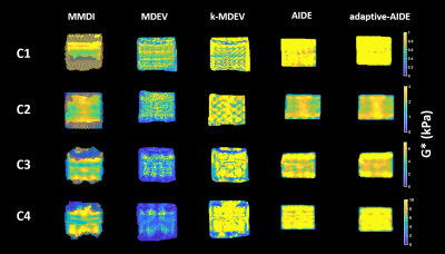 |
51 | Multiresolution MR elastography reconstruction and comparison of different direct inversion algorithms in the liver
Gwenaël Pagé1, Félicia Juléa1, Valérie Paradis2, Valérie Vilgrain1,3, Dominique Valla4, Bernard E. Van Beers1,3, and Philippe Garteiser1
1Laboratory of imaging biomarkers, INSERM UMR 1149, Paris, France, 2Center for research on inflammation, INSERM UMR 1149, Paris, France, 3Department of Radiology, University Hospital Paris Nord, Clichy, France, 4Hepatology service, University Hospital Paris Nord, Clichy, France
In this study we propose using a MR elastography reconstruction method adjust the ratio between wavelength and spatial resolution, by resampling the matrix of shear displacement locally, to optimize the quality of the reconstruction. This reconstruction algorithm was compared to existing direct inversion algorithms in phantoms, healthy volunteers and patients with liver fibrosis. We observed that the algorithm k-MDEV showing the best performance in phantoms and patients did not have the best repeatability. Our proposed method improved the diagnostic performance for advanced fibrosis, compared to classical direct inversion method, without a negative effect on repeatability.
|
||
2239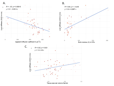 |
52 | Shear stiffness measured by MR elastography correlates with compression modulus in ex vivo rat livers subjected to increasing portal pressure.
Yasmine Safraou1, Karolina Graczynska1, Tom Meyer1, Heiko Tzschätzsch1, Thomas Fischer1, Jürgen Braun1, Ingolf Sack1, and Jing Guo2
1Radiology, Charité – Universitätsmedizin Berlin, Berlin, Germany, 2Radiology, Charité - Universitätsmedizin Berlin, Berlin, Germany
To investigate the effect of perfusion pressure on liver stiffness, 13 ex-vivo rat livers were studied by volumetric MRI, MR elastography (MRE) and diffusion MRI under controlled portal pressures between 0 and 15 cmH2O. Liver volume, tissue-vascular volume fraction, stiffness and bulk modulus decreased while water diffusivity increased with increasing portal pressure. Collectively, our findings suggest that liver softening is caused by a relative increase in fluid volume while an increased vascular pressure, potentially increasing liver stiffness, is of minor importance when tissue can freely expand, as in our scenario.
|
||
2240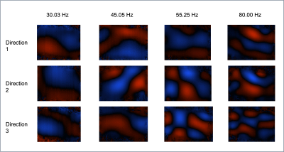 |
53 | Investigating the Repeatability of Multifrequency Magnetic Resonance Elastography applied to a Soft Gelatin Phantom
Owais Siddiqi1, Jessica Winfield1,2, Konstantinos Zormpas-Petridis1, Emma Harris1, Anand Ramkumar1, Antonio Candito1, Steffen Görner3, Christina Messiou1,2, Matthew Blackledge1, and Imogen Thrussell1,2
1Division of Radiotherapy and Imaging, The Institute of Cancer Research, London, United Kingdom, 2Department of Radiology, The Royal Marsden NHS Foundation Trust, London, United Kingdom, 3Charité - Universitätsmedizin Berlin, Corporate Member of Freie Universität Berlin, Humboldt-Universität zu Berlin, and Berlin Institute of Health, Berlin, Germany
New measures of response are required that can capture the inherent heterogeneity and the heterogeneous changes observed in soft tissue tumours. Shear-wave Magnetic Resonance Elastography (MRE) measures stiffness of tissue by measuring the propagation of externally applied shear waves. In this study we explore the test-retest repeatability of Multifrequency MRE (MMRE), acquired using an echo-planar imaging readout applied to a soft gelatin phantom used to mimic the stiffness of soft-tissue tumours. Our results suggest MMRE is highly repeatable and supports further investigation in using MMRE to measure response in soft-tissue tumours.
|
||
2241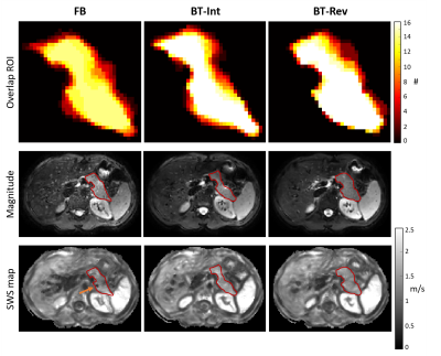 |
54 | Breathing task paradigm to improve the quality of pancreatic Magnetic Resonance Elastography
Nienke P.M. Wassenaar1, Anne-Sophie van Schelt1, Eric M. Schrauben1, Rémi van der Woude2, Jamila E. de Jong2, Jules L. Nelissen1, Hanneke W.M. van Laarhoven3, Jing Guo4, Ingolf Sack4, Jurgen H. Runge1, Aart J. Nederveen1, and Jaap Stoker1
1Radiology and Nuclear Medicine, Amsterdam University Medical Centers, University of Amsterdam, Amsterdam, Netherlands, 2University of Twente, Enschede, Netherlands, 3Department of Medical Oncology, Cancer Center Amsterdam, Amsterdam University Medical Centers, University of Amsterdam, Amsterdam, Netherlands, 4Department of Radiology, Charité - Universitätsmedizin Berlin, corporate member of Freie Universität Berlin, Humboldt-Universität zu Berlin, and Berlin Institute of Health, Berlin, Germany
Free-breathing acquisition of pancreatic MR elastography can potentially introduce errors in stiffness reconstruction. In this study, breathing tasks are introduced during interleaved and reversed slice order MRE acquisition to determine if the quality of MRE increases. The shear-wave speed and octahedral shear strain signal-to-noise-ratio in the pancreas did not significantly change when using breathing tasks. However, the stability of the pancreas location over time increases when using breathing tasks combined with reversed slice ordering as well as the octahedral shear strain signal-to-noise-ratio in the whole abdomen. Future research should focus on comparing shear-wave speed reproducibility of both MRE methods.
|
||
2242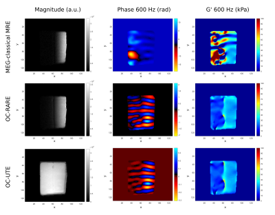 |
55 | MR Elastography of short T2 samples with Optimal Control-based RF pulses demonstrated on Achilles tendon
Pilar Sango-Solanas1, Kevin Tse-Ve-Koon1, Eric Van Reeth1,2, Cyrielle Caussy3,4, and Olivier Beuf1
1Univ Lyon, INSA-Lyon, Inserm, UCBL1, CNRS, CREATIS, UMR 5220, U1294, F-69621, Lyon, France, 2CPE Lyon, Département Sciences du Numérique, Lyon, France, 3Univ Lyon, CarMen Laboratory, INSERM, INRA, INSA Lyon, Université Claude Bernard Lyon 1,69495, Lyon, France, 4Hospices Civils de Lyon, Département Endocrinologie, Diabète et Nutrition, Hôpital Lyon Sud, 6949, Lyon, France
Magnetic Resonance Elastography (MRE) allows the quantitative characterization of mechanical properties of tissues based on the properties of shear wave propagation. MRE uses mostly oscillating motion encoding gradients (MEGs) to encode motion. Their presence involves long echo times, limiting the application of MEGs to very short T2 tissues. RF pulses designed with an optimal control (OC) algorithm applied with a constant gradient can simultaneously perform slice selection and motion encoding, enabling short echo times. In this study, we used OC pulses to mechanically quantify by MRI for the first time a very short T2 tissue such as the Achilles tendon.
|
||
2243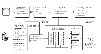 |
56 | Quantitative server-based analysis of MR elastography inversion methods on a phantom and the in vivo human kidney Video Permission Withheld
Tom Meyer1, Stephan Rodrigo Marticorena Garcia1, Heiko Tzschätzsch1, Helge Herthum2, Mehrgan Shahryari1, Jürgen Braun2, Prateek Kalra3,4, Arunark Kolipaka3,4, and Ingolf Sack1
1Department of Radiology, Charité - Universitätsmedizin Berlin, Berlin, Germany, 2Institute of Medical Informatics, Charité - Universitätsmedizin Berlin, Berlin, Germany, 3Department of Biomedical Engineering, The Ohio State University, Columbus, OH, United States, 4Department of Radiology, The Ohio State University, Columbus, OH, United States
Magnetic resonance elastography (MRE) depicts the viscoelastic properties of soft tissues for diagnosis of various diseases such as tumors or fibrosis. However, different MRE inversion methods yield different results. This lack of comparability of different MRE inversion results hinders the dissemination of advanced reconstruction methods. Therefore, we here introduce an extensible, open-access web platform that offers multiple inversion techniques for multifrequency, three-dimensional MRE to promote comparison of values. We demonstrate the utility of the platform in phantom data and in vivo free-breathing multifrequency MRE data of the kidneys of healthy volunteers.
|
||
2244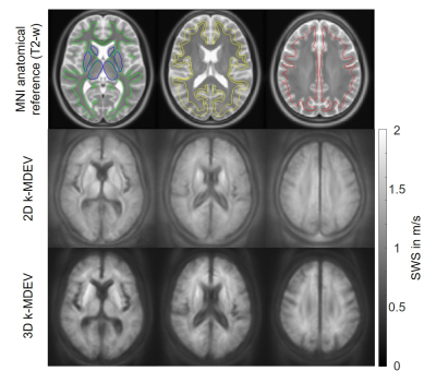 |
57 | 2D or 3D MR elastography of the brain? Answers from a 1-year follow-up study in healthy volunteers.
Helge Herthum1, Heiko Tzschätzsch1, Mehrgan Shahryari1, Bernhard Kreft1, Steffen Görner1, Stefan Hetzer2, Jürgen Braun1, and Ingolf Sack1
1Charité Universitätsmedizin Berlin, Berlin, Germany, 2Berlin Center for Advanced Neuroimaging, Berlin, Germany
MR elastography(MRE) measures in vivo soft-tissue mechanical parameters which are sensitive to various diseases. Robust and reliable measurements are critical for clinical translation; however, the variability of reported values, especially for MRE in the brain, remains a challenge. We here introduce an optimized setup for high-resolution brain MRE and compare the consistency of values obtained from 2D- and 3D-analysis in a group of healthy subjects studied in the follow-up of a year. Both 2D- and 3D-MRE values agreed very well between the two measurements. Intersubject variability was lower in 2D than in 3D while intrasubject variability was lower in 3D.
|
||
2245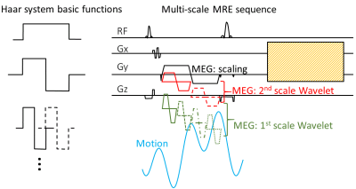 |
58 | Initial Study on Imaging Cyclic Tissue Motion with Motion Encoding Gradients in the Shape of Wavelet Basic Functions
Yuan Le1, Jun Chen1, Phillip J. Rossman1, Bradley D. Bolster Jr.2, Stephan Kannengiesser3, and Richard L. Ehman1
1Radiology, Mayo Clinic, Rochester, MN, United States, 2MR Collaborations, Siemens Medical Solutions USA, Inc., Salt Lake City, UT, United States, 3MR Application Predevelopment, Siemens Healthcare GmbH, Erlangen, Germany The goal of this study was to better detect cyclic motion with multiple frequencies in tissue. A novel MRE technique was developed using motion encoding gradient (MEG) pulses of multiple scales (multi-scale MRE). These MEG pulses were designed and timed to approximate a complete basis of Haar wavelet system. Our hypothesis was that a set of orthogonal MEG pulses can capture the tissue motion more efficiently and tissue motion can be reconstructed by a simple inverse transform. Initial tests were performed on a phantom and a volunteer. |
||
2246 |
59 | Impact of bowel preparation methods on pancreatic Magnetic Resonance Elastography
Anne-Sophie van Schelt1, Nienke P.M. Wassenaar1, Eric M. Schrauben1, Jules L. Nelissen1, Jing Guo2, Ingolf Sack2, Jaap Stoker1, Aart J. Nederveen1, and Jurgen H. Runge1
1Radiology and Nuclear Medicine, Amsterdam UMC location AMC, Amsterdam, Netherlands, 2Department of Radiology, Charité – Universitätsmedizin Berlin, Berlin, Germany
Pancreas magnetic resonance elastography (MRE) allows non-invasice estimation of tissue stiffness for multiple pathophysiological diseases. MRE reproducibility and the effect of bowel-preparation (butylscopolaminebromide and drinking water) for increased MRE data quality were assessed for the pancreas. Shear wave speed and MRE quality were determined for the pancreas, liver and kidneys. Intrasession and intersession reproducibility showed a coefficient-of-variation of 6.59% and 13.10%. Repeated measures ANOVA showed no significant difference in SWS of all organs (f=3/11,p>0.05). Bowel-preparation methods for pancreatic MRE do not increase MRE quality. Drinking water increases the MRE quality for kidneys and liver whilst not altering the measured SWS.
|
||
2247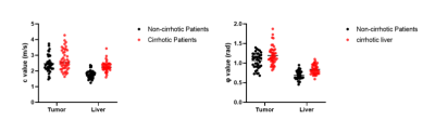 |
60 | Tumor-liver biomechanical interaction investigated by multifrequency MR elastography in patients with HCC
Jiahao Zhou1, Ruokun Li2, Yikun Wang2, Jing Guo3, Ingolf Sack3, and Fuhua Yan2
1radiology, Ruijin Hospital, Shanghai Jiaotong University School of Medicine, Shanghai, China, 2Ruijin Hospital, Shanghai Jiaotong University School of Medicine, Shanghai, China, 3Department of Radiology, Charité–Universitätsmedizin Berlin, Berlin, Germany, Berlin, Germany
Tumor-liver biomechanical interaction investigated by multifrequency MR elastography in patients with HCC
|
||
2248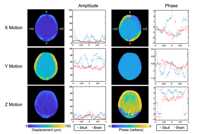 |
61 | Quantifying Relative Displacement and Phase at the Skull-Brain Interface using MR Elastography
Alexa M. Diano1, Grace McIlvain1, Andrew K. Knutsen2, Suhas Vidhate3, Dzung L. Pham2, and Curtis L. Johnson1
1Biomedical Engineering, University of Delaware, Newark, DE, United States, 2Henry M. Jackson Foundation, Bethesda, MD, United States, 3National Institutes of Health, Bethesda, MD, United States This study uses a novel multishot spiral magnetic resonance elastography (MRE) pulse sequence to simultaneously measure skull and brain motion in vivo in a single acquisition. Results in a healthy volunteer and a fat-water phantom showed a reduced transmission of motion from the skull to the brain in all three directions of motion. Additionally, the skull and brain appear to be out of phase in motion in both the x (left-right) and z (superior-inferior) directions. This research provides a basis for future studies quantifying skull-brain coupling including with multiple actuation frequencies and excitation directions. |
||
2249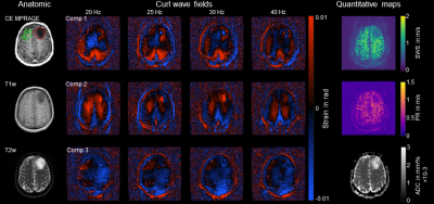 |
62 | Characterization of tumor mechanical properties in glioma patients using 3D phase-gradient inversion in multifrequency MR elastography Video Permission Withheld
Mehrgan Shahryari1, Pablo Gottheil2, Elisabeth Gertrud Hain3, Helge Herthum4, Heiko Tzschätzsch1, Tom Meyer1, Josef Alfons Käs2, Eberhard Siebert5, Vincent Prinz6,7, and Ingolf Sack1
1Department of Radiology, Charité - Universitätsmedizin Berlin, corporate member of Freie Universität Berlin, Humboldt-Universität zu Berlin, and Berlin Institute of Health, Berlin, Germany, 2Faculty of Physics and Earth Sciences, Peter Debye Institute, Leipzig University, Leipzig, Germany, 3Department of Neuropathology, Charité - Universitätsmedizin Berlin, corporate member of Freie Universität Berlin, Humboldt-Universität zu Berlin, and Berlin Institute of Health, Berlin, Germany, 4Institute of Medical Informatics, Charité - Universitätsmedizin Berlin, corporate member of Freie Universität Berlin, Humboldt-Universität zu Berlin, and Berlin Institute of Health, Berlin, Germany, 5Institute of Neuroradiology, Charité - Universitätsmedizin Berlin, corporate member of Freie Universität Berlin, Humboldt-Universität zu Berlin, and Berlin Institute of Health, Berlin, Germany, 6Department of Neurosurgery, Charité - Universitätsmedizin Berlin, corporate member of Freie Universität Berlin, Humboldt-Universität zu Berlin, and Berlin Institute of Health, Berlin, Germany, 7Department of Neurosurgery, University Hospital Frankfurt, Frankfurt am Main, Germany
Clinical MRI is an important method for the delineation and characterization of gliomas for resection planning and therapy monitoring in neurosurgery. Here, we used a 3D curl- and phase-gradient based inversion algorithm in multifrequency magnetic resonance elastography (MRE) to generate maps of the mechanical tumor properties, wave speed and wave damping. Overall, our method improved the spatial resolution of MRE maps in glioma patients and allowed precise measurement of tumor mechanical properties. In the future, the method could help with characterization, surgical planning and treatment monitoring of neuro tumors.
|
||
2250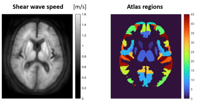 |
63 | Brain stiffness correlates with hematocrit
Jakob Seja1, Bernhard Kreft2, Helge Herthum2, Mehrgan Shahryari3, Jürgen Braun2, Ingolf Sack3, and Stefan Hetzer1
1Berlin Center for Advanced Neuroimaging, Charité - Universitätsmedizin Berlin, Berlin, Germany, 2Institute of Medical Informatics, Charité - Universitätsmedizin Berlin, Berlin, Germany, 3Radiology department, Charité - Universitätsmedizin Berlin, Berlin, Germany
The viscosity of blood scales with the concentration of red blood cells (hematocrit) and potentially influences the viscoelasticity of the perfused tissue. In this study, cerebral MR elastography (MRE) was applied to a group of healthy subjects to analyze the interrelation between mechanical properties of brain tissue and the hematocrit. We found a highly significant positive correlation between in-vivo mechanical properties of cerebral tissue and hematocrit level (p<0.0001).
|
||
2251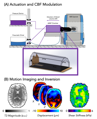 |
64 | Acute Lower Body Negative Pressure Changes Human Brain Stiffness in vivo Measured with MR Elastography
Mary K Kramer1, Grace McIlvain1, Faria Sanjana2, Fiona Horvat2, Christopher R Martens2, and Curtis L Johnson1
1Biomedical Engineering, University of Delaware, Newark, DE, United States, 2Kinesiology and Applied Physiology, University of Delaware, Newark, DE, United States
Lower body negative pressure (LBNP) is a non-invasive physiological method known to decrease cerebral blood flow. This technique was used to study the brain’s response to altered cerebrovascular hemodynamics and showed that, on average, brain stiffness decreases by 2.28% in response to ten minutes of LBNP application and decreased cerebral blood flow. By decreasing and increasing cerebral blood flow by turning the LBNP on and off repeatedly in 10-minute blocks, brain stiffness was shown to increase by 3.02% on average from the initial application of LBNP to the final application of LBNP.
|
||
2252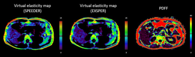 |
65 | Feasibility evaluation of virtual elastography based on diffusion-weighted imaging (vMRE) at 1.5 Tesla
Alicia Palomar Garcia1, Valentin H. Prevost2, Alba Iruela Sanchez1, Wolter de Graaf3, and Bruno Triaire2
1Canon Medical Systems Spain and Portugal, Barcelona, Spain, 2Canon Medical Systems Corporation, Tochigi, Japan, 3Canon Medical Systems Europe, Zoetermeer, Netherlands
This study evaluated the feasibility of performing virtual elastography based on diffusion-weighted MRI (vMRE) on a 1.5T system with two different acceleration methods, SPEEDER and EXSPER. Proton density fat fraction (PDFF) imaging was also acquired. The vMRE and PDFF measurements were consistent with healthy values that can be found in the literature. This study demonstrated the feasibility of performing vMRE at 1.5T for the estimation of overall liver elasticity. SPEEDER and EXSPER vMRE implementations provided consistent stiffness estimations and both would be adequate for conducting future studies at 1.5T systems on patients.
|
||
 Back to Meeting Home
 Back to the Program-at-a-Glance
The International Society for Magnetic Resonance in Medicine is accredited by the Accreditation Council for Continuing Medical Education to provide continuing medical education for physicians.
 Back to Meeting Home
 Back to the Program-at-a-Glance
 View the Presentation