Digital Poster
CEST
Joint Annual Meeting ISMRM-ESMRMB & ISMRT 31st Annual Meeting • 07-12 May 2022 • London, UK

| Computer # | ||||
|---|---|---|---|---|
2073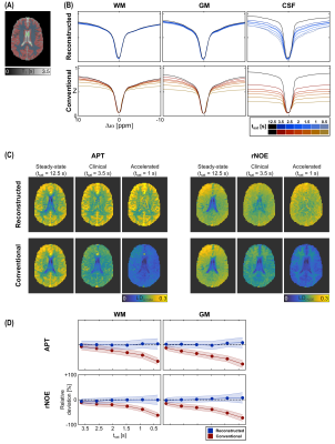 |
63 | Steady-state CEST-MRI using a reduced saturation period: Application to volumetric APT and rNOE brain imaging at 3T
Johannes Breitling1, Andreas Korzowski1, Mark E. Ladd1,2,3, Peter Bachert1,2, and Steffen Goerke1
1Division of Medical Physics in Radiology, German Cancer Research Center (DKFZ), Heidelberg, Germany, 2Faculty of Physics and Astronomy, University of Heidelberg, Heidelberg, Germany, 3Faculty of Medicine, University of Heidelberg, Heidelberg, Germany
In this study, we demonstrate the feasibility of obtaining steady-state 3D CEST contrasts of the human brain at 3T using a reduced saturation period. To this end, we applied our previous approach, which utilizes the analytical equation of the CEST signal, to an optimized clinical protocol using a fast volumetric snapshot-CEST acquisition. Measurements on model solutions and a volunteer demonstrated the feasibility with almost identical APT and rNOE contrasts for saturation durations as short as 1 second. Consequently, this facilitates quantitative CEST-MRI in humans within a reasonable and clinically relevant time frame.
|
||
2074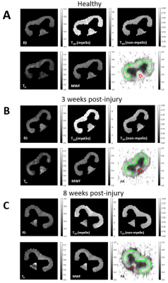 |
64 | Modelling inhomogeneous magnetization transfer in myelin and intra-/extra-cellular water in normal and injured rat spinal cords
Michelle H Lam1,2, Andrew Yung2, Jie Liu3, Wolfram Tetzlaff3, and Piotr Kozlowski1,2,3,4
1Physics and Astronomy, University of British Columbia, Vancouver, BC, Canada, 2UBC MRI Research Centre, University of British Columbia, Vancouver, BC, Canada, 3International Collaboration on Repair Discoveries, Vancouver, BC, Canada, 4Radiology, University of British Columbia, Vancouver, BC, Canada
An MR imaging technique called inhomogeneous magnetization transfer (ihMT) could potentially be used for quantitative myelin imaging by using T1D filtering to filter out the short dipolar relaxation time (T1D) components. However, to show that ihMT with T1D filtering is myelin-specific, we need to confirm that myelin has the longest T1D in nerve tissue. Here, we combined ihMT and myelin water imaging (MWI) to separate the ihMT signal in myelin water from intra-/extra-cellular water, which we fitted using a four-pool model with dipolar order reservoirs. Our model allowed us to connect myelin with myelin water and measure myelin’s T1D.
|
||
2075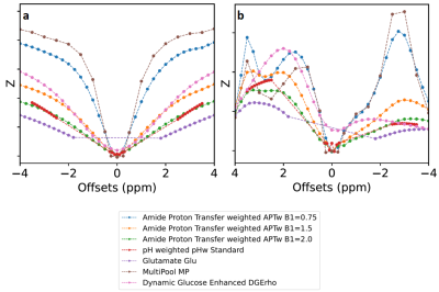 |
65 | comprehenCEST: a clinically feasible CEST protocol to cover all existing CEST preparations
Lukas Kamm1, Kai Herz2, Maria Sedykh1, Moritz Fabian1, and Moritz Zaiss1,2
1Neuroradiology, Friedrich-Alexander-University Erlangen-Nürnberg, University Hospital, Erlangen, Germany, 2Magnetic Resonance Center, Max-Planck-Institute for Biological Cybernetics, Tübingen, Germany
In a clinical setting, it often has to be decided, which CEST sequences to pick of a collection, due to measurement time limitations. Therefore, eventually not all of the desired contrasts can be measured. In this work, we comprehensively combine seven CEST protocols in a single measurement, use a fast single-shot 3D gradient echo readout and remove redundant regions of the experimental parameter space. Hereby, we can half the total measurement time from 35 to 17 minutes. With further parameter reduction, a total measurement time of 10 minutes is conceivable. This allows to design powerful hypothesis generating clinical pilot studies.
|
||
2076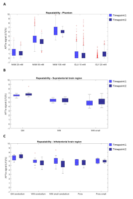 |
66 | Repeatability of APTw imaging at 7T
Iris Obdeijn1,2, Lejla Alic2, Maarten Lequin1,3, Sabine Plasschaert3, Wybe van der Kemp1, Hans Hoogduin1, Dennis Klomp1, Jannie Wijnen1, and Evita Wiegers1
1Department of Radiology, University of Medical Centre Utrecht, Utrecht, Netherlands, 2Magnetic Detection and Imaging Group, Technical Medical Centre, University of Twente, Enschede, Netherlands, 3Department of paediatric neuro-oncology, Princess Maxima Centre, Utrecht, Netherlands
APTw imaging is a potential imaging biomarker to assess treatment effects in brain tumours, especially at high field MRI (7T) due to improved signal-to-noise-ratio enabling the assessment of APTw values in heterogenous tumours. Embedding of APTw imaging in clinical decision making requires insight in the repeatability of APTw imaging. Therefore, we evaluated the repeatability of APTw imaging at 7T by using a phantom and in vivo in the human brain subjects. Repeatable and specific APTw maps were obtained at 7T, which facilitate the potential of detecting metabolic changes in brain tumours due to treatment.
|
||
2077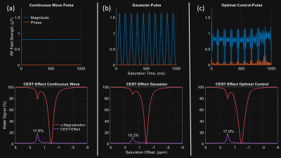 |
67 | Optimization of Pulsed Chemical Exchange Saturation Transfer MRI by Optimal Control
Clemens Stilianu1, Christina Graf1, Clemens Diwoky2, Martin Soellradl1, Armin Rund3, Moritz Zaiss4, and Rudolf Stollberger1
1Medical Engineering, TU Graz, Graz, Austria, 2647 Institut für Molekulare Biowissenschaften, University of Graz, Graz, Austria, 3Institute for Mathematics and Scientific Computing, University of Graz, Graz, Austria, 4Department High-field Magnetic Resonance, Max Planck Institute Tübingen, Tübingen, Germany
We optimized CEST saturation pulse train using an optimal control framework and validated the simulation results on a preclinical scanner that allowed also Gold Standard continuous wave saturation. The optimized pulses almost reached the same saturation as the continuous wave pulse while addressing the allowed duty cycle and SAR limitations of clinical scanners. In measurements, the optimized pulses outperform state-of-the-art Gaussian pulses by over $$$47\%$$$.
|
||
2078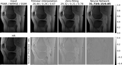 |
68 | Super-Resolution for CEST MRI
Lukas Folle1, Katharina Tkotz2, Andrzej Liebert2, Fasil Gadjimuradov1, Lorenz Kapsner2, Moritz Fabian3, Sebastian Bickelhaupt2, David Simon4, Arnd Kleyer4, Gerhard Krönke4, Frank Roemer2, Moritz Zaiss3,5, Armin Nagel2,6, and Andreas Maier1
1Computer Science, Friedrich-Alexander Universität Erlangen-Nürnberg, Erlangen, Germany, 2Institute of Radiology, University Hospital Erlangen, Friedrich-Alexander-Universität Erlangen-Nürnberg (FAU), Erlangen, Germany, 3Institute of Neuroradiology, University Hospital Erlangen, Friedrich-Alexander-Universität Erlangen-Nürnberg (FAU), Erlangen, Germany, 4Department of Internal Medicine 3, University Hospital Erlangen, Friedrich-Alexander-Universität Erlangen-Nürnberg (FAU), Erlangen, Germany, 5Magnetic Resonance Center, Max-Planck-Institute for Biological Cybernetics, Tübingen, Germany, 6Division of Medical Physics in Radiology, German Cancer Research Center (DKFZ), Heidelberg, Germany
The resolution of chemical exchange saturation transfer (CEST) magnetic resonance imaging is limited by physical constraints. To visualize metabolic processes of small structures using CEST in patients knees, an increased resolution is necessary. In this work, we compared trilinear interpolation and zero-filling to neural network-based approaches to estimate a high-resolution image given the corresponding low-resolution data. We could show that a substantial quantitative improvement using neural networks could be achieved for unsaturated images while maintaining a comparable CEST contrast. Generalization of the method to brain CEST MRI was achieved without retraining of the network.
|
||
2079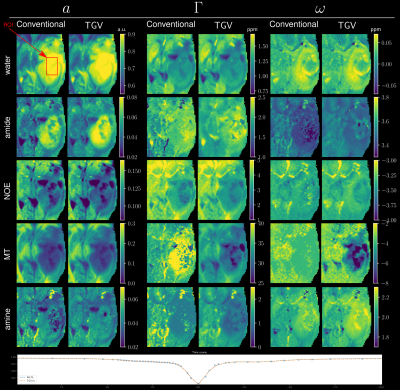 |
69 | Robust Lorentzian fitting of CEST spectra utilizing joint spatial regularization
Oliver Maier1, Moritz Simon Fabian2,3, Angelika Barbara Mennecke2, Manuel Schmidt2, Arnd Dörfler2, Kristian Bredies4, Moritz Zaiss2,3, and Rudolf Stollberger1,5
1Institute of Medical Engineering, Graz University of Technology, Graz, Austria, 2Department of Neuro-radiology, University Clinic of Erlangen, Erlangen, Germany, 3Friedrich-Alexander-University, Erlangen, Germany, 4Institute of Mathematics and Scientific Computing, University of Graz, Graz, Austria, 5BioTechMed Graz, Graz, Austria
Chemical Exchange Saturation Transfer (CEST) enables characterization of tissue in vivo by measuring a z-spectrum. Quantitative separation of individual contributions to this spectrum is performed by fitting several Lorentzian shaped lines to the acquired data. Current implementations of the fitting procedure suffer from poor SNR and ambiguity of peaks lying close to each other. To this end, we propose to include a joint spatial regularization to the fitting process to counter the poor SNR and thus improve separation of individual peaks in the final reconstruction.
|
||
2080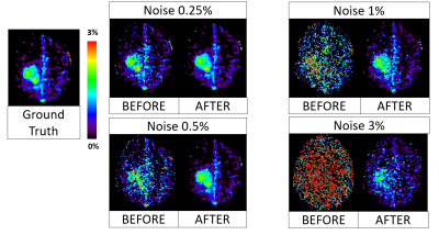 |
70 | Principal Component selections and filtering by spatial information criteria for multi-acquisition CEST MRI denoising
Stefano Casagranda1, Christos Papageorgakis1, Feriel Romdhane2, Eleni Firippi1, Timothé Boutelier1, Laura Mancini3,4, Moritz Zaiss5, Sotirios Bisdas3,4, and Dario Livio Longo2
1Department of Research & Innovation, Olea Medical, La Ciotat, France, 2Institute of Biostructures and Bioimaging (IBB), National Research Council of Italy (CNR), Torino, Italy, 3Lysholm Department of Neuroradiology,, University College of London Hospitals NHS Foundation Trust, London, United Kingdom, 4Institute of Neurology UCL, London, United Kingdom, 5Department of Neuroradiology, University Clinic Erlangen, Friedrich-Alexander Universität Erlangen-Nürnberg (FAU), Erlangen, Germany
This work provides a new denoising methodology for multi-acquisition magnetic resonance images (MRI) based on principal components analysis (PCA). We are proposing a new principal component selection criterion that identifies spatial information in the extracted component coefficients, leading to a better preservation of anatomical structures and pathological information. In addition, our adaptive filtering step allows us to further denoise the MRI data, rejecting persistent spatial noise from the extracted component coefficients. In our investigations the proposed method outperformed the eigenvalue based selection criteria on Amide Proton Transfer weighted CEST data.
|
||
2081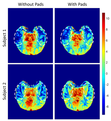 |
71 | Repeatability of B1 Inhomogeneity Correction of Volumetric (3D) Glutamate CEST via High-Permittivity Dielectric Padding at 7T
Paul Samuel Jacobs1, Ravi Prakash Reddy Nanga1, Abigail Cember1, Dylan M Tisdall2, Sandhitsu Das3, John Detre1,3, David Roalf4, and Ravinder Reddy1
1CAMIPM, Radiology, University of Pennsylvania, Philadelphia, PA, United States, 2Radiology, University of Pennsylvania, Philadelphia, PA, United States, 3Department of Neurology, University of Pennsylvania, Philadelphia, PA, United States, 4Department of Psychiatry, University of Pennsylvania, Philadelphia, PA, United States
Dielectric pads were used to improve the contrast of volumetric (3D) gluCEST via enhanced homogenization of B1 fields. Effective placement of the pads required additional coverage of the face due to their physical size to achieve the saturated B1 strength necessary to recover the gluCEST signal from the anterior portions of the brain. The origin of gluCEST's need for high B1 is discussed, and examples of 3D gluCEST images acquired with and without the use of the dielectric pads are provided and repeatability of the 3D gluCEST in the presence of pad placement was determined.
|
||
2082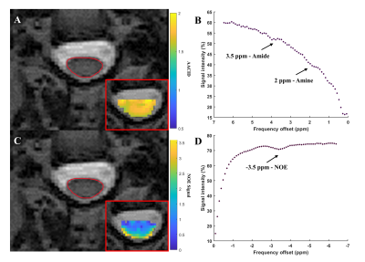 |
72 | Optimization of pH-Weighted Contrasts in the Spinal Cord using Chemical Exchange Saturation Transfer (CEST) MRI
Alicia Cronin1,2, Patrick Liebig3, Sarah Detombe4, Neil Duggal1,4, and Robert Bartha1,2
1Medical Biophysics, Western University, London, ON, Canada, 2Centre for Functional and Metabolic Mapping, Robarts Research Institute, London, ON, Canada, 3Siemens Healthcare GmbH, Erlangen, Germany, 4Clinical Neurological Sciences, University Hospital, London Health Sciences Centre, London, ON, Canada
Degenerative cervical myelopathy (DCM) is a degenerative disease of the spine that leads to compression and neurological dysfunction. Recovery after surgery can be impacted by hypoxia in the cord, however the magnitude of this effect is currently unknown. Chemical Exchange Saturation Transfer (CEST) can produce contrast related to tissue pH, an indicator of hypoxia, but the method works best at ultra-high fields. Performing CEST in the spinal cord is also complicated by respiratory and cardiac motion and cerebrospinal fluid flow. The purpose of this work was to optimize pH-weighted CEST imaging in the human spinal cord at 3 Tesla.
|
||
2083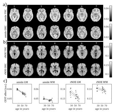 |
73 | CEST effects change with age
Angelika Barbara Mennecke1, Katrin Khakzar1, Alexander German1, Kai Herz2, Moritz Fabian1, Andrzej Liebert3, Armin M. Nagel3, Frederik B. Laun3, Manuel Schmidt1, Jürgen Winkler4, Arnd Dörfler1, and Moritz Zaiss1
1Neuroradiology, University Hospital Erlangen, Friedrich-Alexander-Universität Erlangen-Nürnberg (FAU), Erlangen, Germany, 2Max Planck Institute for Biological Cybernetics, Tübingen, Germany, 3Radiology, University Hospital Erlangen, Friedrich-Alexander-Universität Erlangen-Nürnberg (FAU), Erlangen, Germany, 4Neurology, University Hospital Erlangen, Friedrich-Alexander-Universität Erlangen-Nürnberg (FAU), Erlangen, Germany
The amide and rNOE CEST amplitudes are negatively correlated with age. Most prominently, the mean gray matter amide CEST amplitude decreases on average by 0.22 % per year of age in the cohort of our study.
|
||
2084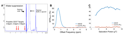 |
74 | Quantitative imaging of gene therapy delivery vehicles using CEST-NMR/MRI
Bonnie Lam1, Mark Velasquez1, Aaron J. Velasquez-Mao1, Kevin Godines1, Wissam AlGhuraibawi1, and Moriel Vandsburger1
1Bioengineering, University of California, Berkeley, Berkeley, CA, United States
We hypothesized that AAV2 capsids may generate endogenous CEST contrast similar to LRP. We tested this using NMR CEST under varying pH, density, biological transduction stage, across serotypes and mixed biological media. Subsequent experiments determined the pH-dependent exchange rate and optimized CEST saturation schemes for AAV contrast detection at 7T. The results of this study reveal that AAV capsids generate endogenous CEST contrast around 0.6-0.8ppm. Exchangeable protons are likely from serine and threonine residues on the AAV capsid. Based on calculated fast exchange rates, an optimized CEST saturation scheme generated robust CEST contrast in mixed biological media phantoms at 7T.
|
||
2085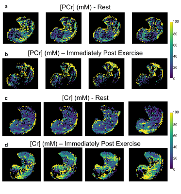 |
75 | Quantitative 3D Mapping of Cr and PCr Concentrations at 3T using Snapshot AREX CEST MRI
Or Perlman1, Jaume Coll-Font1,2, Kai Herz3,4, Moritz Zaiss3,5, Christopher Nguyen1,2,6, and Christian T. Farrar1
1Athinoula A. Martinos Center for Biomedical Imaging, Department of Radiology, Massachusetts General Hospital and Harvard Medical School, Charlestown, MA, United States, 2Cardiovascular Research Center, Cardiology Division, Massachusetts General Hospital, Charlestown, MA, United States, 3Magnetic Resonance Center, Max Planck Institute for Biological Cybernetics, Tübingen, Germany, 4Department of Biomedical Magnetic Resonance, University of Tübingen, Tübingen, Germany, 5Department of Neuroradiology, Friedrich-Alexander Universität Erlangen-Nürnberg (FAU), University Hospital Erlangen, Erlangen, Germany, 6Health Science Technology, Harvard-MIT, Cambridge, MA, United States
The dynamics of creatine and phosphocreatine distributions provide an important means for assessing metabolic function. While CEST-weighted MRI allows for the imaging of metabolite alterations, it is affected by semisolid-MT, spillover, and T1 contributions, and is typically analyzed on a two-dimensional image, given the inherently long acquisition times. Here, we performed 3D quantitative mapping of Cr and PCr concentrations in the human calf muscle at 3T, using a rapid snapshot-CEST protocol followed by apparent exchange-dependent relaxation (AREX) analysis. Significant (p<0.001) changes in the concentrations of both creatine and phosphocreatine were measured during exercise, in agreement with the literature.
|
||
2086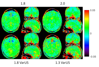 |
76 | Whole Brain 1.3 mm isotropic APTw CEST at 3T in under 2 minutes Video Permission Withheld
Patrick Liebig1, Maria Sedykh2, Kai Herz3,4, Yi-Cheng Hsu5, Moritz Fabian2, Angelika Mennecke2, Manuel Schmidt2, Arnd Doerfler2, and Moritz Zaiss2,3
1Siemens Healthcare GmbH, Erlangen, Germany, 2Department of Neuroradiology, University Hospital Erlangen, Friedrich-Alexander-Universität Erlangen-Nürnberg (FAU), Erlangen, Germany, 3Magnetic Resonance Center, Max-Planck-Institute for Biological Cybernetics, Tuebingen, Germany, 4Department of Biomedical Magnetic Resonance, University of Tübingen, Tuebingen, Germany, 5MR Collaboration, Siemens Healthineers Ltd., Shanghai, China CEST MRI suffers from long acquisition times and/or limited volume coverage. Here we suggest using a spatio-temporal variable density Poisson disk design for whole brain APTw snapshot CEST. This enabled us to acquire 1.3 mm isotropic whole brain APTw CEST maps in under 2 minutes. All the reconstruction and necessary adjustment steps were implemented within the scanner software, enabling a push-button B0-corrected CEST measurement. |
||
2087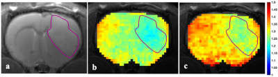 |
77 | Intracellular Acidification Monitored by Chemical Exchange Saturation Transfer MRI: Effect of Cariporide in Rat C6 Brain Tumor
Maryam Mozaffari1, Nivin Nystrom2, Alex Li2, Miranda Bellyou2, Timothy Scholl1, and Robert Bartha1
1Medical Biophysics, Western Univeristy-Robarts Reserch Institute, London, ON, Canada, 2Western Univeristy-Robarts Reserch Institute, London, ON, Canada
Our objective was to acidify rat C6 gliomas by inhibiting NHE1 with cariporide and to monitor the pH changes with AACID-CEST MRI. AACID-CEST MRI was successfully used to monitor changes in tumor pHi over time after cariporide injection. Our results showed a pH decrease in both the tumor and the contralateral tissue following cariporide injection. CEST-MRI measurement of tumor response pH could help to enhance the efficacy of this treatment paradigm in different human malignancies.
|
||
2088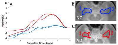 |
78 | Development of CEST MRI for the characterization of Alzheimer’s Disease
Fang Frank Yu1, Natalie M Bell1, Elizabeth M Davenport1,2, Spencer Bowen1,2, Brendan James Kelley3, Ivan Dimitrov2,4, Jochen Keupp5, and Elena Vinogradov1,2
1Radiology, University of Texas Southwestern Medical Center, Dallas, TX, United States, 2Advanced Imaging Research Center, University of Texas Southwestern Medical Center, Dallas, TX, United States, 3Neurology, University of Texas Southwestern Medical Center, Dallas, TX, United States, 4Philips Healthcare, Gainseville, GA, United States, 5Philips Research, Hamburg, Germany
Alzheimer’s Disease (AD) is the leading cause of dementia, afflicting over 5 million Americans and 45 million people worldwide. We are developing CEST protocol for the characterization of abnormal protein accumulation and pathological molecular changes in AD. Our results indicate increased CEST in all frequency ranges, with the maximum around 2ppm. While preliminary, this data demonstrates CEST MRI is capable of clear differentiation between patients with AD and healthy individuals.
|
||
 Back to Meeting Home
 Back to the Program-at-a-Glance
The International Society for Magnetic Resonance in Medicine is accredited by the Accreditation Council for Continuing Medical Education to provide continuing medical education for physicians.
 Back to Meeting Home
 Back to the Program-at-a-Glance
 View the Presentation