Digital Poster
23Na
Joint Annual Meeting ISMRM-ESMRMB & ISMRT 31st Annual Meeting • 07-12 May 2022 • London, UK

| Computer # | ||||
|---|---|---|---|---|
1274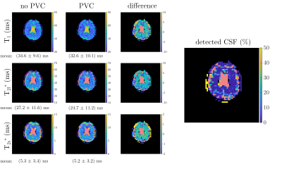 |
70 | Correction of CSF Partial Volume Bias in 23Na Magnetic Resonance Fingerprinting
Fabian J Kratzer1, Sebastian Flassbeck1,2,3, Sebastian Schmitter1,4, Tobias Wilferth5, Peter Bachert1, Mark E Ladd1, and Armin M Nagel1,5
1Medical Physics in Radiology, German Cancer Research Center (DKFZ), Heidelberg, Germany, 2Center for Biomedical Imaging, New York University, New York, NY, United States, 3Center for Advanced Imaging Innovation and Research, New York University, New York, NY, United States, 4Physikalisch Technische Bundesanstalt (PTB), Braunschweig and Berlin, Germany, 5Institute of Radiology, University Hospital Erlangen, Friedrich-Alexander-Universität Erlangen Nürnberg (FAU), Erlangen, Germany
This work investigates the bias in the quantified relaxation times due to partial volume contributions of CSF for 23Na magnetic resonance fingerprinting (MRF) and reference methods. In simulations, a CSF contribution of only 10% resulted in an overestimation of 17% (31%) in T2l* in brain tissue for MRF (the reference). Further, a simple approach for CSF bias correction for 23Na MRF is proposed. This reduced the average absolute T1 deviation in brain tissue from 15% to 4%. The deviation in T2l* (T2s*) was reduced from 17% (35%) to 9% (5%). Finally, the correction was applied to in vivo MRF data.
|
||
1275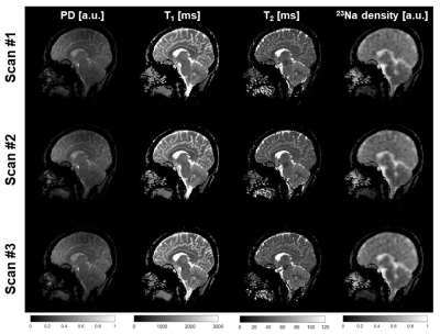 |
71 | Repeatability of simultaneous 3D 1H MRF/23Na MRI in brain at 7T
Gonzalo G Rodriguez1, Zidan Yu1,2, Lauren O'Donnell1, Liz Calderon1, Martijn A Cloos3,4, and Guillaume Madelin1,2
1Center for Biomedical Imaging, Department of Radiology, New York University School of Medicine, New York, NY, United States, 2Vilcek Institute of Graduate Biomedical Sciences, NYU Langone Health, New York, NY, United States, 3Centre for Advanced Imaging, The University of Queensland, Brisbane, Australia, 4ARC Training Centre for Innovation in Biomedical Imaging Technology, The University of Queensland, Brisbane, Australia
In this work, we assess the repeatability of proton density, T1, T2 and sodium density maps measured with simultaneous 3D 1H MRF/23Na MRI in the brain at 7T. We scanned seven healthy subjects three times each. The coefficients of variation (CV) were in the range 1-3% for mean values in GM, WM and CSF, and the intra-class correlation (ICC) was in range 0.44-0.99.
|
||
1276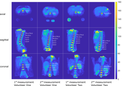 |
72 | Quantitative Sodium MRI of the Intervertebral Discs at 7T
Anna K. Scheipers1,2, Daniel Peach3, Armin M. Nagel1,4, Mark E. Ladd1,2,5, and Tanja Platt1
1Medical Physics in Radiology, German Cancer Research Center (DKFZ), Heidelberg, Germany, 2Faculty of Physics and Astronomy, Ruprecht Karl University of Heidelberg, Heidelberg, Germany, 3Radiology, German Cancer Research Center (DKFZ), Heidelberg, Germany, 4University Hospital Erlangen, Institute of Radiology, Friedrich-Alexander-Universität Erlangen-Nürnberg (FAU), Erlangen, Germany, 5Faculty of Medicine, Ruprecht Karl University of Heidelberg, Heidelberg, Germany 23Na-MRI offers the possibility for non-invasive quantification of the sodium concentration in-vivo. The obtained functional information can provide interesting insights into the degeneration state of the human intervertebral discs1 (IVDs). The presented work thus aimed to quantify the tissue sodium concentration (TSC) in the human intervertebral discs via 23Na-MRI at 7T and to compare the obtained concentrations for two healthy volunteers after the course of one year. The estimate average concentration for all IVDs of both volunteers was found to be (96.5±8.8)mM and (113.6±9.5)mM, respectively. |
||
1277 |
73 | 2D 23Na imaging using half-VERSE pulses
Marco L. Wittrich1,2, Armin M. Nagel1,3, Sebastian Schmitter1,4, Peter Bachert1,2, Mark E. Ladd1,2,5, and Fabian J Kratzer1,2
1Medical Physics in Radiology, German Cancer Research Center (DKFZ), Heidelberg, Germany, 2Faculty of Physics and Astronomy, University of Heidelberg, Heidelberg, Germany, 3Institute of Radiology, University Hospital Erlangen, Friedrich‐Alexander‐Universität Erlangen‐Nürnberg (FAU), Erlangen-Nürnberg, Germany, 4Physikalisch-Technische Bundesanstalt (PTB), Braunschweig and Berlin, Germany, 5Faculty of Medecine, University of Heidelberg, Heidelberg, Germany In this work, a 2D radial 23Na sequence with half-VERSE pulses was developed to reduce SAR and achieve ultra-short TEs. Simulations and measurements of the slice profiles showed good agreement between full- and half-VERSE pulses with a maximal deviation in the FWHM of 7%. In measurements, the TEmin was reduced from 1.41ms to 0.1ms by the use of half- instead of full-VERSE pulses. This resulted in an SNR gain of up to 18% in phantom, 7% in brain and 26% in calf measurements. Last, rapid single-slice 23Na images (2.9x2.9x12mm3) were obtained in a clinically feasible acquisition time of 2:56min. |
||
1278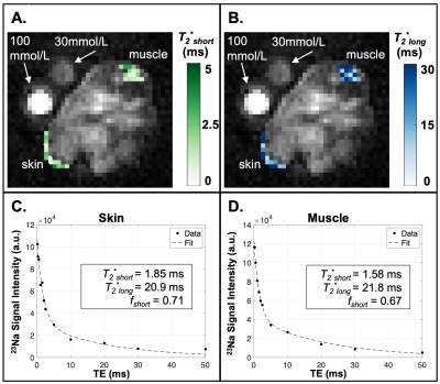 |
74 | Sodium 23Na-MRI relaxometry and tissue sodium content at 15.2T in a murine model of lymphangiogenesis
Shannon L Taylor1,2, Kevin D Harkins2,3, Daniel C Colvin2,3, Joseph M Rutkowski4, Mark D Does1,2,5, John C Gore1,2,3,6,7, and Rachelle L Crescenzi1,2,3
1Biomedical Engineering, Vanderbilt University, Nashville, TN, United States, 2Vanderbilt University Institute of Imaging Science, Vanderbilt University Medical Center, Nashville, TN, United States, 3Radiology and Radiological Sciences, Vanderbilt University Medical Center, Nashville, TN, United States, 4Medical Physiology, Texas A&M University College of Medicine, Bryan, TX, United States, 5Electrical Engineering and Computer Science, Vanderbilt University, Nashville, TN, United States, 6Molecular Physiology and Biophysics, Vanderbilt University, Nashville, TN, United States, 7Physics and Astronomy, Vanderbilt University, Nashville, TN, United States
To investigate questions regarding the physiology of salt storage in skin and muscle and its relationship with lymphangiogenesis, we are developing sodium 23Na-MRI protocols for a mouse model of controllable lymphangiogenesis in adipose tissue. We acquired images at 15.2T from a UTE center-out sequence and quantified sodium longitudinal and bi-exponential transverse relaxation times and tissue sodium content (TSC) in the skin and muscle. Baseline TSC was reduced in animals undergoing lymphangiogenesis compared to littermates, while 23Na-relaxometry was similar. These results will be used for protocol development in this animal model to study sodium and lymphatic physiology.
|
||
1279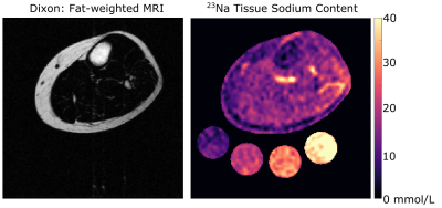 |
75 | Multi-nuclear 1H/23Na Sodium MRI reveals relationship between intermuscular adipose tissue and tissue sodium content
Michael Pridmore1,2, Jorge Gamboa3, Michelle Ormseth3,4, Annette Oeser3, C. Michael Stein3, and Rachelle Crescenzi1,2,5
1Department of Radiology and Radiological Sciences, Vanderbilt University Medical Center, Nashville, TN, United States, 2Institute of Imaging Science, Vanderbilt University Medical Center, Nashville, TN, United States, 3Department of Medicine, Vanderbilt University Medical Center, Nashville, TN, United States, 4U.S. Department of Veterans Affairs, Tennessee Valley Healthcare System, Nashville, TN, United States, 5Biomedical Engineering, Vanderbilt University Medical Center, Nashville, TN, United States
Adipose tissue depositions between muscle compartments, known as intermuscular adipose tissue (IMAT), occurs in various conditions such as with obesity and aging. Tissue sodium content (TSC) measured by sodium magnetic resonance imaging increases with aging and may be associated with insulin resistance. In this study, we found a correlation between IMAT volume percentage and muscle TSC (r=0.52, p=0.01). Additionally, mean TSC in the IMAT region was higher than mean TSC in total muscle (p<0.001). The relationship between tissue sodium content and IMAT opens avenues for understanding the mechanisms of sodium storage and fat depositions in tissue compartments of the leg.
|
||
1280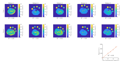 |
76 | Assessing the repeatability of tissue sodium concentration and characterizing the bi-exponential T2* decay signal in the calf
Ben Prestwich1 and Susan Francis1
1Sir Peter Mansfield Imaging Centre, University of Nottingham, Nottingham, United Kingdom The repeatability of sodium (23Na) MRI measures must be understood to allow for its application in clinical studies of patient groups or response to therapy. A series of 23Na scans were performed on the calf of healthy subjects, this included a 3D GRE 23Na scan and phase sensitive B1 mapping to measure tissue sodium concentration (TSC). Subjects were scanned twice for assessment of repeatability of measures. A multi echo UTE scan was used to measure the T2* decay. TSC was shown to have a coefficient of variation of 16±4 % between visits and the bi-exponential decay curve was characterized. |
||
1281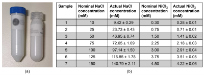 |
77 | Towards standardising quantification in 23Na-MRI by synthetic polyacrylamide gel phantoms
Samuel Rot1,2, Aaron Oliver-Taylor3, Xavier Golay3,4, Bhavana Solanky1, and Claudia AM Gandini Wheeler-Kingshott1,5,6
1NMR Research Unit, Queen Square MS Centre, Department of Neuroinflammation, UCL Queen Square Institute of Neurology, Faculty of Brain Sciences, University College London, London, United Kingdom, 2Department of Medical Physics and Biomedical Engineering, University College London, London, United Kingdom, 3Gold Standard Phantoms, London, United Kingdom, 4Department of Brain Repair and Rehabilitation, UCL Queen Square Institute of Neurology, Faculty of Brain Sciences, University College London, London, United Kingdom, 5Department of Brain & Behavioural Sciences, University of Pavia, Pavia, Italy, 6Brain Connectivity Centre Research Department, IRCCS Mondino Foundation, Pavia, Italy
Accuracy in quantitative 23Na (sodium) MRI is impacted by the quality of the signal calibration phantom. Agarose phantoms, the current benchmark, exhibit various unfavourable qualities. To move towards standardisation in 23Na-MRI, the objective was to develop a synthetic, polymer-based calibration phantom for traceable, reliable and accurate quantification in 23Na-MRI. Crosslinked polyacrylamide gel (PAG) was selected as the most suitable choice, for its bi-exponential 23Na T2 decay. Here, we present the T1, T2 properties and 23Na-MRI data of prototype PAG phantoms at different 23Na concentrations. PAG emerges as a reliable choice to replace the ubiquitous agarose gel phantom.
|
||
1282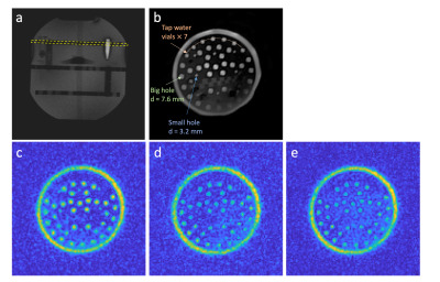 |
78 | 2D ultra-short TE imaging for 23Na MRI using half-pulse excitation
Chengchuan Wu1, Leigh A. Johnston1, and Yasmin Blunck1
1Department of Biomedical Engineering, The University of Melbourne, Parkville, Australia
This work implements 2D ultra-short TE (UTE) imaging for 23Na using half-pulse excitation. The sequence was examined in numerical simulations and phantom experiments at 7T. 2D UTE 23Na imaging is shown to be less prone to partial volume effects than conventional radial 3D sodium imaging and able to produce high in-plane resolution 23Na images.
|
||
1283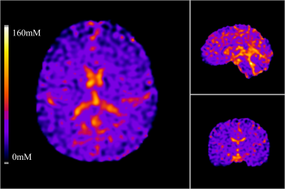 |
79 | Variability in Brain Sodium Total Sodium Content (TSC) Mapping Quantification Methods
Cameron Nowikow1,2, Paul Polak1,2,3, and Michael D Noseworthy1,2,4
1School of Biomedical Engineering, McMaster University, Hamilton, ON, Canada, 2Imaging Research Centre, St. Joseph's Healthcare, Hamilton, ON, Canada, 3Radiology, University of Colorado Anschutz Medical Campus, Aurora, CO, United States, 4Electrical and Computer Engineering, McMaster University, Hamilton, ON, Canada
Throughout the literature, the methods of quantifying total sodium concentration (TSC) brain maps vary. Some studies use a two calibration phantom approach, some use a one calibration phantom approach, and some use anatomical references to quantify the TSC maps. This abstract investigates the variability of using one method versus another to see if a chosen method will inherently bias the resultant maps. It was found that no bias is provided by one method versus another as they provide no significant variance to the data.
|
||
1284 |
80 | Evaluation of 23Na relaxation times and concentrations in the patellar knee cartilage at 3T
Benedikt Kamp1, Miriam Frenken1, Jan M. Henke1,2, Daniel B. Abrar1, Armin M. Nagel3,4, Lena V. Gast3, Georg Oeltzschner5,6, Lena M. Wilms1, Sven Nebelung1, Gerald Antoch1, Hans-Jörg Wittsack1, and Anja Müller-Lutz1
1University Dusseldorf, Medical Faculty, Department of Diagnostic and Interventional Radiology, Dusseldorf, Germany, 2University Dusseldorf, Medical Faculty, Clinic of Nuclear Medicine, Dusseldorf, Germany, 3Institute of Radiology, University Hospital Erlangen, Friedrich-Alexander-Universität Erlangen-Nürnberg 14 (FAU), Erlangen, Germany, 4Division of Medical Physics in Radiology, German Cancer Research Center (DKFZ), Heidelberg, Germany, 5Russell H. Morgan Department for Radiology and Radiological Science, The Johns Hopkins University 18 School of Medicine, Baltimore, MD, United States, 6F. M. Kirby Research Center for Functional Brain Imaging, Kennedy Krieger Institute, Baltimore, MD, United States
23Na relaxation times and concentrations were measured in patellar cartilage with a clinical 3T MRI scanner. Because of the low resolution in 23Na imaging, a focus was set on reducing the influence of synovial fluid. To estimate T1 a biexponential two-compartment fitting model was applied. In T2* measurements, an inversion pulse was used to suppress the signal from synovial fluid. 23Na parameters were successfully determined and are in good accordance to literature results measured at higher field strengths.
|
||
1285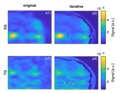 |
81 | An iterative algorithm for resolving high-resolution 23Na Multi-Quantum Coherences MRI from prior 1H constraints
Christian Licht1, Lothar R. Schad1, and Stanislas Rapacchi2
1Computer Assisted Clinical Medicine, Heidelberg University, Mannheim, Germany, 2CRMBM, Aix Marseille University, CNRS, Marseille, France
Sodium (23Na) MRI offers great potentials to be a clinical marker for disease states. Besides the single quantum signal, the triple quantum signal of 23Na could provide novel and complementary information. 3D volumetric 23Na multi-quantum coherences imaging, however, suffers from poor signal-to-noise ratios, which limit spatial resolution. Finer structures such as tissue boundaries are therefore barely observable. In addition, partial volume effects become more severe and diminish image quality as well as diagnostic applicability. The purpose of this work is to develop a reconstruction framework that addresses these shortcomings of 23Na multi-quantum coherences imaging by utilizing 1H prior constraints.
|
||
1286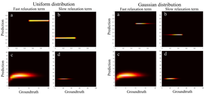 |
82 | Fast 23Na-T2* relaxation parameter estimation in the human brain
Frank Riemer1, Marco Reisert2, Bhavana S Solanky3, Claudia AM Wheeler-Kingshott3,4,5, Golay Xavier3, Renate Grüner1,6, and Ivan I Maximov7
1MMIV, Haukeland University Hospital, Bergen, Norway, 2University Medical Center, Freiburg, Germany, 3UCL Queen Square Institute of Neurology, London, United Kingdom, 4Brain Connectivity Centre, IRCCS Mondino Foundation, Pavia, Italy, 5Department of Brain and Behavioural Sciences, University of Pavia, Pavia, Italy, 6Department of Physics and Technology, University of Bergen, Bergen, Norway, 7Western Norway University of Applied Sciences, Bergen, Norway
Due to quadrupolar interactions, 23Na exhibits a bi-exponential T2. Previous approaches to estimate T2* have relied on simple least squares approaches or treating it as an inverse problem. Here we present a fast method based on Bayesian estimation and illustrate it on an in vivo dataset.
|
||
 Back to Meeting Home
 Back to the Program-at-a-Glance
The International Society for Magnetic Resonance in Medicine is accredited by the Accreditation Council for Continuing Medical Education to provide continuing medical education for physicians.
 Back to Meeting Home
 Back to the Program-at-a-Glance
 View the Presentation