Digital Poster
2H & Other Non-Protons
Joint Annual Meeting ISMRM-ESMRMB & ISMRT 31st Annual Meeting • 07-12 May 2022 • London, UK

| Computer # | ||||
|---|---|---|---|---|
1351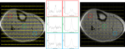 |
57 | Double Quantum Filtered 2H Measurements in Human Subjects Following Deuterium Oxide Loading
Robin Damion1,2,3, Daniel Cocking3,4, Hester Franks1, Daniel Wilkinson5,6, Matthew Brook2,6,7, Dorothee Auer1,2,3, and Richard Bowtell2,3,4
1School of Medicine, University of Nottingham, Nottingham, United Kingdom, 2NIHR Nottingham Biomedical Research Centre/Nottingham Clinical Research Facilities, University of Nottingham, Nottingham, United Kingdom, 3Sir Peter Mansfield Imaging Centre, University of Nottingham, Nottingham, United Kingdom, 4School of Physics & Astronomy, University of Nottingham, Nottingham, United Kingdom, 5Division of Medical Sciences and Graduate Entry Medicine, University of Nottingham, Nottingham, United Kingdom, 6MRC-Versus Arthritis Centre for Musculoskeletal Ageing Research, University of Nottingham, Nottingham, United Kingdom, 7School of Life Sciences, University of Nottingham, Nottingham, United Kingdom
Double-quantum filtered (DQF) deuterium spectra were obtained on a 3T scanner from lower leg and forearm muscles of volunteers whose deuterium abundance was increased by approximately 100 times through ingestion of deuterium oxide. Quadrupolar splitting frequencies of approximately 20 – 40 Hz were measured throughout the various muscle groups of the lower leg, with some regions showing little or no splitting. Although the DQF sequence considerably reduces the signal intensity, it has the advantage that it removes any isotropic signal component and therefore reveals an anisotropic component that might have been obscured.
|
||
1352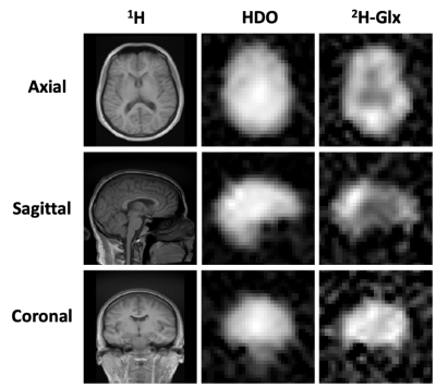 |
58 | Deuterium Metabolic Imaging for 3D Mapping of Human Brain Metabolism at 3T
Philip M. Adamson1, Keshav Datta2, Ron Watkins2, Lawrence Recht3, Ralph Hurd2, and Daniel Spielman2
1Department of Electrical Engineering, Stanford University, Palo Alto, CA, United States, 2Department of Radiology, Stanford University, Palo Alto, CA, United States, 3Department of Neurology, Stanford University, Palo Alto, CA, United States
Deuterium metabolic imaging (DMI) is an emerging modality for investigating glucose metabolism with particular application for assessing the Warburg effect in tumors. Although high field systems, e.g., 7T, provide maximal signal-to-noise ratio (SNR), implementation on widely available 3T scanners could have immediate clinical impact. Using a birdcage 2H RF coil, modified gradient filter, and spherical k-space sampling, we demonstrate high-quality 3T whole-brain DMI of healthy adults following the oral ingestion of deuterated glucose. Results from these experiments provide critical data needed to explore DMI SNR, spatial resolution, and imaging time tradeoffs.
|
||
1353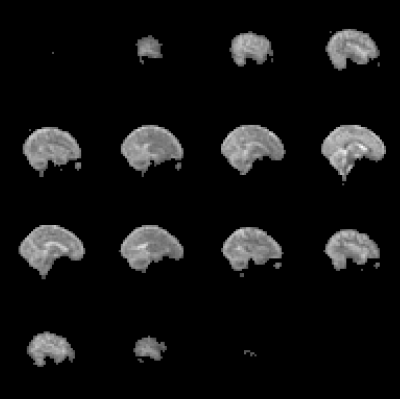 |
59 | Measuring Deuterium Relaxation Times in Human Brain at 7T following D2O Loading
Daniel Cocking1,2, Robin Damion2,3,4, Hester Franks5, Daniel Wilkinson6,7, Matthew Brook4,6,8, Dorothee Auer2,3,4, and Richard Bowtell1,2,4
1School of Physics and Astronomy, University of Nottingham, Nottingham, United Kingdom, 2Sir Peter Mansfield Imaging Centre, University of Nottingham, Nottingham, United Kingdom, 3Mental Health and Clinical Neuroscience, School of Medicine, University of Nottingham, Nottingham, United Kingdom, 4NIHR Nottingham Biomedical Research Centre/Nottingham Clinical Research Facilities, Queen's Medical Centre, Nottingham, United Kingdom, 5Division of Cancer and Stem Cells, School of Medicine, University of Nottingham, Nottingham, United Kingdom, 6MRC-Versus Arthritis Centre for Musculosketal Ageing Research, University of Nottingham, Nottingham, United Kingdom, 7Division of Medical Sciences and Graduate Entry Medicine, School of Medicine, University of Nottingham, Nottingham, United Kingdom, 8School of Life Sciences, University of Nottingham, Nottingham, United Kingdom Deuterium magnetic resonance measurements are of growing interest in the field of metabolic imaging. Four subjects were loaded with D2O to ~1.5% enrichment over a 6-week period for a parallel study of immune cell proteomics. We report T1 and T2* relaxation times of deuterium in HDO measured from cerebral spinal fluid (CSF), white matter (WM) and grey matter (GM) in vivo, using a 7T Philips Achieva scanner with a dual-tuned 2H/1H birdcage coil. 2H T1 values are significantly shorter than corresponding values for 1H in H2O, due to the quadrupolar 2H relaxation, whilst T2* values are comparable to 1H values. |
||
1354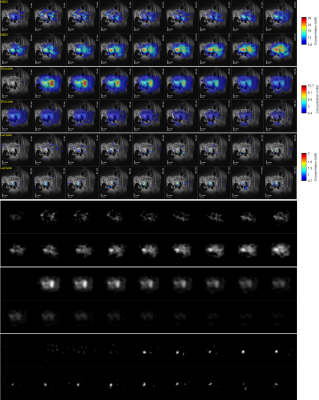 |
60 | 2H multi-echo bSSFP highlights pancreatic ductal adenocarcinomas and distinguishes them from pancreatitis in murine models
Elton Tadeu Montrazi1, Qingjia Bao2, Dana Peters3, Keren Sasson1, Lilach Agemy1, Avigdor Scherz1, and Lucio Frydman1
1Weizmann Institute of Science, Rehovot, Israel, 2Chinese Academy of Sciences, Wuhan, China, 3Yale School of Medicine, New Haven, CT, United States
Pancreatic ductal adenocarcinoma (PDAC) is among the deadliest of cancers. We recently showed that deuterium metabolic imaging (DMI) can serve as a potential method for PDAC’s detection; we also demonstrated that multi-echo balanced steady-state free precession (ME-bSSFP) can greatly increase DMI’s SNR per unit time compared to CSI acquisitions. This study synergizes these aspects, to provide high resolution maps on the metabolic kinetic of PDAC and pancreatitis mouse models. ME-bSSFP at 15.2T showed that lactate accumulated in different kinds of tumors. In general, glucose was also uptaken by the tumors, and HDO concentrated in them.
|
||
1355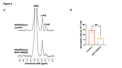 |
61 | Deuterated α-ketoglutarate has the potential to detect the isocitrate dehydrogenase 1 mutation in low-grade gliomas. Video Permission Withheld
Celine Taglang1, Georgios Batsios1, Meryssa Tran1, Anne-Marie Gillespie1, Sabrina Ronen1, and Pavithra Viswanath1
1Radiology, University of California San Francisco, San Francisco, CA, United States 2H-MRS is a novel method of imaging flux through metabolic pathways. Here, we show that 2H-MRS can be used to probe the isocitrate dehydrogenase 1 mutation (IDHmut), which catalyzes production of the oncometabolite 2-hydroxyglutarate (2-HG) in low-grade gliomas. Our results indicate that 2-HG production from [3,3’-2H]-α-KG can be specifically observed in IDHmut cells. Importantly, treatment with an IDHmut inhibitor, which is in clinical trials for low-grade glioma patients, results in a significant reduction in 2-HG production from [3,3’-2H]-α-KG in IDHmut cells. Our results point to the potential of [3,3’-2H]-α-KG as a probe of glioma IDHmut status and response to therapy. |
||
1356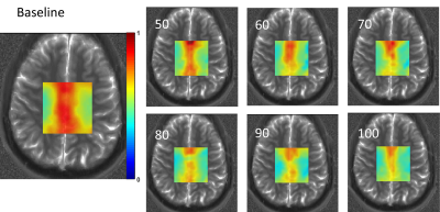 |
62 | Prior knowledge fitting of 2H-labeled glutamate in the 1H MRS spectrum to map neural metabolism following ingestion of deuterated glucose
Neil Wilson1, Abigail Cember1, Laurie Rich1, Puneet Bagga2, Ravi PR Nanga1, Sophia Swago3, Deepa Thakuri1, Mark Elliott1, Mitchell Schnall1, John Detre4, and Ravinder Reddy1
1Department of Radiology, University of Pennsylvania, Philadelphia, PA, United States, 2Diagnostic Imaging, St Jude Children's Research Hospital, Memphis, TN, United States, 3Department of Bioengineering, University of Pennsylvania, Philadelphia, PA, United States, 4Department of Neurology, University of Pennsylvania, Philadelphia, PA, United States
Deuterium metabolic imaging (DMI) has brought about a renewed interest in the application of 2H-labeled substrates to map metabolism in vivo, yet deuterium spectroscopy remains challenging. We have shown that metabolism of deuterium-labeled glucose can be observed in the proton spectrum through a reduction in signal of downstream metabolites in a technique called quantitative exchange label turnover MRS (qMRS). Here, we show that prior knowledge fitting that includes unlabeled as well as deuterium-labeled forms of glutamate can be used to map neural metabolism reliably with qMRS.
|
||
1357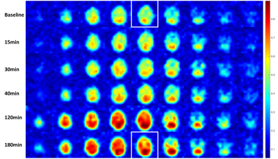 |
63 | Deuterium metabolic imaging of a healthy human brain after oral glucose uptake on a clinical 3T MRI scanner
Michael Vaeggemose1,2, Rolf F. Schulte3, Christoffer Laustsen2, Esben Søvsø Szocska Hansen2, and Nikolaj Bøgh2
1GE Healtcare, Broendby, Denmark, 2MR Research Centre, Department of Clinical Medicine, Aarhus University, Aarhus N, Denmark, 3GE Healtcare, Munich, Germany
Deuterium metabolic imaging (DMI) has emerged as a novel biomarker comparable to 18FDG PET by adding quantitative metabolic kinetic capabilities at high field (>3T). The main barrier for widespread clinical adoption, is the availability of high field clinical scanners. The aim of this study is to determine if DMI for neurological applications is feasible on a clinical 3T MRI scanner. Our findings suggests that it is indeed feasible, and that metabolic breakdown product (Glutamine/Glutamate) can be quantified around 120 min after ingestion.
|
||
1358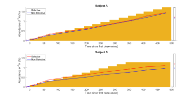 |
64 | Monitoring water uptake in the human body during oral loading of heavy water using deuterium magnetic resonance
Daniel Cocking1,2, Robin Damion2,3,4, Hester Franks5, Daniel Wilkinson6,7, Dorothee Auer2,3,4, Matthew Brook4,6,8, and Richard Bowtell1,2,4
1School of Physics and Astronomy, University of Nottingham, Nottingham, United Kingdom, 2Sir Peter Mansfield Imaging Centre, University of Nottingham, Nottingham, United Kingdom, 3Mental Health and Clinical Neuroscience, School of Medicine, University of Nottingham, Nottingham, United Kingdom, 4NIHR Nottingham Biomedical Research Centre/Nottingham Clinical Research Facilities, Queen's Medical Centre, Nottingham, United Kingdom, 5Division of Cancer and Stem Cells, School of Medicine, University of Nottingham, Nottingham, United Kingdom, 6MRC-Versus Arthritis Centre for Musculosketal Ageing Research, University of Nottingham, Nottingham, United Kingdom, 7Division of Medical Sciences and Graduate Entry Medicine, School of Medicine, University of Nottingham, Nottingham, United Kingdom, 8School of Life Sciences, University of Nottingham, Nottingham, United Kingdom Deuterium magnetic resonance imaging and spectroscopy at 7T has been used to follow the deuterium concentration in the brain over an ~eight-hour period while subjects orally-loaded with D2O to 100x natural abundance. Changes in deuterium concentration of ~ 0.1% can be readily monitored in 2H spectra acquired in 1 minute and images acquired in 7.5 minutes. The change in deuterium concentration estimated from 2H spectra is in agreement with the value calculated from cumulative D2O dose and body mass and the signal changes measured from ROIs in the brain have similar time-courses, with relative signal strengths dictated by T2*-weighting. |
||
1359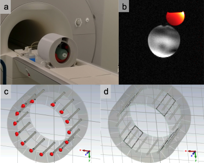 |
65 | Performance of a TEM 2H/1H 7T head coil
Minghao Zhang1, Jabrane Karkouri1, Daniel Atkinson1, Brandon Tramm2, Scott Schillak2, and Christopher T. Rodgers1
1Wolfson Brain Imaging Centre, University of Cambridge, Cambridge, United Kingdom, 2Virtumed LLC, Minneapolis, MN, United States Deuterium metabolic imaging (DMI) has been proposed as a means to probe glucose uptake and metabolism. It is expected that DMI will reveal important changes in cancer and neurodegeneration. We constructed a dual-tuned 7T 2H/1H array coil and assessed appropriate RF power limits by EM modelling. The coil performance has been assessed with phantom scans on a D2O-enriched agar phantom. We are now ready to start human in vivo scanning. |
||
1360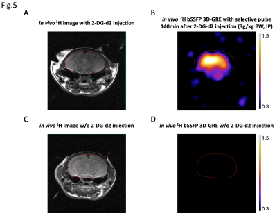 |
66 | An old tracer learns new tricks: Imaging cerebral glucose uptake with deuterated 2-deoxy-d-glucose-2,2-d2
Xiao Gao1,2,3, Jeremy Gordon2, Kai Qiao2, Ilona Polvoy2, Tanner Nickles2,3, David Wilson2, and Myriam M. Chaumeil1,2,3
11 Department of Physical Therapy and Rehabilitation Science, University of California, San Francisco, San Francisco, CA, United States, 2Department of Radiology and Biomedical Imaging, University of California, San Francisco, San Francisco, CA, United States, 3UC Berkeley-UCSF Bioengineering Program, University of California, San Francisco, San Francisco, CA, United States
2-deoxy-d-glucose (2-DG) is a glucose analog widely used in FDG-PET imaging as a biological tracer of glucose uptake. This study innovatively redirects its application in 2H NMR by using a deuterated version of 2-DG (2-DG-d2) and explores its imaging potential in mapping glucose uptake without ionizing radiation. A workflow is described here regarding multi-band RF pulse design and bSSFP sequence optimization, which enables a rapid 2H imaging with high sensitivity. Both in vitro and in vivo imaging results have validated the pulse sequence’s specificity and sensitivity of detecting 2-DG-d2 with high spatial resolution, inspiring its further implementation in the future.
|
||
1361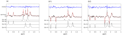 |
67 | STEAM-based proton observed carbon edited (POCE) 13C magnetic resonance spectroscopy without decoupling at 7T
Ying Xiao1,2, Daniel Wenz1, and Lijing Xin1
1Animal imaging and technology (CIBM), EPFL, Lausanne, Switzerland, 2Laboratory for Functional and Metabolic Imaging (LIFMET), EPFL, Lausanne, Switzerland 1H-[13C] NMR spectroscopy is a powerful tool to study metabolic information in human brain. At 7T, the proton-observed carbon-edited (POCE) method provides high sensitivity and improved spectral resolution. However, the application in human brain has more constraints due to high RF power deposition at 7T. In this study, we investigated the performance of the STEAM-based POCE sequence without decoupling to reduce the RF power. The phantom experiments and the Monte Carlo simulations demonstrated high-quality 1H-[13C] MR spectra and the accurate quantification of 13C-labeled glutamate and glutamine without decoupling at 7T. |
||
1362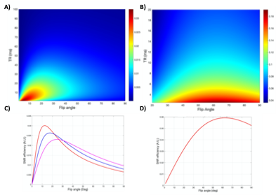 |
68 | Optimization of a Phase-Cycled bSSFP for Improved SNR-efficiency in Fluorine-19 MRI at 3T
Lawrence Michael Lechuga1, Paul Begovatz2, Bruce D Collick3, and Sean B Fain4
1Medical physics, University of Wisconsin Madison, Madison, WI, United States, 2Medical Physics, University of Wisconsin, Madison, Madison, WI, United States, 3Radiology, University of Wisconsin, Madison, Madison, WI, United States, 4Radiology, University of Iowa, Iowa City, WI, United States The focus of this work is on the optimization of fluorine-19 enabled SPGR, bSSFP, and phase cycled bSSFP (bSSFP-C) pulse sequence parameters to improve signal acquisition efficiency for perfluoropolyether (PFPE) at 3T with a multi-channel coil. Bloch simulations and subsequent experimental validation were performed to determine the parameters to maximize the SNR-efficiency for each sequence. Acquisitions of the optimized SPGR, bSSFP, and bSSFP-C were then assessed for their achieved SNR-efficiency. bSSFP and bSSFP-C demonstrated increased sensitivity compared to SPGR. Feasibility of bSSFP-C with parallel imaging demonstrated the ability to reduce scan times by 2-fold without compromising the qualitative image quality. |
||
1363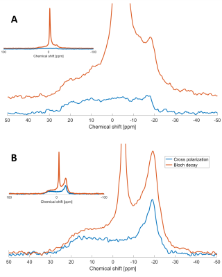 |
69 | 1H-31P Cross-polarization: A new frontier to study myelin in white matter
Alex Ensworth1,2, Cariad-Arianna Knight1, Piotr Kozlowski1,2,3,4, Cornelia Laule1,2,3,5, Alex L. MacKay1,3,4, and Carl A. Michal1
1Physics and Astronomy, University of British Columbia, Vancouver, BC, Canada, 2International Collaboration on Repair Discoveries (ICORD), University of British Columbia, Vancouver, BC, Canada, 3Radiology, University of British Columbia, Vancouver, BC, Canada, 4UBC MRI Research Centre, University of British Columbia, Vancouver, BC, Canada, 5Pathology & Laboratory Medicine, University of British Columbia, Vancouver, BC, Canada Phosphorus (31P) nuclear magnetic resonance spectroscopy (NMR) has the potential to play a valuable role in the characterization of myelin due to the concentration of 31P present in myelin phospholipids. This study demonstrates the feasibility of detecting 31P through cross polarization (CP) and magnetization transfer (MT) from hydrogen in preserved murine spinal cord and fresh porcine brain. The results demonstrate the detection of myelin phospholipids through these NMR techniques and provide compelling proof of concept for the potential applicability of MT-CP in 31P MRI for non-ambiguous myelin characterization in vivo. |
||
1364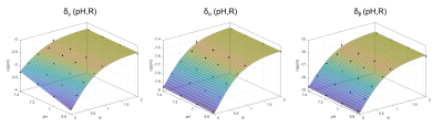 |
70 | A dictionary-based approach for the determination of pH values using 31P MRS
Vanessa L. Franke1,2, Johannes Breitling1, Renate Bangert1, Mark E. Ladd1,2,3, Peter Bachert1,2, and Andreas Korzowski1
1Division of Medical Physics in Radiology, German Cancer Research Center (DKFZ), Heidelberg, Germany, 2Faculty of Physics and Astronomy, University of Heidelberg, Heidelberg, Germany, 3Faculty of Medicine, University of Heidelberg, Heidelberg, Germany
The determination of tissue pH under varying cellular conditions by means of 31P MRS is challenging. We propose a concept for a dictionary-based approach for pH estimation under different chemical conditions. As proof-of-concept, we demonstrate its feasibility for a limited subset of conditions using the chemical shifts of ATP and their dependence on pH and magnesium. For the presented subset, the estimated pH values are in good agreement with the prepared values. However, for application to in vivo data, an extension of the dictionary to include other influences, e.g. of other ions, is required and the subject of ongoing work.
|
||
1365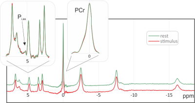 |
71 | Functional 31P MRS in the visual cortex at 9.4 T
Rolf Pohmann1 and Klaus Scheffler1,2
1Magnetic Resonance Center, Max Planck Institute for Biological Cybernetics, Tübingen, Germany, 2Biomedical Magnetic Resonance, University of Tübingen, Tübingen, Germany
Even after a large number of studies with varying outcome, the effect of brain activation on the 31P spectrum is still unclear. Here, the SNR gain expected from a field strength of 9.4 T is used to determine changes in the metabolite peaks during a 5 min stimulus. In spite of the high SNR and spectral quality, no significant variations were found. Especially the amplitude and linewidth of the PCr-peak did not change, and, while the signal of extracellular inorganic phosphate was well visible after averaging over all subjects, no variations in its amplitude or position were observed.
|
||
1366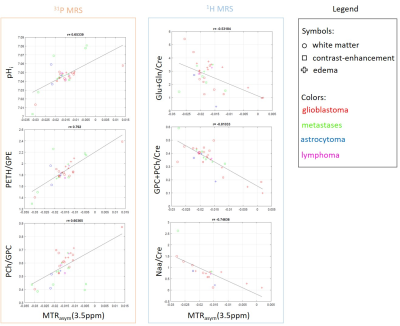 |
72 | Metabolic information in APT-CEST - A combined 1H and 31P MRS study
Jan-Ruediger Schuere1, Eike Steidl1, Elisabeth Neuhaus2, Manoj Shrestha3, Elke Hattingen1, and Ulrich Pilatus1
1Neuroradiology, Goethe University Hospital Frankfurt, Frankfurt am Main, Germany, 2Goethe University Hospital Frankfurt, Frankfurt am Main, Germany, 3Brain Imaging Center, Goethe University, Frankfurt am Main, Germany
In this work, APT-CEST imaging in combination with 1H and 31P spectroscopy was performed to investigate 17 patients with different brain tumors. A synergistic analysis should help for a better understanding of the APT-CEST contrasts such as MTRasym with regard to tumor metabolism and further physiological relationships such as the pH-sensitivity.
|
||
 Back to Meeting Home
 Back to the Program-at-a-Glance
The International Society for Magnetic Resonance in Medicine is accredited by the Accreditation Council for Continuing Medical Education to provide continuing medical education for physicians.
 Back to Meeting Home
 Back to the Program-at-a-Glance
 View the Presentation