Digital Poster
New Ideas in Coils I: Metamaterials & Dielectrics
Joint Annual Meeting ISMRM-ESMRMB & ISMRT 31st Annual Meeting • 07-12 May 2022 • London, UK

Digital Poster
New Ideas in Coils I: Metamaterials & Dielectrics
| Computer # | ||||
|---|---|---|---|---|
2177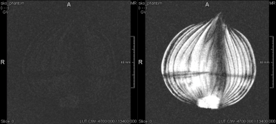 |
79 | Smart MetaBox: Metamaterial arrangement for volume-homogeneous SNR enhancement
Dennis Philipp1,2, Endri Stoja3, Simon Konstandin1, Robin Wilke1, Diego Betancourt3, Thomas Bertuch3, Juergen Jenne1,4, Reiner Umathum1,4, and Matthias Guenther1,2
1Fraunhofer MEVIS, Bremen, Germany, 2University of Bremen, Bremen, Germany, 3Fraunhofer FHR, Wachtberg, Germany, 4DKFZ, Heidelberg, Germany
A metamaterial arrangement that encloses a volume "metaBox" is shown to yield a significant and volume-homogeneous SNR enhancement in 3T MRI. Due to the integration of non-linear components, the structure self-detunes in Tx whilst being resonant in Rx. Fine-tuning capabilities are included in two different prototypes via (i) a manually trimmable capacitor and (ii) a Bluetooth-controlled digital capacitor that allows for a wireless interface. On-bench characterization as well as MRI experiments verify the functionality of the device. Thus, the metaBox is ideally suited for, e.g., imaging of extremities.
|
||
2178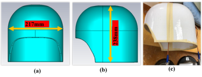 |
80 | Parametric Study of High Dielectric Constant Helmet Characterization
Parisa Lotfi Poshtgol1, Soo Han Soon2, Michael Lanagan3, Eugene Furman4, Xinlian Chen5, Xin Li2, Xiao-Hong Zhu2, Wei Chen2, and Qing X Yang1
1Center for NMR Research, Departments of Neurosurgery and Radiology, Pennsylvania State University, Hershey, PA, United States, 2Center for Magnetic Resonance Research, University of Minnesota, Minneapolis, MN, United States, 3Department of Engineering Science and Mechanics, Pennsylvania State University, State college, PA, United States, 4Materials Research Institute, Pennsylvania State University, State college, PA, United States, 5Department of Biomedical Engineering, South China University of Technology, Guangzhou, China Helmets conformal to the human head with high or ultra-high dielectric constant (HDC/uHDC) materials have been shown to enhance the SNR of entire human head along with a receive array. However, the dielectric resonance modes (DRMs) of the helmet have not been studies systematically. This investigation aimed to; 1) describe the characteristics of the DRMs and their RF field distributions; 2) establish the relationships between DRMs and the basic geometric parameters of the helmet using numerical modeling. Understand the DRMs of large uHDC structures are fundamentally important for future applications of uHDC materials for RF field reach. |
||
2179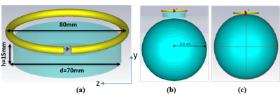 |
81 | Effect of Dielectric Resonator (DR) Modes on RF-Magnetic Field
Parisa Lotfi Poshtgol1, Navid Pourramzan Gandjii1, Michael Lanagan2, Eugene Furman3, Soo Han Soon4, Xinlian Chen5, Saber Soltani6, Xiao-Hong Zhu4, Christopher T. Sica1, Hannes M Wiesner4, Wei Chen4, kamil Ugurbil4, and Qing X Yang1
1Departments of Neurosurgery and Radiology, College of Medicine, Pennsylvania State University, Hershey, PA, United States, 2Department of Engineering Science and Mechanics, Pennsylvania State University, State college, PA, United States, 3Materials Research Institute, Pennsylvania State University, State college, PA, United States, 4Center for Magnetic Resonance Research, University of Minnesota, Minneapolis, MN, United States, 5Department of Biomedical Engineering, South China University of Technology, Guangzhou, China, 6Department of Electrical Engineering, The Pennsylvania State University, State college, PA, United States Dielectric resonators with Ultra-High Dielectric Constant (uHDC) materials possess intrinsic resonance modes that have been used for RF transmission and reception. The resonance modes and conditions of the uHDC discs were studied through computer simulations. We compared the B1+ field and RX-sensitivity when employing cylindrical uHDC discs at both resonant and non-resonant conditions. We showed in this work, to achieve the best focusing effect into phantom, the design with uHDC material should operate around the first TE01δ mode and lower than the second mode TE01δ with the highest permittivity. |
||
2180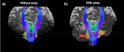 |
82 | Improving Diffusion MRI in the Posterior Fossa Using a Wireless RF Array at 7T
Akbar Alipour1, Ameen Al Qadi1, Gaurav Verma1, Alan C Seifert1, and Priti Balchandani1
1BioMedical Engineering and Imaging Institute (BMEII), Icahn School of Medicine at Mount Sinai, Manhattan, NY, United States
Ultra-high field (UHF ≥7T) MRI scanners can provide stronger signals than standard field strengths, which boosts signal-to-noise ratio (SNR) for improved diffusion MRI (dMRI). However, at 7T, wavelength effects cause highly inhomogeneous $$$B_1^+$$$ in the human brain, with lower transmit efficiency in the cerebellum and temporal lobes manifesting as signal dropouts in these regions. Recently, we reported a simple approach of using a wireless radiofrequency (RF) array to improve transmit efficiency and signal sensitivity at 7T focusing on the posterior fossa. Here we demonstrate the feasibility and effectiveness of using the RF array for in-vivo dMRI at 7T.
|
||
2181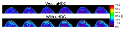 |
83 | Evaluation of an Ultra High Dielectric Constant (uHDC) Package for Enhancement of a Knee Phased Array
Christopher T Sica1, Parisa Lofti Poshtgol2, Sebastian Rupprecht3, and Qing X Yang2
1Radiology, Pennsylvania State University College of Medicine, Hershey, PA, United States, 2Neurosurgery, Pennsylvania State University College of Medicine, Hershey, PA, United States, 33HyQ Research Solutions, LLC, College Station, TX, United States
Prior work has explored the use of dielectric materials to enhance imaging of the brain and spine with rectangular blocks. One area that has not been examined to date is knee imaging, where space constraints limit the use of blocks. In this work we evaluate the performance of an ultra-high dielectric constant (uHDC) curved plate (εr ~ 4500) for knee imaging in a clinical array at 3T. SNR gains of 9 to 71% percent were observed across the patella and patellar cartilage. Transmit efficiency increased up to 100% close to the material, with an increase in transmit inhomogeneity.
|
||
2182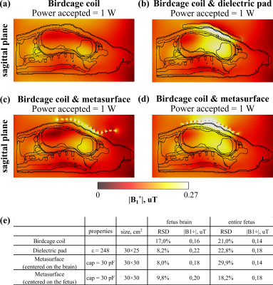 |
84 | Metasurface-based pad for improving fetal imaging at 3T
Viktor Puchnin1, Evgeniy Koreshin1, Anna Kalugina1, Aleksander Efimtcev1,2, Irina Mashchenko2, Wyger Brink3, and Alena Shchelokova1
1School of Physics and Engineering, ITMO University, Saint Petersburg, Russian Federation, 2Federal Almazov North-West Medical Research Center, Saint Petersburg, Russian Federation, 3Department of Radiology, C.J. Gorter Center for High‐Field MRI, Leiden University Medical Center, Leiden, Netherlands
Radiofrequency magnetic field homogeneity improvement and SAR reduction are two essential tasks for fetal MRI at 3T. We demonstrate for the first time that the metasurface-based pad can effectively improve the transmit efficiency while reducing SAR within the entire fetus and fetus brain. The metasurface is assembled from the metal wires loaded with capacitors. Numerical studies of a pregnant woman voxel model in the 9th month covered with the proposed metasurface centered within fetus brain (body) showed 25% (28%) transmit efficiency improvement and up to 9% (2.8%) increased magnetic field homogeneity, while the whole-body SAR was reduced by 1.2 (1.6)-fold.
|
||
2183 |
85 | Actively coupled dielectric pads for adaptive B1+ field shimming
Paulina Šiurytė1, Friso de Boer1, Can Akgun2, and Sebastian Weingärtner1
1Imaging Physics, TU Delft, Delft, Netherlands, 2Microelectronics, TU Delft, Delft, Netherlands
MRI at high and ultra high field strengths suffers from B1+ inhomogeneities, but B1+ shimming with multiple transmit coils is only available at top-line scanners. Dielectric pads allow for localized B1+ changes, but need to be specifically designed for the scan and anatomy. In this work we explore the use of an actively switchable dielectric device to allow for spatial B1+ correction without the need for multiple transmit coils. Our phantom experiments at 3T show that coupling mini-pockets with barium titanate slurry enables various field configurations that are switchable, localized and significant in magnitude.
|
||
2184 |
86 | Investigation of MR visibility control of water-based (CaTiO3) dielectric padding with iron oxide contrast agent in 7T human brain MRI Video Permission Withheld
Seulki Yoo1,2, Ji Seong Barg1, Bo-yong Park3,4, and Seung-Kyun Lee1,2
1Department of Biomedical Engineering, Sungkyunkwan university, Suwon, Korea, Republic of, 2Intelligent Precision Healthcare Convergence, Sungkyunkwan University, Suwon, Korea, Republic of, 3Department of Data Science, Inha University, Incheon, Korea, Republic of, 4Center for Neuroscience Imaging Research,Institute for Basic Science, Suwon, Korea, Republic of
Ultra-high-field MRI provides improved signal-to-noise ratio (SNR) and tissue contrast. However, at the same time, it exhibits stronger RF field inhomogeneity compared to lower-field MRI. Although such inhomogeneity can be mitigated by customized RF coils and parallel transmit methodology, it is often difficult to translate these to clinical use. Recently, a simple and low-cost high dielectric-constant padding has been proposed to improve the quality of 7T brain images in many sequences. Here, we propose a novel approach to suppress the MR visibility of a water-based dielectric padding by controlled addition of iron oxide contrast agent in 7T human brain MRI.
|
||
2185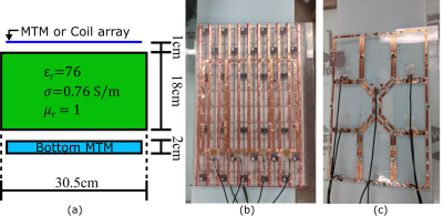 |
87 | Hybrid 3T 8-channel Receive Array Using a Metamaterial Slab and Companion Loop Elements: Comparison to a Conventional Array
Léo Rémillard1, Adam Mitchell Maunder1,2, Ashwin Iyer1, and Nicola De Zanche2,3
1Electrical and Computer Engineering, University of Alberta, Edmonton, AB, Canada, 2Oncology, University of Alberta, Edmonton, AB, Canada, 3Medical Physics, Cross Cancer Institute, Edmonton, AB, Canada
Metamaterials (MTMs) have been used as passive elements to improve the receive sensitivity in MRI by enhancing the field locally, but not connected directly as receive elements. We present the design and construction of a novel 2D transmission line based MTM employed as a 4-port element combined with an additional 4-element coil array. Simulation and measurement of receive sensitivity are compared to those of a conventional 8-element coil array covering the same region. The receive sensitivity in a human torso sized phantom is found to be equivalent, while the MTM slab additionally enhances the transmit performance.
|
||
2186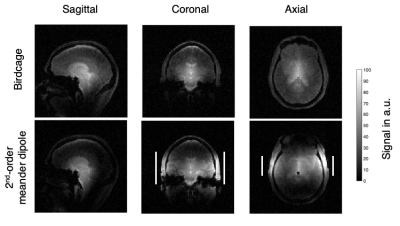 |
88 | 2nd-order meander dipoles for 7T head coil passive RF shimming
Tania del Socorro Vergara Gomez1,2, Roxane Mamberti2, Marc Dubois3, Elodie Georget3, Tryfon Antonakakis3, Alexandre Vignaud4, Frank Kober1, Stefan Enoch2, and Redha Abdeddaim2
1Aix Marseille Univ, CNRS, CRMBM, Marseille, France, 2Aix Marseille Univ, CNRS, Centrale Marseille, Institut Fresnel, Marseille, France, 3Multiwave Imaging, Marseille, France, 4CEA, DRF/Joliot/Neurospin, Université Paris-Saclay & CNRS, Gif-sur-Yvette Cedex, France
Active and passive RF shimming approaches have been proposed to diminish the B1+ inhomogeneities commonly found in a birdcage coil at 7T. In a previous work, we have introduced a meander dipole based on two 3rd-order Hilbert fractals as an alternative solution for passive shimming. In this work we optimized their design using six 2nd-order Hilbert fractals. Simulations and in vivo MRI at 7T using these structures showed strong B1+ enhancement in the area between the two meander dipoles whereas B1+ was reduced in regions below and above the fractal pads.
|
||
2187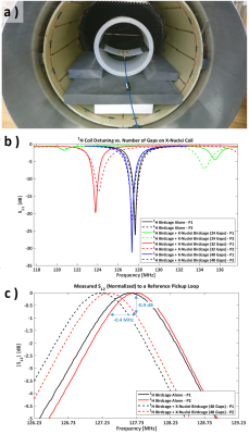 |
89 | Coupling study between two concentric whole-body MRI birdcage coils: towards a whole-body X-nucleus coil
Juan Diego Sánchez Heredia1, Wenjun Wang2, Vitaliy Zhurbenko2, and Jan Henrik Ardenkjær-Larsen1
1Department of Health Technology, Technical University of Denmark - DTU, Kgs. Lyngby, Denmark, 2Department of Electrical Engineering, Technical University of Denmark - DTU, Kgs. Lyngby, Denmark The feasibility of integrating a whole-body X-nucleus birdcage in a wide-bore clinical 3T MR scanner is evaluated. The challenge is that this extension coil has to be tightly fit into the scanner bore, having a diameter just 16 mm smaller than the concentric 1H coil. Different configurations are proposed and evaluated with the purpose of minimizing interaction between the X-nucleus coil and the native 1H body coil of the scanner. Experimental results are in agreement with simulations and demonstrate a 1H coil detuning of 0.4 MHz due to co-integration with X-nucleus birdcage. |
||
 Back to Meeting Home
 Back to the Program-at-a-Glance
The International Society for Magnetic Resonance in Medicine is accredited by the Accreditation Council for Continuing Medical Education to provide continuing medical education for physicians.
 Back to Meeting Home
 Back to the Program-at-a-Glance
 View the Presentation