Digital Poster
Phantoms & System Imperfections I
Joint Annual Meeting ISMRM-ESMRMB & ISMRT 31st Annual Meeting • 07-12 May 2022 • London, UK

Digital Poster
Phantoms & System Imperfections I
| Computer # | ||||
|---|---|---|---|---|
2721 |
85 | Developing 3D-printed phantoms for quality assurance and validation of quantitative DCE–MRI measurements
Muhammad Sulaiman Sarwar1,2, Antoine Vallatos1,3, Cher Hon Lau4, Adam Waldman1,3, Simone Dimartino2, and Michael J Thrippleton1,3
1Centre for Clinical Brain Sciences, The University of Edinburgh, Edinburgh, United Kingdom, 2Institute for Bioengineering, The University of Edinburgh, Edinburgh, United Kingdom, 3Edinburgh Imaging, The University of Edinburgh, Edinburgh, United Kingdom, 4Institute for Materials and Processes, The University of Edinburgh, Edinburgh, United Kingdom Difficulties in engineering accurate biomimetic phantoms make it challenging to validate MRI models. This is particularly true for quantitative vascular permeability measurements using dynamic contrast-enhanced (DCE-) MRI, where no robust controllable phantoms are available for validating novel techniques and harmonising multicentre results. We developed 3D-printed biomimetic vascular permeability phantoms with controllable properties and assessed their ability to interrogate common DCE model quantification approaches. Parameters such as blood flow, vascular permeability, plasma volume and extravascular volume were reproduced by adapted 3D-printed material and flow circuit properties. The resulting phantoms were shown to reproduce realistic DCE-MRI signals observed clinically. |
||
2722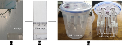 |
86 | Analysis of variability of diffusion tensor imaging measurements using co-electrospun fibre phantom
Fenglei Zhou1,2, Fei Gao3, Longji Xu3, Jing Yang3, Fuxin Ren3, Weibo Chen4, and Geoff JM Parker1,5
1Centre for Medical Image Computing, University College London, London, United Kingdom, 2School of Pharmacy, University College London, London, United Kingdom, 3Department of Radiology,Shandong Provincial Hospital, Cheeloo College of Medicine, Shandong University, Jinan, China, 4Philips Heathcare, Shanghai, China, 5Bioxydyn Limited, Manchester, United Kingdom
Brain diffusion tensor imaging (DTI) measurements are sensitive to pathological changes, but in order for them to be of greatest use it is vital to understand intra-scanner and inter-scanner variations in DTI metrics. Here we demonstrate a physical phantom with variable pore sizes and fibre orientation that can be used to assess the repeatability and reproducibility of DTI measurements across scanners and protocols and present its application in assessing 9 separate scanner/protocol combinations.
|
||
2723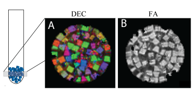 |
87 | Microscopically Anisotropic and Randomly Oriented 3D Printed MRI Phantom for Size, Orientation, and Anisotropy Validation in Diffusion Imaging
Velencia Witherspoon1, Michal Komlosh1,2,3, Dan Benjamini1,2,3, Peter Basser1,3, and Nick Lavrik4
1Section on Quantitative Imaging and Tissue Sciences, Eunice Kennedy Shriver National Institute of Child Health and Human Development, National Institutes of Health, Bethesda, MD, United States, 2Center for Neuroscience and Regenerative Medicine, Uniformed Services University of the Health Sciences, Bethesda, MD, United States, 3The Henry M. Jackson Foundation for the Advancement of Military Medicine (HJF), Bethesda, MD, United States, 4The Center for Nanophase Materials Sciences (CNMS) at Oak Ridge National Laboratory (ORNL), Oakridge, TN, United States
Microscopically anisotropic but globally isotropic DTI MRI phantom was designed and fabricated using a 2 Photon Nanoscribe 3D Printer. The capillary array blocks were measured aligned and randomly oriented to determine the ability of ddPFG and DTI to characterize local anisotropy in a globally isotropic system. These experiments combined computational methods can provide MRI phantoms that can be employed to validate novel or proposed microstructure imaging experiments.
|
||
2724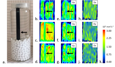 |
88 | Investigation of sodium alginate- and polyvinyl alcohol-based anisotropic hydrogel phantoms for calibration applications by DWI at 0.6 T Video Permission Withheld
Weronika Mazur1,2, Anna Stefańska-Bernatowicz2, Ewa Stodolak-Zych3, and Artur Tadeusz Krzyżak2
1Faculty of Physics and Applied Computer Science, AGH University of Science and Technology, Kraków, Poland, 2Faculty of Geology, Geophysics and Environmental Protection, AGH University of Science and Technology, Kraków, Poland, 3Faculty of Materials Science and Ceramics, AGH University of Science and Technology, Kraków, Poland
Anisotropic hydrogels has been attracting attention for biomedical purposes. These highly-hydrated materials have potential for tissue-mimicking and then, may serve for advanced medical imaging studies. In the work, we present two types of anisotropic hydrogels made from sodium alginate (NA) and polyvinyl alcohol (PVA) in the form of fiber bundle- a single component for larger phantom. We investigated their potential for diffusion-weighted imaging. NA-based phantoms are characterized by highly uniform diffusivity distribution, even higher then water. Single component turned out to be insufficient for the accurate quantification of the microstructure. PVA-based phantom exhibited desirable diffusion properties for tissue-mimicking.
|
||
2725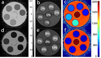 |
89 | Soy lecithin: a phantom material for the adjustment of apparent diffusion coefficient in magnetic resonance imaging
Victor Fritz1, Petros Martirosian1, Jürgen Machann1,2, and Fritz Schick1,2
1Department of Diagnostic and Interventional Radiology, University of Tuebingen, Germany., Section on Experimental Radiology, Tuebingen, Germany, 2Institute for Diabetes Research and Metabolic Diseases of the Helmholtz Centre Munich at the University of Tuebingen, Tuebingen, Germany
The aim of this work was to systematically investigate whether lecithin is suitable for the construction of a diffusion phantom that covers and simulates ADC-values occurring in human tissue. For this, aqueous solutions of different lecithin concentrations (0%-10%) were prepared and measured by DWI. The presented results showed that lecithin is a suitable agent for simulating a wide variety of ADC-values. Even low concentrations showed a strong decrease in the ADC-value of water. However, with increasing concentration, lecithin also showed a strong influence on T2, so that ADC and T2 cannot be set independently from one another in lecithin-based phantoms.
|
||
2726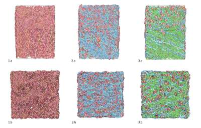 |
90 | Large-scale simulations to create large collections of realistic white matter samples using MEDUSA
Alexis Brullé1, Anas Bachiri1, Christophe Destrieux2, Gilles Wiber3, Thierry Delzescaux 4, Ivy Uszynski1, and Cyril Poupon1
1BAOBAB/NeuroSpin, Université Paris-Saclay, CNRS, CEA, Gif-sur-Yvette, France, 2iBrain U1253, Université de Tours, CHU Bretonneau, INSERM, Tours, France, 3DSSI, DIF, DAM, CEA, Bruyères-le-Châtel, France, 4LMN, MIRCen, Université Paris-Saclay, CNRS, CEA, Fontenay-aux-Roses, France
In this work, we demonstrate that the MEDUSA simulator can be used to create huge collections of brain white matter tissue microstructure samples in the frame of large-scale HPC simulation campaigns and how these Big Data could serve the design of novel computational models able to decode white matter microstructure.
|
||
2727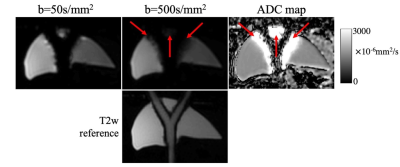 |
91 | Fabrication of an Anthropomorphic, In-Vitro Liver Flow Phantom for Use in Motion-Robust MRI Sequence Validation Experiments
James Rice1,2, Ruiqi Geng2,3, Diego Hernando2,3,4,5, and Alejandro Roldan-Alzate1,2
1Mechanical Engineering, University of Wisconsin-Madison, Madison, WI, United States, 2Radiology, University of Wisconsin-Madison, Madison, WI, United States, 3Medical Physics, University of Wisconsin-Madison, Madison, WI, United States, 4Electrical and Computer Engineering, University of Wisconsin-Madison, Madison, WI, United States, 5Biomedical Engineering, University of Wisconsin-Madison, Madison, WI, United States
A major limitation of standard diffusion-weighted imaging (DWI) in the various organs, including the liver, is the presence of artifacts, bias, and poor reproducibility resulting from physiologic cardiovascular-induced motion. Recent efforts to develop motion-robust DWI sequences show promise, but there is an important unmet need for highly controlled validation in motion phantoms. This study presents the fabrication of an anthropomorphic liver flow phantom that can produce compressive motion in the liver to generate and control artifact seen in DW images. This phantom may enable improved validation of motion-robust DWI and other motion-robust applications.
|
||
2728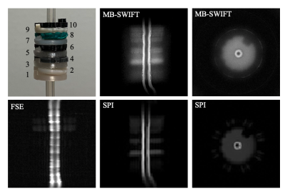 |
92 | MR-visibility and relaxation properties of 3-D printed FDM polymers
Teemu Tuomainen1, Antti Paajanen1, and Mikko J. Nissi1,2
1Department of Applied Physics, University of Eastern Finland, Kuopio, Finland, 2Research Unit of Medical Imaging, Physics and Technology, University of Oulu, Oulu, Finland
Increasing availability and affordability of 3-D printers utilizing fused deposition modeling (FDM) have enabled their widespread use especially in pre-clinical MRI, allowing in-house design and manufacturing of elaborate coil housings, animal holders, sample holders, etc. Here, we investigated ten common and broadly available FDM polymers for their MRI-visibility at 9.4T. We utilized ultra-short echo time imaging sequences, specifically, single-point-imaging (SPI) and multi-band SWIFT to measure the relaxation properties of the different polymers. The findings support the selection of appropriate printing materials when either visibility or invisibility for ultra-short echo time imaging is required.
|
||
2729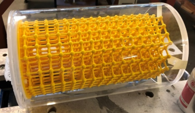 |
93 | Comparing geometric accuracy in MRI using a 3D printed distortion phantom
Alex Barrett1 and Marc Rea1
1Medical Physics, Clatterbridge Cancer Centre NHS Foundation Trust, Liverpool, United Kingdom
Distortion in MRI can have a significant impact on diagnosis and radiotherapy treatment planning. We have 3D printed a large, low cost phantom that we used along with a CT ground truth to measure 6 MR scanners geometric accuracy. We found significantly more distortion for the 3T and mobile scanners tested which we conclude means they would require a more careful evaluation of distortion if they are to be used for radiotherapy applications and that care should be taken for all scanners where specialist radiotherapy applications such as stereotactic radiosurgery are used.
|
||
2730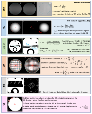 |
94 | A protocol for the acceptance tests of an ultra-high field MR system, in compliance with new standard IEC 62464-1:2018
Laura Biagi1,2, Paolo Cecchi2, and Michela Tosetti1,2
1Laboratory of Medical Physics and Magnetic Resonance, IRCCS Stella Maris, Pisa, Italy, 2Imago7 Research Foundation, Pisa, Italy
The European Standard IEC 62464-1:2018 [1] describes the methodologies to carry out acceptance and constancy tests of an MR scanner. Its come into effect extends its applicability in the range of ultra-high field (UHF) tomographers. Here, we present a protocol, with an effective time of acquisition of about 11 hours, developed for the testing of a 7T MR system, in order to address all the requirements of the standard for the evaluation of essential image quality parameters. The protocol provided for the test of two different coil configurations, using proper phantoms.
|
||
2731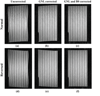 |
95 | B0 Inhomogeneity Characterization and Correction on an open bore MRI-Linac
Shanshan Shan1,2, Paul Liu1,2, David Waddington1,2, Bin Dong2, Mingyan Li3, Fangfang Tang3, Gary Liney2, Feng Liu3, Paul Keall1,2, and Brendan Whelan1
1ACRF Image X Institute, University of Sydney, Sydney, Australia, 2Ingham Institute For Applied Medical Research, Sydney, Australia, 3School of Information Technology and Electrical Engineering, University of Queensland, Brisbane, Australia
MRI-Linac systems require high fidelity geometric information to localize and track tumors during radiotherapy treatments. B0 field inhomogeneity causes image distortions and can provide inaccurate tumor anatomy. Here, we develop a high-order spherical harmonic method to correct for B0 inhomogeneity induced geometric distortions. Experimental data acquired from a 1T open bore MRI-Linac was used to validate the proposed method.
|
||
2732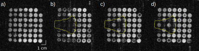 |
96 | Convection triggers local MR signal loss during proton beam irradiation of liquid water phantoms
Juliane Peter1,2, Sebastian Gantz1,2, Leonhard Karsch1,2, Jörg Pawelke1,2, and Aswin Hoffmann1,2,3
1OncoRay – National Center for Radiation Research in Oncology, Faculty of Medicine and University Hospital Carl Gustav Carus, Technische Universität Dresden, Helmholtz-Zentrum Dresden-Rossendorf, Dresden, Germany, 2Helmholtz-Zentrum Dresden-Rossendorf, Institute of Radiooncology – OncoRay, Dresden, Germany, 3Department of Radiotherapy and Radiation Oncology, Faculty of Medicine and University Hospital Carl Gustav Carus, Technische Universität Dresden, Dresden, Germany
MRI-based proton beam visualisation in water has proven feasible in exploratory irradiation experiments performed on a first research prototype in-beam MRI system. Beam-induced convection was hypothesised to be implicated into MR signal loss observed within the beam volume. In this study, this hypothesis was tested in liquid water-filled phantoms by suppression of convection-induced motion using mechanical barriers and temperature control of water expansivity. In absence of convection-induced motion, no beam-induced signal changes occurred, supporting the hypothesis that convection triggers local MR signal loss during proton beam irradiation. The elucidation of the exact mechanism of convection-induced signal loss requires further investigation.
|
||
 Back to Meeting Home
 Back to the Program-at-a-Glance
The International Society for Magnetic Resonance in Medicine is accredited by the Accreditation Council for Continuing Medical Education to provide continuing medical education for physicians.
 Back to Meeting Home
 Back to the Program-at-a-Glance
 View the Presentation