Digital Poster
Phantoms & System Imperfections II
Joint Annual Meeting ISMRM-ESMRMB & ISMRT 31st Annual Meeting • 07-12 May 2022 • London, UK

Digital Poster
Phantoms & System Imperfections II
| Computer # | ||||
|---|---|---|---|---|
2807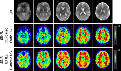 |
75 | Evaluation of the REFILL dynamic distortion correction method for fMRI
Simon Daniel Robinson1,2,3, Beata Bachrata3, Korbinian Eckstein3, Barbara Dymerska4, Saskia Bollmann2, Steffen Bollmann5, Shota Hodono2, Martijn Cloos2, Monique Tourell2, Jin Jin6, Kieran O'Brien6, David Reutens2, Siegfried Trattnig3, Christian Enzinger1, and Markus Barth5
1Department of Neurology, Medical University of Graz, Graz, Austria, 2Centre for Advanced Imaging, University of Queensland, Brisbane, Australia, 3Department of Biomedical Imaging and Image-guided Therapy, Medical University of Vienna, Vienna, Austria, 4UCL Queen Square Institute of Neurology, University College London, London, United Kingdom, 5School of Electrical Engineering and Information Technology, University of Queensland, Brisbane, Australia, 6Siemens Healthineers, Brisbane, Australia
We evaluate the performance of a recently-proposed dynamic distortion correction (DDC) method for fMRI at 7T, with a task which generates field fluctuations. The Reverse-Encoded First Image and Low resoLution reference scan (REFILL) method generates fieldmaps from the phase of standard, single-echo EPI from fMRI time series, using coil sensitivity information from a fast reference scan and removing other, non-B0-related contributions to the phase, which are calculated from a readout-reverse EPI volume. In contrast to conventional static distortion correction (SDC) with a GE-based fieldmap, REFILL captured dynamic changes to the field, leading to an accurate correction and increased tSNR.
|
||
2808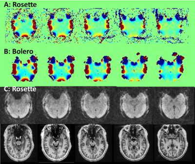 |
76 | B0 shimming in human brain using rosette encoding at 7T Video Not Available
Jullie W Pan1, Chan H Moon2, and Hoby P Hetherington3,4
1Radiology, University of Missouri Columbia, Columbia, MO, United States, 2Radiology, University of Pittsburgh, Pittsburgh, PA, United States, 3Resonance Research Inc., Billerica, MA, United States, 4University of Missouri Columbia, Columbia, MO, United States
With increasing use of ultra-high field, there is a need for fast and efficient field mapping. Towards this purpose, the non-Cartesian circular rosette trajectory is advantageous due to its low gradient demands and its efficiency due to the simultaneous acquisition of spatial and field encoding information. We describe the implementation of the circular rosette trajectory for field mapping at 7T in the human brain and compare this to the highly accurate although slow 5 echo Bolero field map.
|
||
2809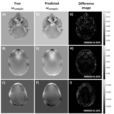 |
77 | REconstruction of MR images acquired in highly inhOmogeneous fields using DEep Learning (REMODEL 2.0)
Marina Manso Jimeno1,2, John Thomas Vaughan Jr.1,2, and Sairam Geethanath2
1Department of Biomedical Engineering, Columbia University, New York, NY, United States, 2Columbia Magnetic Resonance Research Center, New York, NY, United States The trade-off of a shorter MRI magnet design is having to compromise image quality due to reduced field homogeneity. Moreover, the sample induces unique field perturbations during the scan that can only be corrected if the exact field map is known. Here we proposed a DL model that synthesizes sample-specific field maps based on artifact-corrupted acquired images and the system’s field distribution. In the retrospective study, we obtained a mean NRMSE of 0.04 during testing between model output and the true field maps and found that using the model output for correction significantly reduced the artifacts on the images. |
||
2810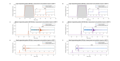 |
78 | Towards gradient perturbation correction in diffusion weighted imaging based on the gradient system transfer function
Hannah Scholten1 and Herbert Köstler1
1Department of Diagnostic and Interventional Radiology, University of Würzburg, Würzburg, Germany
Eddy current induced field perturbations can cause artifacts in diffusion weighted imaging (DWI). Such perturbations may be calculated and corrected for by the gradient system transfer function (GSTF), provided the gradient system behaves linear and time-invariant. We discovered that the linearity assumption can be violated by gradients with high zeroth order moments. We therefore propose a modified measurement scheme for the GSTF including higher gradient moments to better approximate their behavior. Our approach substantially reduces errors in the predicted gradient time courses. We may thus enable the application of GSTF-based correction methods to DWI in the future.
|
||
2811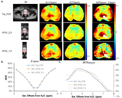 |
79 | Reduced field of view CEST for high performance prostate imaging at 3T human scanner
Xubin Chai1,2, Chuyu Liu1, Chu Wang1, Rong Xue2, and Xiaolei Song1
1Center for Biomedical Imaging Research, Department of Biomedical Engineering, Tsinghua University, Beijing, China, 2State Key Laboratory of Brain and Cognitive Science, Institute of Biophysics, Chinese Academy of Sciences, Beijing, China
For imaging small-sized organs like the prostate, reduced-field-of-view (rFOV) technique is useful for shortening scan time, increasing resolution and reducing artifacts caused by field inhomogeneity and motion. Herein on a 3T clinical scanner, rFOV CEST was obtained using an off-resonance saturation preparation (seconds long), followed by readouts at the crossing section of a 90 deg excitation slab and a 180 deg refocus slab that had an angle in between. For saturation powers of 0.7 uT and 2 uT, CEST contrast maps and quantitative curves suggested the rFOV-CEST outperformed the Traditional-FOV acquisitions, which has potential for prostate CEST imaging at 3T.
|
||
2812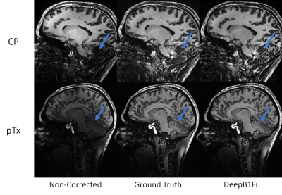 |
80 | Generalized Framework for Homogeneous Ultra-High-Field Brain Imaging Video Permission Withheld
Patrick Liebig1, Juergen Herrler2, Raphael Tomi-Tricot3,4,5, Sydney Williams6, Belinda Ding-Yuan3,6, Majd Hlou7, Venkata Chebrolu8, Fasil Gadjimuradov7,9, Tom Hilbert10,11,12, Tobias Kober10,11,12, Rene Gumbrecht1, Robin Martin Heidemann1, Thomas Benkert7, Chris Rodgers13, David Andrew Porter6, Iulius Dragonu3, Armin Nagel14,15, and Shaihan Malik4,5
1Siemens Healthcare GmbH, Erlangen, Germany, 2Department of Neuroradiology, University Hospital Erlangen, Friedrich-Alexander-Universität Erlangen-Nürnberg (FAU), Erlangen, Germany, 3MR Research Collaborations, Siemens Healthcare Limited, London, United Kingdom, 4Department of Biomedical Engineering, School of Biomedical Engineering and Imaging Sciences, King’s College London, St. Thomas’ Hospital, London, United Kingdom, 5Center for the Developing Brain, School of Biomedical Engineering and Imaging Sciences, King’s College London, St. Thomas’ Hospital, London, United Kingdom, 6Imaging Centre of Excellence, University of Glasgow, Glasgow, United Kingdom, 7MR Applications Predevelopment, Siemens Healthcare GmbH, Erlangen, Germany, 8Siemens Medical Solutions USA, Inc., Rochester, MN, United States, 9Department of Computer Science, Friedrich-Alexander-Universität Erlangen-Nürnberg (FAU), Erlangen, Germany, 10Advanced Clinical Imaging Technology, Siemens Healthcare AG, Lausanne, Switzerland, 11Department of Radiology, Lausanne University Hospital and University of Lausanne, Lausanne, Switzerland, 12LTS5, École Polytechnique Fédérale de Lausanne (EPFL), Lausanne, Switzerland, 13Wolfson Brain Imaging Centre, University of Cambridge, Cambridge, United Kingdom, 14Institute of Radiology, University Hospital Erlangen, Friedrich-Alexander-Universität Erlangen-Nürnberg (FAU), Erlangen, Germany, 15Medical Physics in Radiology, German Cancer Research Center (DKFZ), Heidelberg, Germany 7T MRI is affected by inhomogeneous transmit and receive B1 field that can impede the inherent gains in signal-to-noise ratio. pTx provides excellent results in correcting the transmit field and showed feasibility in a clinical setting as well1,2. Although multiple algorithms have been developed to correct for the receive profile or signal homogeneities in general, each algorithm has its own shortcomings. Here, we suggest combining prospective correction of the transmit field by pTx with a deep learning network to retrospectively correct for the remaining signal inhomogeneities (mainly receive field variations) in a generalized fashion. |
||
2813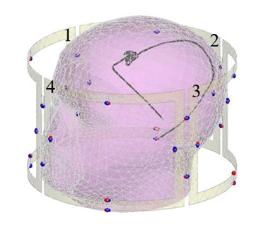 |
81 | Safety of 4-channel parallel RF transmission MRI at 3 T: Effects of system uncertainty and system failure pertinent to deep brain stimulation
Benson Yang1,2, Chih-Hung Chen2, and Simon J Graham1,3
1Physical Sciences, Sunnybrook Research Institute, Toronto, ON, Canada, 2Electrical and Computer Engineering, McMaster University, Hamilton, ON, Canada, 3Medical Biophysics, University of Toronto, Toronto, ON, Canada
Parallel radiofrequency transmission (pTx) technology continues to demonstrate its potential at addressing MRI safety concerns relating to patients with implanted deep brain stimulation devices. One promising technique involves electromagnetic simulation that determine optimized pTx inputs to generate a safe mode of imaging surrounding the implanted device. However, in practice, instrumentation uncertainty can impact the ability of the pTx system to accurately produce the desired signals. The present work studied the safety implications of system uncertainty and system failure in a 4-channel pTx platform. The preliminary results showed that in a worst-case scenario, temperature elevations that exceed MRI guidelines are possible.
|
||
2814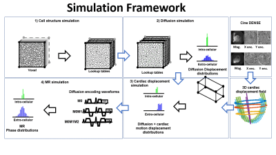 |
82 | Cardiac motion modeling using DENSE imaging to study cardiac diffusion tensor MRI
Kevin Moulin1,2, Tanjib Rahman 3, Magalie Viallon1,2, Pierre Croisille1,2, and Luigi Perotti3
1CREATIS, Lyon, France, 2Department of Radiology, University Hospital Saint-Etienne, Saint Etienne, France, 3Department of Mechanical and Aerospace Engineering, University of Central Florida, Orlando, FL, United States Cardiac Diffusion Tensor imaging (cDTI) is a powerful non-contrast technique that can characterize cellular structure in vivo. Despite motion-compensated diffusion encoding designs, cDTI remains sensitive to unwanted cardiac motion, particularly during diastole. The lack of knowledge of the detailed interaction between real cardiac motion and motion-compensated gradients prevents more advanced diffusion encoding designs. The objective of this work is to provide a new simulation framework to study the interaction between cardiac displacements estimated in vivo from DENSE imaging and motion-compensated diffusion encoding waveforms. |
||
2815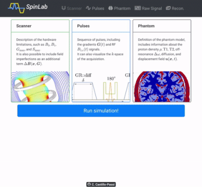 |
83 | MRIsim.jl: A framework for end-to-end spin-level MRI simulations with GPU acceleration
Carlos Castillo-Passi1,2,3, Ronal Coronado2,3,4, Sergio Uribe2,3,5, and Pablo Irarrazaval1,2,3,4
1Institute of Biological and Medical Engineering, Pontificia Universidad Católica de Chile, Santiago, Chile, 2Biomedical Imaging Center, Pontificia Universidad Católica de Chile, Santiago, Chile, 3Millennium Nucleus in Cardiovascular Magnetic Resonance, Cardio MR, Santiago, Chile, 4Electrical Engineering, Pontificia Universidad Católica de Chile, Santiago, Chile, 5Radiology Department, School of Medicine, Pontificia Universidad Católica de Chile, Santiago, Chile
In this work, we proposed a GPU-enabled MRI simulation framework written in Julia. The primary purpose of this simulator is to evaluate imaging pipelines from the acquisition to the reconstruction. To showcase this, we simulated two variations of an MRF sequence with radial acquisitions and then reconstructed using two methods. We demonstrated that this framework aids users to quickly write and test state-of-the-art MRI techniques and showed its potential to compare imaging pipelines in quantitative mapping.
|
||
2816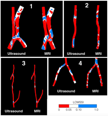 |
84 | Image-based computer simulations of haemodynamics and blood cells distributions: A comparison between MRI and ultrasound
Vasit Sirilapanan1, Yongmann M Chung1, Richard Harrison2, Clare Cameron3, Michael Lynn4, Charles Hutchinson5, and Farhan Ahmed4
1School of Engineering, University of Warwick, Coventry, United Kingdom, 2School of Psychology and Clinical Language Sciences, University of Reading, Reading, United Kingdom, 3University of Herefordshire, Hatfield, United Kingdom, 4Department of Radiology, Royal Berkshire Hospital, Reading, United Kingdom, 5Medical School, University of Warwick, Coventry, United Kingdom
MRI-based CFD simulations are compared with ultrasound-based CFD to ascertain the capability of ultrasound.
|
||
 Back to Meeting Home
 Back to the Program-at-a-Glance
The International Society for Magnetic Resonance in Medicine is accredited by the Accreditation Council for Continuing Medical Education to provide continuing medical education for physicians.
 Back to Meeting Home
 Back to the Program-at-a-Glance
 View the Presentation