Digital Poster
RF Pulse Design, Parallel Transmission & B1 Shimming I
Joint Annual Meeting ISMRM-ESMRMB & ISMRT 31st Annual Meeting • 07-12 May 2022 • London, UK

Digital Poster
RF Pulse Design, Parallel Transmission & B1 Shimming I
| Computer # | ||||
|---|---|---|---|---|
2864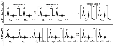 |
109 | Acceleration Methods to Acquire Fast 3D Multi-Channel Absolute B1+ Maps at 7T for 8-Channel pTx
James L. Kent1, Ladislav Valkovic2,3, Iulius Dragonu4, and Aaron T. Hess1
1Wellcome Centre for Integrative Neuroimaging, FMRIB, Nuffield Department of Clinical Neurosciences, University of Oxford, Oxford, United Kingdom, 2Oxford Centre for Clinical Magnetic Resonance Research, University of Oxford, Oxford, United Kingdom, 3Department of Imaging Methods, Institute of Measurement Science, Slovak Academy of Sciences, Bratislava, Slovakia, 4Siemens Healthineers, Frimley, United Kingdom Ultra-high field MRI with parallel transmit offers significant advantages but B1 transmit maps must be acquired to utilize its full potential. Acquiring absolute B1+ maps for each channel is a time-consuming process and limits the application of UHF to clinical practice. We compare two methods of accelerating the transmit mapping procedure to obtain fast volumetric multi-channel maps for an 8-channel transmit array. Both methods make use of a low-rank reconstruction (TxLR) from undersampled data and a modified pre-saturation TurboFLASH (satTFL) B1+ mapping sequence in combination with either Fourier encoding or a B1TIAMO style acquisition. |
||
2865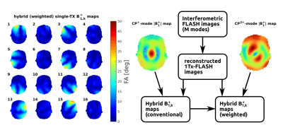 |
110 | Interferometric, Hybrid, and Weighted $$$B_1^+$$$ Mapping for Expedited RF Calibration of Parallel Transmit Ultrahigh-Field MRI
Dario Bosch1,2, Fabian Müller1, and Klaus Scheffler1,2
1Magnetic Resonance Center, MPI for Biological Cybernetics, Tuebingen, Germany, 2Department for Biomedical Magnetic Resonance, University Tuebingen, Tuebingen, Germany
With an increasing number of transmit channels, faster methods for acquiring $$$B_1^+$$$ maps on pTx MRI systems grow in importance. We compared different interferometry modes for accurate absolute $$$B_1^+$$$ mapping using satTFL. We also introduced weighted hybrid $$$B_1^+$$$ mapping as an attempt to improve hybrid $$$B_1^+$$$ mapping. In this, a second absolute $$$B_1^+$$$ map was introduced as an attempt to further shorten measurement duration while keeping acceptable accuracy. From the tested interferometry modes, phase-cycling performed best in both absolute and weighted mapping. Weighted hybrid mapping exceeded conventional hybrid mapping substantially and could therefore be sufficiently accurate for many applications.
|
||
2866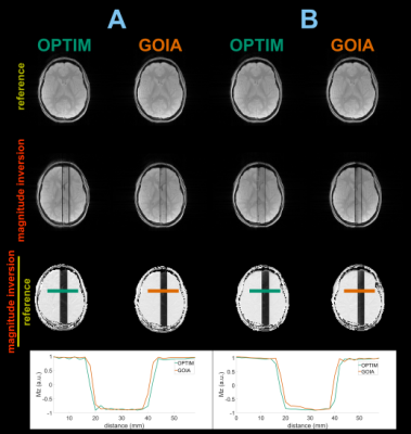 |
111 | Improved B1+- and resonance offset-robust slice-selective inversion pulses
Christina Graf1, Adam Berrington2, Martin Soellradl1, Armin Rund3, and Rudolf Stollberger1
1Institute of Medical Engineering, Graz University of Technology, Graz, Austria, 2Sir Peter Mansfield Imaging Centre, School of Physics and Astronomy, University of Nottingham, Nottingham, United Kingdom, 3Institute for Mathematics and Scientific Computing, University of Graz, Graz, Austria
In this work, an optimal control framework and its application for the development and improvement of $$$B_1^+-$$$ and $$$\Delta B_0-$$$robust, slice-selective inversion pulses is introduced. The optimized RF pulse shows superior performance compared to adiabatic, state of the art pulses through simulations, phantom experiments, and in vivo measurements. Measurements on a 3T MR system revealed improved slice-profile quality of the optimized pulse, especially for low $$$B_1^+$$$ and high $$$\Delta B_0$$$. Since the performance is independent of the field strength, its application in ultrahigh-field spectroscopy seems straightforward.
|
||
2867 |
112 | Calibration of saturation prepared turbo FLASH B1+ maps by actual flip angle imaging at 7T.
Jan Sedlacik1,2,3,4, Raphael Tomi-Tricot1,2,3,5, Tom Wilkinson1,2,3, Pip Bridgen1,2,3, Franck Mauconduit6, Alexis Amadon6, Sharon Giles1,2,3, Joseph V Hajnal1,2,3, and Shaihan J Malik1,2,3
1Centre for the Developing Brain, School of Biomedical Engineering & Imaging Sciences, King’s College London, London, United Kingdom, 2Biomedical Engineering Department, School of Biomedical Engineering & Imaging Sciences, King’s College London, London, United Kingdom, 3London Collaborative Ultra high field System (LoCUS), London, United Kingdom, 4Radiology Department, Great Ormond Street Hospital for Children, London, United Kingdom, 5MR Research Collaborations, Siemens Healthcare Limited, Frimley, United Kingdom, 6Paris-Saclay University, CEA, CNRS, BAOBAB, NeuroSpin, Gif-sur-yvette, France
A fast and accurate B1+mapping method is essential for making proper use of a parallel transmit array with high number of coil elements in a clinical setting. The saturation-prepared turbo FLASH method offers this capability and is implemented in the fully automated pre-scan adjustments of the currently only clinical ultra-high magnetic field strength MRI system. However, some parallel transmit methods require an even higher accuracy. This can be achieved by calibrating the fast B1+measurements with a more accurate but longer B1+mapping method.
|
||
2868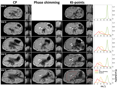 |
113 | Whole-liver flip angle shimming at 7T using eight-channel parallel transmission kt-points pulses with FPE-DREAM B1+ mapping
Bobby Runderkamp1, Wietske van der Zwaag2, Thomas Roos2, Gustav Strijkers3, Matthan Caan3, and Aart Nederveen1
1Radiology & Nuclear Medicine, Amsterdam UMC, Amsterdam, Netherlands, 2Spinoza Center for Neuroimaging, Amsterdam, Netherlands, 3Biomedical Engineering and Physics, Amsterdam UMC, Amsterdam, Netherlands
Liver MRI could benefit from the increased SNR at 7T but suffers from severe flip angle inhomogeneity. In this work, eight-channel parallel transmission is used for flip angle shimming, comparing circularly polarized transmission and phase shimming with a kt-points pulse. Fourier phase-encoded DREAM is used for artifact-free B1+ mapping. With kt-points, the nominal flip angle of 8˚ could be achieved with homogeneous signal over the entire liver, while phase shimming only delivers a flip angle of approximately 4˚ with less homogeneity. Future work is directed towards use of universal slab-selective kt-points pulses to make 7T liver MRI clinically feasible.
|
||
2869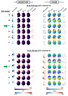 |
114 | Inter-coil applicability of trained neural networks for B1+ prediction in parallel-transmit.
Alix Plumley1 and Emre Kopanoglu1
1Cardiff University, Cardiff, United Kingdom
Motion-resolved parallel-transmit (pTx) B1-maps can be predicted using neural networks, facilitating online pulse re-design – a prospective solution to motion. However, networks require large training-datasets. Since different pTx coils inherently produce different B1-distributions, it is unclear whether coil-specific training-datasets are needed. Here, we train networks on simulated data from one coil-model and test on 6 differently-sized coil-models. While performance was optimal for the coil on which networks were trained, B1-prediction yielded lower error than that caused by motion in ≥91% of magnitude, and ≥55% of phase evaluations for 5 out of the 6 models, demonstrating some generalisability across coils.
|
||
2870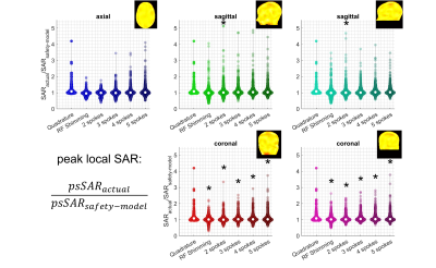 |
115 | Head Position Related SAR Uncertainty Depends on Slice Orientation and Pulse Complexity
Emre Kopanoglu1
1CUBRIC / Psychology, Cardiff University, Cardiff, United Kingdom
Safety models on scanners are unaware of the actual patient position while excitation pulses are inherently position dependent. This study investigates the effect of this positional mismatch on SAR estimation for axial, coronal and sagittal slice orientations. The positional mismatch yields up to 5.2-fold underestimation of peak local SAR. RF shimming and 2-spoke parallel-transmit pulses for axial slice orientation have reduced SAR-sensitivity to positional mismatch with the worst-case underestimation being <2.0-fold whereas a reduced SAR-sensitivity was not observed for coronal and sagittal slices. For extreme head positions not represented in safety-models, axial RF shimming / 2-spokes parallel-transmit pulses maybe beneficial.
|
||
2871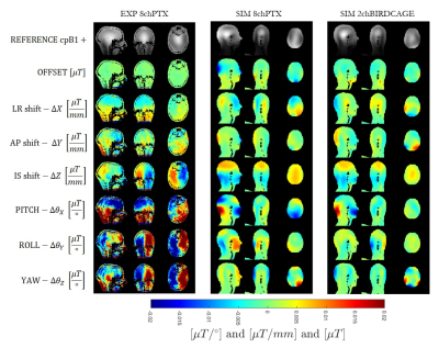 |
116 | Motion-Dependent Low-Order Predictive Linear Model for Patient Safety Assessment at 7T with Technically Feasible Coil Models
Amer Ajanovic1,2, Yannick Brackenier1,2, Raphael Tomi-Tricot1,2, Joseph V Hajnal1,2,3, and Shaihan Malik1,2
1Biomedical Engineering Department, School of Biomedical Engineering and Imaging Sciences, King's College London, LONDON, United Kingdom, 2London Collaborative Ultra high field System (LoCUS), London, United Kingdom, 3Centre for the Developing Brain, School of Biomedical Engineering and Imaging Sciences, King's College London, London, United Kingdom
Establishing a comprehensive model that addresses patient safety at UHF MRI with respect to motion is a non-trivial task because of the underlying computationally expensive frameworks. Using MARIE we computed a large number of pose simulations of 3 feasible coil models (birdcage, 4-channel and 8-channel pTx) in proximity to Duke body model to allow the parameter space to be well explored. It is found that a low order linear model can capture most of the variation in cpB1+. We also observe low variations in cpSAR for 2 realistic coil models across the conditions examined, with 8-channel coil being most robust.
|
||
2872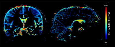 |
117 | Calibration-free parallel transmission for improved CSF mobility imaging at 7T
Philipp Ehses1, Lydiane Hirschler1,2, Eberhard Daniel Pracht1, Katerina Deike-Hofmann1, Vincent Gras3, Franck Mauconduit3, Aurélien Massire4, Nicolas Boulant3, Matthias J. P. van Osch2, and Tony Stoecker1,5
1German Center for Neurodegenerative Diseases (DZNE), Bonn, Germany, 2Radiology, Leiden University Medical Center, Leiden, Netherlands, 3University of Paris-Saclay, CEA, CNRS, BAOBAB, NeuroSpin, Gif sur Yvette, France, 4Siemens Healthcare SAS, Saint-Denis, France, 5Department of Physics and Astronomy, University of Bonn, Bonn, Germany
Better understanding of how the brain cleans itself is of high importance as its failure has been linked to various neurodegenerative diseases. A first step towards elucidating CSF-mediated clearance mechanisms is the measurement of CSF motion in the human brain. In this work, we present an imaging sequence for CSF mobility measurements employing calibration-free parallel transmission. During the imaging session, no pulse calculations or complex B1 shimming procedures are necessary, making this approach feasible for large scale patient studies.
|
||
2873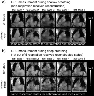 |
118 | Respiration robust universal pulses in the human heart at 7T for shallow and deep breathing
Christoph Stefan Aigner1, Sebastian Dietrich1, Felix Krüger1, Max Lutz1, and Sebastian Schmitter1,2,3
1Physikalisch-Technische Bundesanstalt (PTB), Braunschweig and Berlin, Germany, 2German Cancer Research Center (DKFZ), Medical Physics in Radiology, Heidelberg, Germany, 3University of Minnesota, Center for Magnetic Resonance Research, Minneapolis, MN, United States
This study demonstrates the design and application of a new class of universal pulses (UP) designed for a library of 22 non-respiration resolved (NRR) B1+-maps acquired in-vivo during shallow breathing (SB) and 30 respiration resolved (RR) B1+-maps acquired during deep breathing (DB). The proposed UP was tested on 9 NRR SB (test-cases-SB) and 15 RR DB B1+-maps (test-cases-DB) and was applied experimentally in five volunteers (test-cases-DB) along with tailored pulses in multiple gradient echo measurements. Compared to tailored pulses, UP resulted in a slightly overall decrease of the FA homogeneity but similar image quality across all test-cases.
|
||
2874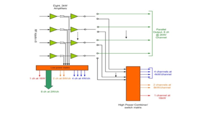 |
119 | Eight Channel, Dual-Matrixed, Parallel Transmit RF Power Amplifier for 1.5T MRI
Thomas Vaughan1, Michael Garwood2, Steve Suddarth3, Lance DelaBarre2, and Dan Myer4
1BME, Radiology, Columbia University, New York, NY, United States, 2University of Minnesota, Minneapolis, MN, United States, 3University of Minneosta, Minneapolis, MN, United States, 4Communications Power Corp., Hauppauge, NY, United States
A new, laboratory grade RF power amplifier that offers eight channels of low, CW power for simultaneous transmit and receive applications, and eight channels of high, peak-pulsed power for conventional MRI is presented. These FPGA controlled, multiple transmit channels can be matrix switched into 1, 2, 4, and 8 channel combinations for a range of 2W, 4W, 8W and 16W CW, and 2kW, 4kW, 8kW and 16kW peak power pulses. Further each channel can be used for independent phase, gain, frequency, space and time for maximum flexibility in RF field modulation. This power amplifier meets our wide ranging experimental requirements.
|
||
 Back to Meeting Home
 Back to the Program-at-a-Glance
The International Society for Magnetic Resonance in Medicine is accredited by the Accreditation Council for Continuing Medical Education to provide continuing medical education for physicians.
 Back to Meeting Home
 Back to the Program-at-a-Glance
 View the Presentation