Digital Poster
MRI Safety I
Joint Annual Meeting ISMRM-ESMRMB & ISMRT 31st Annual Meeting • 07-12 May 2022 • London, UK

| Computer # | ||||
|---|---|---|---|---|
2545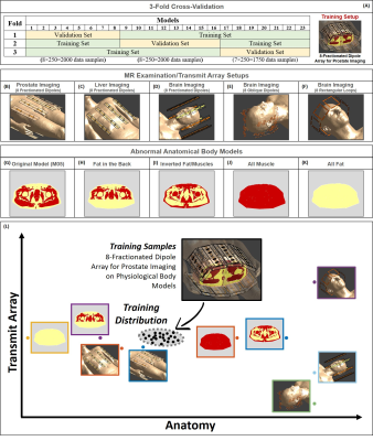 |
77 | Detection of Samples Out of Training Distribution: Rejection of Potential Erroneous Local SAR Predictions
Ettore Flavio Flavio Meliado1,2,3, Alexander A.J. Raaijmakers1,2,4, P.R. A.J. Luijten1, and C.A.T. A.J. van den Berg2,5
1Department of Radiology, University Medical Center Utrecht, Utrecht, Netherlands, 2Computational Imaging Group for MR diagnostics & therapy, Center for Image Sciences, University Medical Center Utrecht, Utrecht, Netherlands, 3Tesla Dynamic Coils BV, Zaltbommel, Netherlands, 4Biomedical Image Analysis, Dept. Biomedical Engineering, Eindhoven University of Technology, Eindhoven, Netherlands, 5Department of Radiotherapy, Division of Imaging & Oncology, University Medical Center Utrecht, Utrecht, Netherlands
Recognize potential hazardous situations far from the modeled MR examination scenarios is crucial for RF safety applications and especially for deep-learning-based approaches. Last year we presented a Bayesian deep-learning approach for local SAR assessment. This approach allowed accurate local SAR estimations and returned reliable feedbacks on the error/uncertainty of the estimates for the MR examination scenario observed during training. However, it also showed the dangers of using the predicted uncertainty to identify out-of-training MR examination scenarios. In this study, we propose a simple approach to detect/reject potential erroneous local SAR predictions due to out-of-training transmit array and/or anatomical variations.
|
||
2546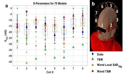 |
78 | Effects of Intersubject Differences on Scattering Parameters, SAR, and B1+ in a 7T 8ch. Head Coil
Benjamin Michael Hardy1,2, Rana Banik1,3, Xinqiang Yan1,4, and Adam W Anderson1,3,4
1Vanderbilt University Institute of Imaging Science, Vanderbilt University, Nashville, TN, United States, 2Department of Physics and Astronomy, Vanderbilt University, Nashville, TN, United States, 3Department of Biomedical Engineering, Vanderbilt University, Nashville, TN, United States, 4Department of Radiology and Radiological Sciences, Vanderbilt University Medical Center, Nashville, TN, United States Shorter wavelengths and higher frequencies at UHF contribute to complicated electric field distributions and higher power absorption in the human head leading to potential safety concerns. Scattering parameters, local and global SAR, and B1+ fields are calculated in an 8-channel surface loop Tx array simulated over 70 head-and-shoulder models of 10 tissue compartments. RF shimming methods and pulse sequences are analyzed over each model to demonstrate local and global SAR variation in a population. Patient proximity, coil loading and design, patient composition, and RF shim weights contribute to the variations in SAR and B1+ experienced by each subject. |
||
2547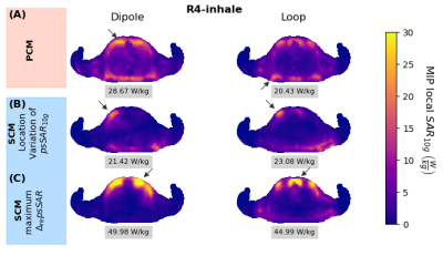 |
79 | Respiratory motion affects peak spatial SAR value and location – impact on safety supervision for 7T cardiac imaging
Natalie Schoen1, Frank Seifert1, Gregory J. Metzger2, Oliver Speck3, Bernd Ittermann1, and Sebastian Schmitter1,2
1Physikalisch-Technische Bundesanstalt (PTB), Braunschweig and Berlin, Berlin, Germany, 2Center for Magnetic Resonance Research, University of Minnesota, Minneapolis, MN, United States, 3Department for Biomedical Magnetic Resonance, Otto-von-Guericke University Magdeburg, Magdeburg, Germany Respiratory motion impacts peak spatial specific absorption rate (psSAR) at cardiac UHF MR imaging as shown previously for individual$$$B_1^+$$$shim vectors. Here two saftey supervision modes were compared: a local SAR control mode (SCM) and a channel-wise power control mode (PCM). Results show amplitude and location changes of the psSAR between inhale and exhale and this effect depends on the coil setup, element type and breathing pattern. Respiratory-induced psSAR variations are lower when applying PCM compared to SCM, while PCM is more conservative with respect to the total power allowed. |
||
2548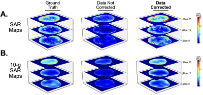 |
80 | Correction Factor for EPT-based Patient-Specific SAR Maps
Jessica A. Martinez1, Alessandro Arduino1, Oriano Bottauscio1, and Luca Zilberti1
1Advanced Materials Metrology and Life Science, Istituto Nazionale di Ricerca Metrologica, Torino, Italy
Specific absorption rate (SAR) values can be obtained using MRI data to calculate the electrical conductivity via Electric Properties Tomography (EPT) and by indirectly obtaining the electric field by the B1+ field. However, only using B1+ will result in incorrect SAR values. In this work, we propose the calculation of a tissue-specific correction factor to mitigate the inaccuracy of SAR.
|
||
2549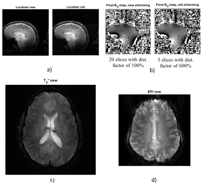 |
81 | An extended version of the restricted SAR protocol allowing safe in vivo testing of RF coils in an unconditional and flexible way
Natalia Dudysheva1,2, Alexis Amadon1, Alexandre Vignaud1, Redha Abdeddaim3, and Franck Mauconduit1
1CEA, NeuroSpin, Paris-Saclay University, CNRS, Gif-sur-Yvette, France, 2Multiwave Imaging, Marseille, France, 3Fresnel Institute, Aix-Marseille University, CNRS, Marseille, France
This work promotes the RF coil elaboration facilitating its in-vivo testing phase. To exclude patient risk, in-vivo studies with each new coil configuration require prior SAR verifications and commission approval lengthening the design process. The so-called “Restricted SAR” (rS) protocol offers a solution to safely use homemade coils in vivo at any development stage without prior validation. This protocol constrains transmitted RF power and automatically respects the SAR safety limits, whatever the SAR distribution. Here, we propose the extended rS protocol with flexible sequence parametrization. This adjustability offers better B0 mapping coverage, optimal B1+-map estimation, and higher spatial image resolutions.
|
||
2550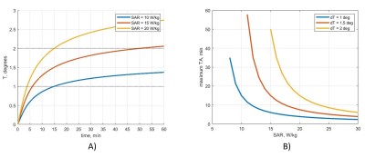 |
82 | New “restricted SAR mode” definition based on a thermal conservative model for relaxed unconditional safe in vivo experiments
Natalia Dudysheva1,2, Nicolas Boulant1, Alexandre Vignaud1, and Franck Mauconduit1
1CEA, NeuroSpin, Paris-Saclay University, CNRS, Gif-sur-Yvette, France, 2Multiwave Imaging, Marseille, France
In this work, we propose to extend the capacities of ultra-low SAR protocol used in coil design. A developed restricted SAR protocol holds the worst-case SAR below the threshold by constraining the total transmitted RF power. Since SAR reflects tissue heating, we exploit a conservative thermal model to derive relaxed SAR constraints depending on acquisition time. This enables to extend the options available for sequences played under this mode, including fat suppression, higher image resolution, or decreased TR.
|
||
2551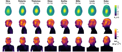 |
83 | Evaluation of SAR and heating in children exposed to a 7T MRI Head Coil
Shaihan J Malik1,2, David J Carmichael1, Jeffrey W Hand1, and Joseph V Hajnal1,2
1Biomedical Engineering Department, School of Biomedical Engineering and Imaging Sciences, King's College London, London, United Kingdom, 2Centre for the Developing Brain, School of Biomedical Engineering and Imaging Sciences, King's College London, London, United Kingdom
Electromagnetic and thermal simulations of a range of models (body mass 14-70 kg) were performed for a 7T (297MHz) birdcage head transmit coil. Head averaged SAR (hdSAR) was found to be the limiting factor for all cases, and a (weak) negative linear correlation with body mass was observed (i.e. hdSAR increases with decreasing body mass). Thermal simulations with fixed blood temperature (assumes good thermoregulation) predict temperature increases remain within guidelines if SAR limits are respected. If blood temperature is allowed to vary then elevated core temperatures are predicted for subjects with smaller body mass even when SAR limits are respected.
|
||
2552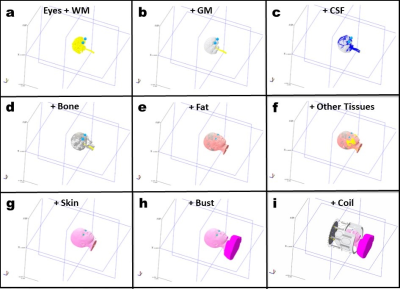 |
84 | Variability of the specific absorption rate in the child’s head using a transmit array head coil at 7T: a simulation study
Maya Delbany1, Michel Luong1,2, Laurent Morel3, Jean-Pierre Adam3, Jean-Christophe Joly3, and Vincent Gras1
1Université Paris-Saclay, CEA, CNRS, BAOBAB, NeuroSpin, 91191, Gif sur Yvette, France, 2CEA, DRF, IRFU, Gif sur Yvette, France, 3CEA, DAM, Gramat, France
In this study, a 3T MRI database is used to create numerical children’s head models in order to study the variability of the specific absorption rate in the head of children at 7T using a transmit array head coil. These numerical models are integrated in a homemade multi-transmit RF coil model and then used to simulate SAR maps in the head using a finite difference time domain solver called GORF.
|
||
2553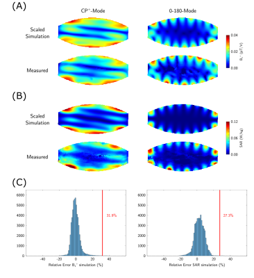 |
85 | A 16-channel transceiver dipole array for 1H body imaging at 10.5T: Validation and VOP enabled imaging in-vivo
Simon Schmidt1, Arcan Erturk1, Xiaoxuan He1, Yigitcan Eryaman1, and Gregory J Metzger1
1Center for Magnetic Resonance Research, University of Minnesota, Minneapolis, MN, United States To realize the increased pTx functionality of a 16-channel transceiver dipole array for body imaging at 10.5T, a validation process was implemented. After quantifying the uncertainty of the RF arrays electromagnetic simulations a total safety factor was determined enabling the implementation of real-time local SAR monitoring on the scanner through use of virtual observation points (VOP). In vivo data was acquired with the VOP enabled power monitoring demonstrating local RF shimming performance with and without local SAR constrained optimizations. |
||
2554 |
86 | Numerical Simulation of Tissue Damage Due to Magnetically Induced Force for Multi-Configuration Passive Medical Devices
Zainul Ihsan1, Michael Schwarz2, Mubashir Husain2, and Gregor Schaefers1,2
1MR:comp GmbH, Gelsenkirchen, Germany, 2MRI-STaR - Magnetic Resonance Institute for Safety, Technology and Research GmbH, Gelsenkirchen, Germany
Numerical simulation was performed to evaluate tissue damage due to magnetically induced (MI) force in 3T Magnetic Resonance (MR) environment for multi-configuration passive medical devices. Static magnetic and mechanical simulations were employed to determine the impact of MI force on the tissue surrounding the implant. The simulation results were used to select the worst-case for passive medical devices and for quantitative assessment of the tissue damage.
|
||
2555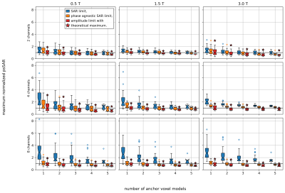 |
87 | On the complexity of pTx systems in SAR assessment
Johannes Petzold1, Bernd Ittermann1, and Frank Seifert1
1Physikalisch-Technische Bundesanstalt (PTB), Berlin, Germany Different methods for SAR control (SAR limit, phase-agnostic SAR limit, amplitude limit) are evaluated in a simulation study of parallel transmit (pTx) body coil systems (2,4, or 8 channels) at different B0 (0.5T, 1.5T, 3T). Criteria are the necessary safety factors to accommodate for subject variability, and the resulting mean(B1+). Due to the high safety factor necessary for a phase-based SAR limit, its mean(B1+) performance becomes effectively the same as the much simpler to implement amplitude limit. |
||
2556 |
88 | Local SAR management strategies for RF shimming in fetal MRI at 3T
Filiz Yetisir1, Esra Abaci Turk1,2, Elfar Adalsteinsson3,4,5, Patricia Ellen Grant1,2,6, and Lawrence L Wald4,7,8
1Fetal-Neonatal Neuroimaging and Developmental Science Center, Boston Children's Hospital, Boston, MA, United States, 2Department of Pediatrics, Boston Children's Hospital, Boston, MA, United States, 3Department of Electrical Engineering and Computer Science, Massachusetts Institute of Technology, Cambridge, MA, United States, 4Harvard-MIT Health Sciences and Technology, Cambridge, MA, United States, 5Institute for Medical Engineering and Science, Massachusetts Institute of Technology, Boston, MA, United States, 6Department of Radiology, Boston Children's Hospital, Boston, MA, United States, 7Athinoula A. Martinos Center for Biomedical Imaging, Harvard Medical School, Boston, MA, United States, 8Department of Radiology, Massachusetts General Hospital, Boston, MA, United States
RF shimming (RFS) improves the transmit field for fetal MRI, however, fetal safety is understudied. Previous simulations studied the SARlocal of the standard imaging mode (CP mode) of each subject to inform the safety of RFS. We evaluated two local SAR management strategies which utilize subject-specific models and use either the individual’s CP mode SARlocal as a limit for RFS or the maximum CP mode SARlocal value across 7 subjects. We evaluated the B1+ performance inside the fetus for each strategy. Using the maximum CP mode SARlocal across the population as the SAR limit greatly improves RFS performance.
|
||
2557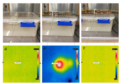 |
89 | Investigation of the Ability of Slot Element Metallization to Reduce SAR and RF Heating due to Associated Components
Dheyaa Alkandari1 and Steven M Wright2
1Electrical Engineering, Kuwait University, Abdullah Al Mubarak Al Sabah, Kuwait, 2Texas A&M University, College Station, TX, United States
Evidence indicates that excess RF power from coil’s associated circuitry could cause injuries to patients. Slot elements have been investigated earlier and demonstrated good potential when used for multi-channel RF coils. Here we investigate the potential of using the extended metallization of slot elements to shield stray electric field generated from RF coil associated circuitry. RF heating experiment was designed to compare the temperature of the phantom with and without using the slot’s extended metallization to shield the associated circuitry. Experimental results demonstrated an observable reduction in the phantom temperature resulting from extending the slots metallization.
|
||
2558 |
90 | MR signal conditioning for an electro-optic conversion with a polarization state modulator
paul nobre1, Gwenaël Gaborit2,3, Raphael Sablong1, Nadege Courjal4, Florent Behague4, Antoine Coste4, Adrien Godet4, Miguel Suarez4, Lionel Duvillaret3, and Olivier Beuf1
1Univ. Lyon, INSA-Lyon, Université Lyon 1, UJM-Saint Etienne, CNRS, Inserm, CREATIS, UMR 5220, U1206, Villeurbanne, France, 2Université de Savoie, IMEP-LAHC, UMR 5130, Le Bourget-du-Lac, France, 3KAPTEOS, Sainte-Hélène-du-Lac, France, 4Institut FEMTO-ST, UMR CNRS 6174, Université Bourgogne Franche-Comté, Besançon, France
The coaxial cables connecting coils to the MRI are subject to many unwanted interactions with RF pulses and with patients’ tissues. Currents flowing on the shielding of the cable can lead to local SAR increase and RF-induced burns. Despite their limitations and the amount of effort to discard them, there is still no satisfying alternative. One of the issue for competing technologies is to safely provide on coil power supply without cables inside the MRI. In this work, we propose to evaluate the feasibility of passive optical conversion and transmission of the signal to the MRI console.
|
||
2559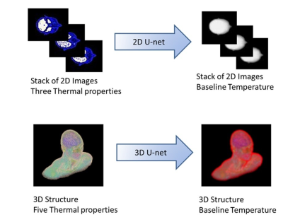 |
91 | Comparison of 2D vs 3D Deep Learning Algorithms to Estimate Temperature Throughout the Human Body
Giuseppe Carluccio1, Eros Montin1, Riccardo Lattanzi1, and Christopher Michael Collins1
1Center for Advanced Imaging Innovation and Research (CAI2R), New York, NY, United States
We developed and compared two different Deep-Learning (DL) based approaches to approximating temperature in subject-specific body models. The first involved use of a 2D U-net to predict temperature throughout the body on a slice-by-slice basis, and the second involved use of a 3D U-net to predict temperature in the 3D body. The 3D approach greatly outperformed the 2D approach, and was very fast.
|
||
 Back to Meeting Home
 Back to the Program-at-a-Glance
The International Society for Magnetic Resonance in Medicine is accredited by the Accreditation Council for Continuing Medical Education to provide continuing medical education for physicians.
 Back to Meeting Home
 Back to the Program-at-a-Glance
 View the Presentation