Digital Poster
MRI Safety II
Joint Annual Meeting ISMRM-ESMRMB & ISMRT 31st Annual Meeting • 07-12 May 2022 • London, UK

| Computer # | ||||
|---|---|---|---|---|
2622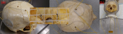 |
63 | Deep brain stimulation patient safety: Effects of patient-based lead trajectories on full-body 3 T MRI
Benson Yang1, Maryam Arianpouya2, Aaron Loh3, Gavin Elias3, Artur Vetkas3, Can Sarica3, Brendan Santyr3, and Simon J Graham1,2
1Physical Sciences, Sunnybrook Research Institute, Toronto, ON, Canada, 2Medical Biophysics, University of Toronto, Toronto, ON, Canada, 3Surgery, University of Toronto, Toronto, ON, Canada
High SAR RF pulse sequences continue to be avoided for patients with implanted deep brain stimulation (DBS) devices, due to the increased risk of injury. Recent studies have shown that DBS lead management can impact electromagnetic behavior and thus, the overall RF heating effects along the implanted device. The present work studied two patient-based DBS lead trajectories using a commercial DBS device with MR-conditional labeling for full-body 3 T MRI, implanted in a novel anthropomorphic phantom. The preliminary results showed temperature elevations that exceed current imaging guidelines remains possible for high SAR RF pulse sequences.
|
||
2623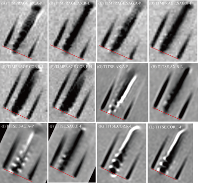 |
64 | What you see is not what you get: localization of Deep Brain Stimulation leads from metal artifacts
Noa Beth Nuzov1, Bhumi Bhusal2, Fuchang Jiang1, Joshua Rosenow3, and Laleh Golestanirad1,2
1Biomedical Engineering, McCormick School of Engineering, Northwestern University, Evanston, IL, United States, 2Radiology, Feinberg School of Medicine, Northwestern University, Chicago, IL, United States, 3Neurosurgery, Feinberg School of Medicine, Northwestern University, Chicago, IL, United States Deep Brain Stimulation (DBS) implants are implanted into target regions of the brain to treat symptoms of many neurological disorders. Postoperative Magnetic Resonance Imaging (MRI) scans are used to localize DBS leads, however, there are large, distorted artifacts surrounding the leads in the images. This study uses MRI phantoms to investigate the true location of a DBS lead relative to its artifact. The results will help researchers and clinicians know the actual locations of the lead tip and contacts from postoperative MRI scans to confirm the implant is correctly targeting the desired brain area. |
||
2624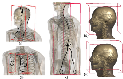 |
65 | Significance of Covering Anatomical Variations for Conservative Assessment of Deposited Power of Leaded Implants in Patients
Lena Kranold1,2, Tolga Goren1, Sunder Rajan3, and Niels Kuster1,2
1IT'IS Foundation, Zurich, Switzerland, 2Department of Information Technology and Electrical Engineering, ETH Zurich, Zurich, Switzerland, 3Center for Devices and Radiological Health (retired), FDA, Silver Spring, MD, United States Anthropomorphic computational phantoms are used to numerically estimate the potential envelope of intensity and distribution of induced E-fields in patients undergoing MR examination. The impact of anatomical variation of the phantom on medical implant risk assessment is demonstrated for generic transfer functions and lead trajectories representing pacemaker, DBS, SCS, and cochlear implant routings. Results showed that even when only one generic transfer function was considered, each of the 12 anatomical phantoms represented the worst-case for at least one configuration. Thus, the widest available range of anatomical phantoms must be examined to define a conservative safety envelope. |
||
2625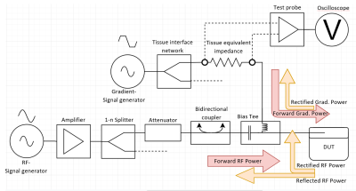 |
66 | Combined RF & Gradient Injection Network for AIMDs with leads
Vincent Hammersen1, Michael Wolff2, Andreas Rennings3, and Gregor Schaefers1,2
1MRI-STaR GmbH, Gelsenkirchen, Germany, 2MR:comp GmbH, Gelsenkirchen, Germany, 3General and Theoretical Electrical Engineering (ATE), University of Duisburg-Essen, Duisburg, Germany
MR safety of AIMD with leads is assessed test procedures defined in ISO/TS10974. This separates the different electromagnetic field interactions into independent test procedures. E.g., malfunction and rectification testing, with gradient and RF injection networks connected to the lead ports of AIMDs. But this does not correspond to "realistic MRI exposures", where a precise combination of RF and magnetic fields is used for imaging. For this reason, a concept is being proposed in which both injection networks are combined to allow a concerted exposure. This allows the simultaneous exposition of any gradient and RF signal combination.
|
||
2626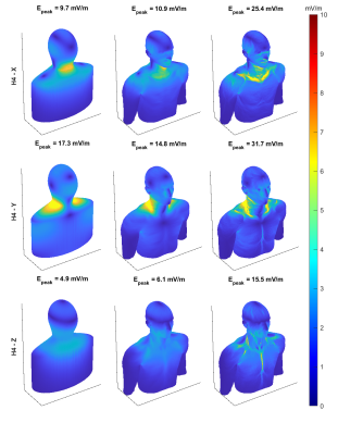 |
67 | Comparison of Gradient Induced Peak E-fields on Simplified and Realistic Body Models
Koray Ertan1,2, Paolo Decuzzi2, Peter B Roemer3, and Brian Rutt1
1Department of Radiology, Stanford University, Stanford, CA, United States, 2Laboratory of Nanotechnology for Precision Medicine, Italian Institute of Technology, Genoa, Italy, 3Roemer Consulting, Lutz, FL, United States
Either peak E-fields on simple geometries with uniform interior properties as suggested by IEC or coupled E-field and neurodynamic simulations on realistic body models can be employed in PNS optimal gradient coil design. Magneto quasi-static simulations were performed to compare the magnitude and location of the peak E-fields in uniform interior body models with smoothed or realistic surface features to fully heterogeneous Virtual Family models. Although peak E-fields are higher on the heterogeneous body models due to complex tissue interfaces, simplified and/or homogeneous models represent a more practical and robust approach predicting population-wide PNS performance.
|
||
2627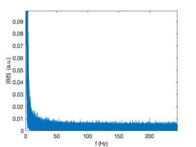 |
68 | Continuous monitoring of MRI staff exposure to stray magnetic field at 1.5 T and 3 T
Angelo Galante1,2,3, Valentino Vito3, Elia Braggio4, Roberta Milanesi4, Vincenzo Giugno5, and Marcello Alecci1,2,3
1Consiglio Nazionale Ricerche - Istituto SPIN, L'Aquila, Italy, 2Istituto Nazionale Fisica Nucleare - Laboratori Nazionali del Gran Sasso, L'Aquila, Italy, 3Università dell'Aquila, L'Aquila, Italy, 4Tecnorad SrL, Verona, Italy, 5Azienda Sanitaria Locale 1 - Abruzzo, L'Aquila, Italy We show how to monitor the MRI staff exposure to the $$$B_0$$$ stray-field. Monitoring is extended to several body districts without interferences with normal hospital operative procedures. The Weighted Peak index is calculated, for different Action Levels limits as from the current EU regulations, to consider the non-sinusoidal nature of exposure. Results show that exposure limits are exceeded even at 1.5 T and their occurrences significantly increase at 3.0 T scanners. |
||
2628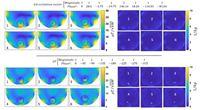 |
69 | Excitation vector optimization for safe parallel transmission MRI of passively conducting implants in the presence of motion
Mostafa Berangi1,2,3, Andre Kuehne1, Helmar Waiczies1, and Thoralf Niendorf1,2,3
1MRI.TOOLS GmbH, Berlin, Germany, 2Berlin Ultrahigh Field Facility (B.U.F.F.), Max Delbrück Center for Molecular Medicine in the Helmholtz Association, berlin, Germany, 3Charité – Universitätsmedizin Berlin, berlin, Germany MRI aided monitoring of (biodegradable) passively conducting implants is challenged by transmission field inhomogeneities and potential elevation of RF power deposition (SAR) in the vicinity of an implant. Small movements of an implant with respect to the RF transceiver constitute another potential risk factor for clinical MRI. Recognizing this challenge and the opportunities, this work uses a multi-objective genetic algorithm (GA) to examine the feasibility of excitation vector optimized parallel transmission in the presence of small variations in implant position/orientation. The GA approach provided excitation vectors that meet the safety guidelines for SAR in close vicinity of a (biodegradable) passively conducting implant and that are immune to small changes in the relative position between the implant and the RF transceiver. Our findings are not limited to the specific implant configurations and experimental setups used in this study but provide the technical foundation to derive a generalized transfer function. |
||
2629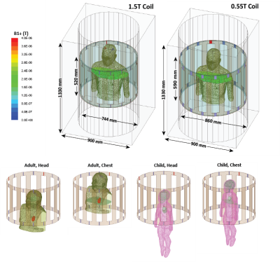 |
70 | When they go high, go low? (Maybe, but not so fast): Comparing RF heating of active implantable medical devices in 0.55 T vs. 1.5 T horizontal scanners
Pia Panravi Sanpitak1, Bhumi Bhusal1, Bach Nguyen1, Fuchang Jiang2, Ehsan Kazemivalipour3,4, Giorgio Bonmassar3,4, Julie Pilitsis5, Joshua Rosenow6, Gregory Webster7, Andrada Popescu8, Daniel Kim1, and Laleh Golestanirad1,2
1Department of Radiology, Northwestern University, Chicago, IL, United States, 2Department of Biomedical Engineering, Northwestern University, Evanston, IL, United States, 3A.A. Martinos Center for Biomedical Imaging, Department of Radiology, Massachusetts General Hospital, Charlestown, MA, United States, 4Harvard Medical School, Boston, MA, United States, 5Department of Neurosciences & Experimental Therapeutics, Albany Medical College, Albany, NY, United States, 6Department of Neurosurgery, Northwestern University, Chicago, IL, United States, 7Division of Cardiology, Department of Pediatrics, Northwestern University, Chicago, IL, United States, 8Department of Medical Imaging, Lurie Children's Hospital, Chicago, IL, United States
Low-field Magnetic Resonance Imaging (MRI) systems are gaining traction, but their impact on radiofrequency (RF) heating of elongated implants is unclear. The RF heating of conductive lead models with realistic trajectories was examined inside a 1.5 T MRI coil and a 0.55 T Wide Bore MRI coil. 0.1g-SAR was recorded at the tips of conductive implanted leads in adult patients with deep brain stimulation (DBS) systems and pediatric patients with cardiac implants, at both chest and head landmarks. In some instances, heating was higher in the 0.55 T coil, so caution must be exercised before promoting low-field coils as "implant-friendly".
|
||
2630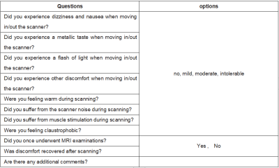 |
71 | Study of Subjective Tolerance during 5T Whole-Body MRI Examinations
Xuchen Zhu1, Wenbo Sun2, Shuo Zhu3,4, Zhenhua Shen1, Lan Lan2, Shihong Han1, Shuheng Zhang1, Zhang Shi3,4, Qi Liu5, Yongquan Ye5, Liang Liang3,4, Haibo Xu2, and Liang Liu1
1United Imaging Healthcare, Shanghai, China, 2Department of Radiology,Zhongnan Hospital of Wuhan University, Wuhan, China, 3Department of Radiology, Zhongshan Hospital, Fudan University, Shanghai, China, 4Shanghai Institute of Medical Imaging, Shanghai, China, 5UIH America, Inc., Houston, TX, United States
Questionnaires on subjective feeling were collected and analyzed from 202 subjects following whole-body MRI examinations at 5T. No intolerable discomfort was reported, indicating whole-body examinations at 5T is safe when necessary safety protocols were followed.
|
||
2631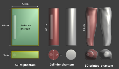 |
72 | Implant heating in muscle-mimicking phantoms: an in silico evaluation of perfusion cooling
Amgad Louka1,2, William Bradfield-Handler2,3, and Blaine Chronik1,2,3
1Medical Biophysics, Western University, London, ON, Canada, 2The xMR Labs, Western University, London, ON, Canada, 3Physics & Astronomy, Western University, London, ON, Canada
We have developed a novel perfusion phantom and this work describes the simulated RF-heating of a 10 cm titanium rod inside this phantom when placed inside the current gold standard (ASTM) phantom, and our geometry/muscle-mimicking phantoms. Simulations show a 30-45% decrease in observed heating due to perfusion cooling, and once these simulations are experimentally validated they could pave the way for improved acceptance criteria and ultimately improved patient access to MRI
|
||
2632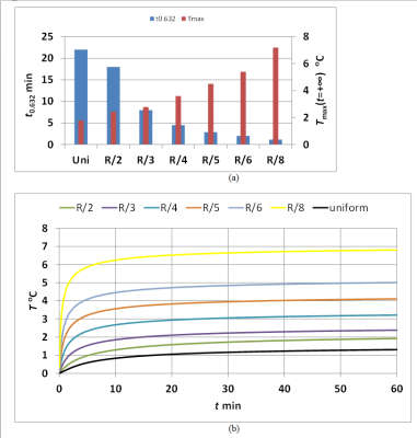 |
73 | Analytical Evaluation of Radio-Frequency Energy-Induced Heating in a Hot Spot without perfusion
Mikhail Kozlov1 and Nicolas Boulant 2
1Max Planck Institute for Human Cognitive and Brain Sciences, Leipzig, Germany, 2Université Paris-Saclay, CEA, CNRS, BAOBAB, NeuroSpin, Gif-Sur-Yvette, France We derived analytical steady-state and transient solutions for a sphere and a SAR source with uniform and the Gaussian distribution located in an infinite medium of homogeneous properties. The results obtained analytically showed that even without taking into account blood perfusion (i) if power deposition is present only in a sphere that encloses 10 gram of human tissue and SAR10g=10W/kg the steady-state temperature rise is below 3°C for power deposition distributions as sharp as Gaussian with σ=R/3=4.33mm, (ii) the sharper the Gaussian distribution of power deposition, the higher Tmax(t=+∞) and the faster it will approach steady-state. |
||
2633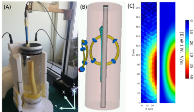 |
74 | Assessment of Femur Metallic Implant Safety at 3T
Amira Trabelsi1,2,3, Megdouda Benamara4, Pierre Jomin1, Stefan Enoch1, Marc Dubois4, Martine Pithioux3, David Bendahan2, and Redha Abdeddaim1
1Aix Marseille Univ, CNRS, Centrale Marseille, Institut Fresnel, Marseille, France, 2Aix Marseille Univ, CNRS, CRMBM, Marseille, France, 3Aix Marseille Univ, CNRS, ISM, Marseille, France, 4Multiwave Imaging, Marseille, France
We assessed electromagnetic behavior of metallic implant used for distal femur fracture in typical MRI situation. |B1+| field, SAR and temperature variations were computed at 3T using a surface coil on human model for different positions of the implant relative to the coil. To validate the simulation, we measured the E-field and compared it to simulated E-field. |B1+| field maps showed an interesting augmentation near the implant. Both global SAR and local SAR levels proved that it is possible to safely image bone repair. However, temperature elevation near the tip of the implant was important and is to be considerate.
|
||
2634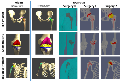 |
75 | Virtual precision surgery for mri implant safety
Oriano Bottauscio1, Alessandro Arduino1, Fabio Baruffaldi2, Mario Chiampi1, Jessica Martinez1, Umberto Zanovello1, and Luca Zilberti1
1Advanced Materials Metrology and Life Science, Istituto Nazionale di Ricerca Metrologica, Torino, Italy, 2IRCCS Istituto Ortopedico Rizzoli, Bologna, Italy
In silico evaluation of heating during an MRI exam for patients with passive implants patients need detailed anatomical models to reproduce in vivo scenarios. A quantification of the influence of the model approximation is fundamental to define the main requirements of the digital model.Here, we analyze the impact of different model approximation in simulating RF and gradient-induced heating caused by orthopedic implants. It was found that, whereas the heating due to switched gradient fields does not strictly require the adoption of advanced virtual surgery, the heating caused by RF is strongly affected by the precision of the digital model.
|
||
2635 |
76 | Dependence of Electromagnetic Exposure on Design and Excitation Positions of 3T Two-Channel Whole-Body Birdcage Coils
Mikhail Kozlov1, Wolfgang Kainz2, and Manuel Murbach3
1Max Planck Institute for Human Cognitive and Brain Sciences, Leipzig, Germany, 2U.S. FDA, CDRH, Office of Science and Engineering Laboratories, Division of Biomedical Physics, Silver Spring, MD, United States, 3EMConsulting, Zurich, Switzerland The safety assessment of RF exposure in MRI heavily relies on electromagnetic simulations. As there are numerous different coil designs on the market, this assessment is typically performed with sets of generic coil geometries to cover the commercially available bore diameter and coil lengths. This abstract investigated the effect of other design parameters, which are currently less considered. Our results show that feed-port positioning and non-ideal tuning of the coil may considerably alter the RF absorption pattern in an oval phantom, and even more so the E-field distribution outside the phantom. |
||
2636 |
77 | MR Safety of Inductively-coupled and Conventional Intra-oral Coils
Agazi Samuel Tesfai1, Simon Reiss1, Thomas Lottner1, Andreas Vollmer2, Michael Bock1, and Ali Caglar Özen 1
1Dept. of Radiology, Medical Physics, Medical Center – University of Freiburg, Faculty of Medicine, University of Freiburg, Freiburg, Germany, 2Department of Oral and Craniomaxillofacial Surgery, Center for Dental Medicine, University Medical Center Freiburg, Freiburg, Germany
Intraoral coils (IOCs) increase the signal-to-noise ratio in dental MRI exams several times over external coils. However, the intraoral coil placement might be associated with safety hazards such as RF heating. This study evaluates the safety of different IOCs using temperature and E-field measurements to detect temperature hotspots. An inductively-coupled and a wired IOC were constructed, E-field hotspots were detected and the temperature increase was measured showing that it remains below 1K over 6min RF exposure.
|
||
 Back to Meeting Home
 Back to the Program-at-a-Glance
The International Society for Magnetic Resonance in Medicine is accredited by the Accreditation Council for Continuing Medical Education to provide continuing medical education for physicians.
 Back to Meeting Home
 Back to the Program-at-a-Glance
 View the Presentation