Digital Poster
Hybrid Systems, Magnets, Gradient Systems & Shims/Shimming II
Joint Annual Meeting ISMRM-ESMRMB & ISMRT 31st Annual Meeting • 07-12 May 2022 • London, UK

Digital Poster
Hybrid Systems, Magnets, Gradient Systems & Shims/Shimming II
| Computer # | ||||
|---|---|---|---|---|
1367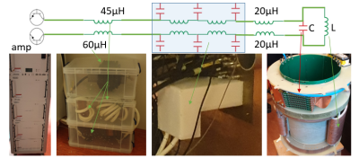 |
73 | Short-TE diffusion-MRI by combining strong gradients with ultrasonic readout
Aris van Ieperen1,2, Chantal Tax1,3, Dennis Klomp1, Bas Vermulst2, Jeroen Siero1,4, Joost van Straalen5, Martijn Heintges5, and Edwin Versteeg1
1Radiology, University Medical Center Utrecht, Utrecht, Netherlands, 2Electromechanics & Power Electronics, Eindhoven University of Technology, Eindhoven, Netherlands, 3CUBRIC, School of Physics and Astronomy, Cardiff University, Cardiff, United Kingdom, 4Spinoza Centre for Neuroimaging, Amsterdam, Netherlands, 5Prodrive Technologies B.V., Son, Netherlands
For high-resolution diffusion-MRI, strong gradients combined with fast readout are desired. In this study, we propose a gradient system architecture for such strong gradients and fast readouts without nerve stimulation by implementing a filter topology that allows for both low and high frequency gradient waveforms. Two gradient inserts with different specifications are realized and two proposed modulation strategies are implemented in the amplifier firmware. Field measurements demonstrated the desired dual-mode capability of the system. With one gradient insert, a gradient amplitude of 300 mT/m and slew rates well above 10.000 T/m/s can be reached with only 500A&330V as amplifier output.
|
||
1368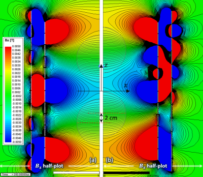 |
74 | Z-Gradient Array Coil Equipped with a Tunable Shield Array for Creating Multiple-Imaging Volumes
Manouchehr Takrimi1 and Ergin Atalar2
1National Magnetic Resonance Research Center, Bilkent University, Ankara, Turkey, 2Department of Electrical and Electronics Engineering, Bilkent University, Ankara, Turkey
A z-gradient array coil equipped with a tunable shield array is proposed to achieve multiple imaging volumes that can be shifted along the coil axis. This is achieved by a set of independently tunable power amplifiers that feed the array elements. The proposed array dynamically provides a proper shield for the main array in which its magnetic profile can be adjusted on the fly. Five multiple-region magnetic profiles are simulated to demonstrate the flexibility of the proposed array: (a) a double-gradient profile; (b) shifted version of (a); (c) highly linear double-gradient profile without shielding; (d) triple-gradient profile; (e) quintuple-gradient profile.
|
||
1369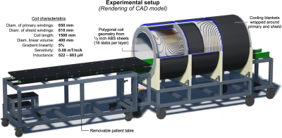 |
75 | Design Analysis and Prototype Construction of PNS Optimized MRI Gradient Coils
Mathias Davids1,2,3, Livia Vendramini1, Natalie Ferris4,5, Valerie Klein1,3, Bastien Guerin1,2, and Lawrence L. Wald1,2,5
1Martinos Center for Biomedical Imaging, Charlestown, MA, United States, 2Harvard Medical School, Boston, MA, United States, 3Computer Assisted Clinical Medicine, Medical Faculty Mannheim, Heidelberg University, Heidelberg, Germany, 4Harvard Graduate Program in Biophysics, Harvard University, Cambridge, MA, United States, 5Harvard-MIT Division of Health Sciences and Technology, Boston, MA, United States
We describe the process of designing and analyzing two body gradient coils with and without PNS optimization suitable for prototype construction and experimental validation. The optimized coil achieves a 51% increase in PNS thresholds at a 15% inductance penalty. Both coils are construction-ready (single continuous wire path) and have realistic and matched design characteristics (actively shielded, torque/force balanced, high field linearity in 40 cm ROL). We are in the process of constructing coil prototypes, with the ultimate goal of experimentally validating their PNS differences.
|
||
1370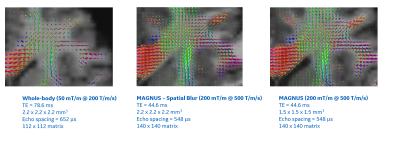 |
76 | Diffusion Imaging comparison of high-performance Gradient system (MAGNUS) with clinical MR system.
H. Douglas Morris1, Nastaren Abad2, Radhika Madhavan3, Chitresh Bhushan2, Ante Zhu2, Luca Marinelli2, Eric Fiveland2, J Kevin DeMarco4,5, Robert S Shih4,5, Maureen Hood4,5, Gail Kohls4, Kimbra Kenny4, Tom K F Foo2, and Vincent B Ho4,5
1Radiology and Radiological Sciences, Uniform Services University of the Health Sciences, Bethesda, MD, United States, 2GE Global Research, Niskayuna, NY, United States, 3GE Healthcare, Niskayuna, NY, United States, 4Walter Reed National Military Medical Center, Bethesda, MD, United States, 5Uniform Services University of the Health Sciences, Bethesda, MD, United States
Comparisons of the multi-shell DWI is described for the USU/GE MAGNUS 3T head-only gradient system and a clinical whole-body GE MR750 3T magnet system. The advantages of high-performance gradients are shown in analysis outcomes of dMRI metrics. Direct comparison is made between identical diffusion pulse sequences and system optimized sequences for each platform. The shorter TE and high-gradient strength of MAGNUS enhance SNR and diffusion imaging response.
|
||
1371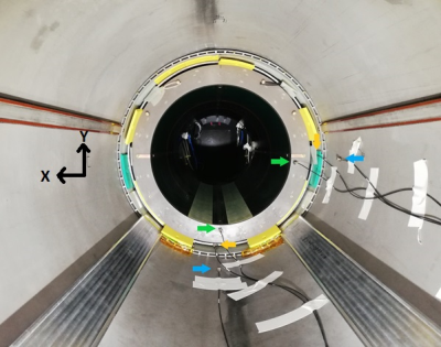 |
77 | Vibration measurements of the SC72 gradient versus field strength in the Iseult magnet
Nicolas Boulant1, Cécile Lerman1, Lionel Quettier2, Olivier Dubois2, Frédéric Molinié2, Peter Dietz3, and Guy Aubert2
1University Paris-Saclay, CEA, CNRS, BAOBAB, NeuroSpin, Gif sur Yvette, France, 2University Paris-Saclay, CEA, Irfu, Gif sur Yvette, France, 3Siemens Healthcare GmbH, Erlangen, Germany
The whole-body Iseult 11.7T CEA magnet has delivered its first images after nearly 20 years of research and development. Before reaching this long-waited step, a gradient coil–magnet interaction test campaign was run for several months at 3T, 7T, 10.2T and 11.7T on the same identical system. It included acoustics, vibrations, magnet safety system voltage and power deposition in the He bath measurements. Some vibration results versus field strength are presented here. Vibration amplitudes are shown to increase less than with B0 field strength.
|
||
1372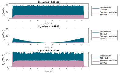 |
78 | Reducing MRI acoustic noise burden with Predictive Noise Cancelling
Paulina Šiurytė1, Joao Tourais1, and Sebastian Weingärtner1
1Imaging Physics, TU Delft, Delft, Netherlands
Acoustic noise in MRI is a main source of patient anxiety, with noise levels reaching up to 130 dB. In this work, a low-cost solution is proposed, combining active noise cancelling and direct sequence noise prediction from the scanner gradient inputs. The prediction is based on the linearity between gradient coil input derivative and corresponding acoustic noise. A proposed Predictive Noise Cancelling demonstrated an in-bore noise reduction up to 10 dB despite the system imperfections.
|
||
1373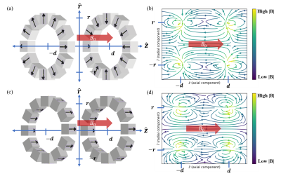 |
79 | Hybrid Superconducting-Permanent Magnet Designs for Short Mid-Field MRI Magnets
Alex C Barksdale1,2, Clarissa Cooley1, and Lawrence Wald1
1MGH, Charlestown, MA, United States, 2MIT, Cambridge, MA, United States
Mid-field (0.5T) MRI is an attractive alternative to higher field strengths for improved cost, accessibility and patient comfort. Magnet length is a major determinant of patient comfort/acceptance but is constrained by escalating wire costs. We present a mid-field magnet design using rare-earth permanent magnets to supplement solenoidal superconducting windings, enabling shorter bore lengths than superconducting windings alone. The optimization problem is formulated and solved using linear programming. For a given superconducting wire length, we demonstrate that adding rare-earth materials can reduce bore lengths by 10% using <250kg of rare-earth material for a 5ppm, 0.5T B0 specification over a 450mm DSV.
|
||
1374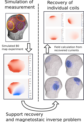 |
80 | A Fast Magnetostaic Inverse Approach for Subject-Specific ∆B0 Shim Coil Calibration
Nicolas Arango1, Jacob White1, and Elfar Adalsteinsson1,2
1Department of Electrical Engineering and Computer Science, Massachusetts Institute of Technology, Cambridge, MA, United States, 2Institute for Medical Engineering and Science, Massachusetts Institute of Technology, Cambridge, MA, United States
For a rigidly positioned ∆B0 shim coil, calibration is a one-time process of acquiring high resolution ∆B0 fields. For subject-specific shim coil position, this approach is too slow. We propose a fast magnetostatic inverse approach for subject-specific calibration by only acquiring few, low resolution fieldmaps each with several active shim coils. Our simulation study, which included a noise model and a measured ∆B 0 test case, suggests a scan-time reduction of afactor of 170, reducing calibration time from over an hour to less than a minute with negligible impact on shim quality.
|
||
1375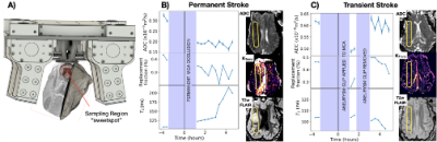 |
81 | Portable magnetic resonance based monitoring of MCA occlusion in an ovine sheep stroke model.
Dion Thomas1, Freya Harrison2, Alice Little1,3, Alex King1, Annabel Jain Sorby-Adams4, Shieak Tzeng2, Renee Turner4, and Sergei Obruchkov1
1Victoria University of Wellington, Wellington, New Zealand, 2University of Otago, Wellington, New Zealand, 3The University of Auckland, Auckland, New Zealand, 4The University of Adelaide, Adelaide, Australia
A custom built single sided magnetic resonance system, generates an external region of homogeneous B0 = 0.2T field, a sweet spot, from which signal can be detected. The device is capable of relaxometry, diffusion and perfusion measurements and was used to continuously monitor progress of an MCA occlusion in a ovine sheep stroke model. The data from the device was compared to image data acquired on a clinical 3T MRI system.
|
||
1376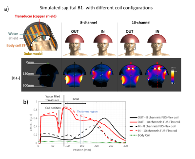 |
82 | Improving image quality and acoustic transparency in MRgFUS at 3T using a low-profile, flexible receive coil array
Isabellle Saniour1, Fraser J.L. Robb2, Victor Taracila2, James Shin1, Vishwas Mishra1, Rena Fukuda1, Jana Vincent2, Henning U. Voss1, Michael G. Kaplitt3, J. Levi Chazen1, and Simone Angela Winkler 1
1Department of Radiology, Weill Cornell Medicine, New York, NY, United States, 2GE Healthcare, Aurora, OH, United States, 3Department of Neurological Surgery, Weill Cornell Medicine, New York, NY, United States
Transcranial MRgFUS has been successfully used to treat a variety of neurodegenerative diseases. However, body coil brain image quality is poor, and a low-signal band artifact may occur in some regions due to RF wave reflections. Further, acoustic coil transparency has not been addressed extensively to date. In this work, we simulate a 10-channel coil design that can significantly increase the SNR by a factor of 20 over the body coil and thus indirectly improve the signal at the region of interests. Acoustic simulations/experiments exhibit transparency of the FUS-Flex coil as high as 97% at 650 kHz.
|
||
 Back to Meeting Home
 Back to the Program-at-a-Glance
The International Society for Magnetic Resonance in Medicine is accredited by the Accreditation Council for Continuing Medical Education to provide continuing medical education for physicians.
 Back to Meeting Home
 Back to the Program-at-a-Glance
 View the Presentation