Digital Poster
New (RF) Devices & High-Field MR I
Joint Annual Meeting ISMRM-ESMRMB & ISMRT 31st Annual Meeting • 07-12 May 2022 • London, UK

Digital Poster
New (RF) Devices & High-Field MR I
| Computer # | ||||
|---|---|---|---|---|
1999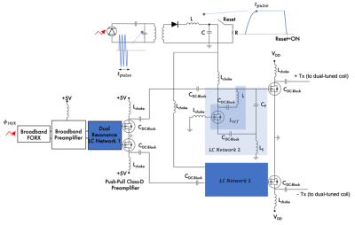 |
94 | Automated tuning of gate driver circuit using eGaN FETs
Natalia Gudino1
1LFMI, NIH, Bethesda, MD, United States
An automated tuning of the dual resonance LC network that forms the gate driver circuitry of a dual tuned RFPA is presented. Through this automated tuning the RFPA could be adapted from the control to operate at the selected 1H and X-nucleus frequencies.
|
||
2000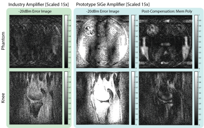 |
95 | A Calibration and Compensation Method for Dynamic Range Recovery of Low-Power SiGe Pre-Amplifiers.
Christopher Vassos1, Fraser Robb2, Shreyas Vasanawala3, John Pauly1, and Greig Scott1
1Electrical Engineering, Stanford University, Stanford, CA, United States, 2GE Healthcare, Aurora, CO, United States, 3Radiology, Stanford University, Stanford, CA, United States
Power consumption reduction is key when exploring wireless MRI arrays. An alternative low-power (5.7mW) SiGe pre-amplifier is presented and its impact on imaging performance evaluated in a benchtop setting. The reduction in power results in a more extreme non-linearity that introduces distortion into the images. It is possible to calibrate and compensate for this distortion through receive-only methods. Applying this compensation to the distorted images results in a restoration of image quality with resulting RMSE comparable to industry standard amplifiers representing a potential power reduction up to 30x.
|
||
2001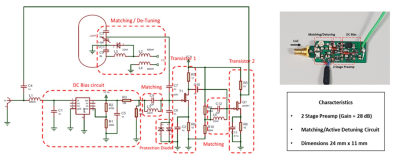 |
96 | Design of a Miniaturized High Impedance Preamplifier for 7T MRI
Saadou Almokdad1, Paul-François Gapais1,2, Michel Luong3, Alexis Amadon1, Elodie Georget2, and Eric GIACOMINI 1
1CEA Saclay/DRF/JOLIOT/NEUROSPIN/BAOBAB/METRIC, Gif-sur-Yvette, France, 2Multiwave Imaging SAS, Marseille, France, 3CEA Saclay/DRF/IRFU/DACM//LISAH, Gif-sur-Yvette, France
Coil arrays enhance human body performance for an MRI measurement both in speed and signal-to-noise ratio. However, size and cabling of such arrays can deteriorate the performance of the imaging, or put at risk the safety of the patient. A miniaturized preamplifier is proposed (dimensions 24mm x 11mm), to be placed directly onto the receive coil. The design needs to preserve a good performance (noise figure ≤ 1dB, gain ≈ 28dB), and provide high impedance to minimize the coupling to nearby coils.
|
||
2002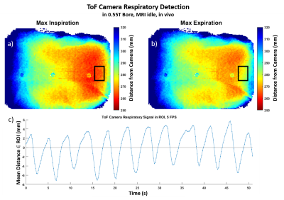 |
97 | Compatibility of a 3D Time-of-Flight Video Camera for Motion Capture with a 0.55T MRI System
Douglas Brantner1, Leanna Pancoast1, Jerzy Walczyk1, Roy Wiggins1, Ryan Brown1, and Christopher Collins1
1Center for Advanced Imaging Innovation and Research, New York University, New York, NY, United States
We evaluated compatibility of a 3D Time-of-Flight (ToF) video camera with a 0.55T MRI system. Initial tests showed severe MR image artifacts when the ToF camera was operating, and USB communication with a computer outside the MR suite was interrupted during MR image acquisition. With adequate conductive shielding of the ToF camera and typical MR imaging bandwidths, MR images could be acquired with no measurable effects from the ToF camera, and with no effects of the MRI system on ToF signal acquisition.
|
||
2003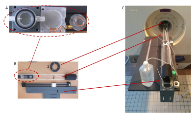 |
98 | 3D MRI characterization of 3D printed tumor tissue models using a plastronic MR-Bioreactor : Preliminary results
Jean-Lynce Gnanago1, Tony Gerges1, Laura Chastagnier2, Emma Petiot2, Vincent Semet1, Philippe Lombard1, Christophe Marquette2, Michel Cabrera1, and Simon Auguste Lambert1
1Université de Lyon, INSA Lyon, Université Claude Bernard Lyon 1, Ecole Centrale de Lyon, CNRS, Ampère UMR5005, Villeurbanne, France, 23d.FAB, Univ Lyon Université Lyon1 CNRS, INSA, CPE-Lyon ICBMS UMR 5246, Villeurbanne, France
Tissue engineering for regenerative medicine have been developing for a few decades now and the number of applications is increasing to tackle the shortage of organ donors. To date, only few systems can allow both monitoring and 3D characterization of tissue constructs during their growth. In this study, we decided to focus on following the Apparent Diffusion Coefficient (ADC) known to be a marker of cell density and built a MR-Bioreactor to probe the ADC of a growing tissue. In this preliminary work, we were able to follow the cell density of a tumor tissue model using our dedicated MR-bioreactor.
|
||
2004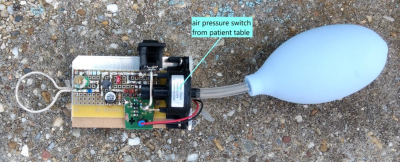 |
99 | A Wireless Pilot Tone Based Patient Squeeze-Ball (P2TSB)
Mario Bacher1, Peter Speier2, and Jan Bollenbeck3
1MR R&D CFP, Siemens Healthineers, Erlangen, Germany, 2MR DL CARD, Siemens Healthineers, Erlangen, Germany, 3MR R&D HW&SYS CRX, Siemens Healthineers, Erlangen, Germany
We developed a prototype wireless patient alarm-ball based on Pilot Tone technology which could speed up patient setup time and increase patient comfort. The use of Pilot Tone as a means of signal transmission enables easy and cost-effective use of this concept on existing scanners with only a minimal software upgrade.
|
||
2005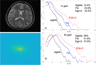 |
100 | Assessing Universal Code Compression to Lower Wireless MRI Data Rates
Greig Scott1
1Electrical Engineering, Stanford University, Stanford, CA, United States
For wireless MRI data links employing simple ASK or DPSK binary modulation, there is a need to compress the k-space data to lower data rates and use less spectral bandwidth. We explored Fibonacci and Exp-Golomb universal codes for lossless compression. Our results indicate that compression can reduce rates to 1/3 to 1/4 of the original levels. Actual compression will depend on signal gain, as well as k-space trajectories. These results suggest the development of an MRI codec for future wireless or fiber optic RF coil array data links.
|
||
2006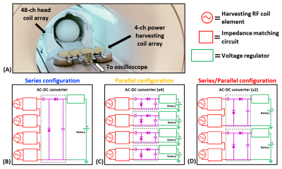 |
101 | Wireless Power Harvesting of the B1 Field During MR Image Acquisition for Pulse Charging of MR-Compatible Batteries
Jonathan Cuthbertson1,2, Trong-Kha Truong1,2, Fraser Robb3, Allen W. Song1,2, and Dean Darnell1,2
1Medical Physics Graduate Program, Duke University, Durham, NC, United States, 2Brain Imaging and Analysis Center, Duke University, Durham, NC, United States, 3GE Healthcare, Aurora, OH, United States A 4-channel power harvesting coil array was developed to allow the energy emitted from RF transmit pulses within the scanner bore during imaging to be converted into DC voltage pulses for recharging MR-compatible batteries, regardless of the scan parameters or imaging pulse sequence. Proof-of-concept experiments in a phantom show that this power harvesting coil array was able to provide energy to a battery during GRE image acquisition for various flip angles and additionally during GRE-EPI, DTI, and MPRAGE image acquisitions. |
||
2007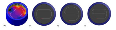 |
102 | Novel Compact Dual-Band E-Field Generator for MR Safety Testing of Active Implantable Medical Devices
Lena Kranold1,2, Myles Capstick1, Tolga Goren1, and Niels Kuster1,2
1IT'IS Foundation, Zurich, Switzerland, 2Department of Information Technology and Electrical Engineering, ETH Zurich, Zurich, Switzerland A compact dual-band E-field generator for MRI implant safety testing has been developed to produce diverse, well-defined tangential incident E-fields along defined implant test routings for efficient validation of the transfer function of AIMD. The MITS medical implant test system consists of two pairs of electrodes whose relative amplitude and phase are controlled to create E-field conditions at the frequencies corresponding to 1.5 T and 3 T MRI. Simulations of different exposure settings were validated by measurements of the resulting field distributions with an E-field vector probe along the routing paths. |
||
2008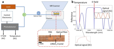 |
103 | Dual Mode Acousto-optic RF Safety Sensor for Electric Field and Temperature Measurement at 1.5T MRI
Yusuf Samet Yaras1, Lee W Bradley1, Dursun Korel Yildirim2, Ozgur Kocaturk3, John Oshinski4,5, and F. Levent Degertekin1
1Mechanical Engineering, Georgia Institute of Technology, ATLANTA, GA, United States, 2Division of Intramural Research, National Heart Lung and Blood Institute, National Institutes of Health, Bethesda, MD, United States, 3Institute of Biomedical Engineering, Bogazici University, Istanbul, Turkey, 4Wallace H. Coulter Department of Biomedical Engineering, Georgia Institute of Technology, Atlanta, GA, United States, 5Department of Radiology and Imaging Sciences, Emory University, Atlanta, GA, United States
Electric field and temperature sensors are essential tools for RF safety assessment of implants during magnetic resonance imaging. In this work, an acousto-optic sensor was used for both local tangential electric field and temperature measurements. The inherent electric field sensitivity of the piezoelectric crystal mechanically coupled to a fiber Bragg grating provides the electric field information while the thermal sensitivity of FBG is used for temperature measurement for dual mode sensing. The sensor was used to measure the electric field concentration around a reference implant with high SNR as well as the temperature rise at the tip of the implant.
|
||
2009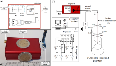 |
104 | Wireless reference implant and communication methodology to assess and investigate RF safety and pTx mitigation strategies for AIMDs
Berk Silemek1, Frank Seifert1, Bernd Ittermann1, and Lukas Winter1
1Physikalisch-Technische Bundesanstalt (PTB) Braunschweig and Berlin, Berlin, Germany
Parallel transmission (pTx) systems can substantially improve the RF-safety of active implantable medical devices (AIMDs) in MRI. Here, a wireless reference implant is presented to test pTx mitigation strategies of RF induced implant heating. It is demonstrated that the proposed hardware and communication workflow can measure the sensor Q matrix and use this information to mitigate RF induced heating. The proposed setup enables conceptualization and further testing of a safety strategy relying on an implant communicating with a pTx capable MRI to improve RF safety without major restrictions in MR imaging performance.
|
||
2010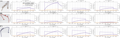 |
105 | Mitigation of RF-induced heating on realistic deep brain stimulator lead trajectories by wireless sensor Q-matrix and parallel transmission
Berk Silemek1, Frank Seifert1, Rüdiger Brühl1, Bastien Guerin2,3, Reiner Montag1, Bernd Ittermann1, and Lukas Winter1
1Physikalisch-Technische Bundesanstalt (PTB) Braunschweig and Berlin, Berlin, Germany, 2Athinoula A. Martinos Center for Biomedical Imaging, Massachusetts General Hospital, Boston, MA, United States, 3Harvard Medical School, Boston, MA, United States
Measurement-based pTx mitigations on realistic DBS lead trajectories are presented based on small, embedded sensors in the implant casing. Sensor Q-matrix measurements are performed and transmitted wirelessly from the implant to the pTx console. Three different DBS implant trajectories at four different locations have been tested showing successful pTx mitigations of RF induced heating in in-vitro. The methodology is conceptually appealing to be investigated further towards a safety strategy, where an implant communicates with a pTx capable MR scanner in order to improve MR safety and MR imaging performance.
|
||
 Back to Meeting Home
 Back to the Program-at-a-Glance
The International Society for Magnetic Resonance in Medicine is accredited by the Accreditation Council for Continuing Medical Education to provide continuing medical education for physicians.
 Back to Meeting Home
 Back to the Program-at-a-Glance
 View the Presentation