Digital Poster
New (RF) Devices & High-Field MR II
Joint Annual Meeting ISMRM-ESMRMB & ISMRT 31st Annual Meeting • 07-12 May 2022 • London, UK

Digital Poster
New (RF) Devices & High-Field MR II
| Computer # | ||||
|---|---|---|---|---|
2089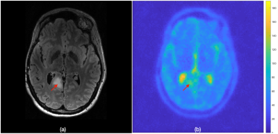 |
79 | Sodium Coil dedicated Universal Sensitivity Map for scan time reduction of 7T MR in-vivo Sodium Imaging
Sanghoon Kim1, Sai Merugumala1, and Alexander Lin1
1Radiology, BWH Center for Clinical Spectroscopy, Boston, MA, United States
Sodium studies have shown subtle pathophysiologic changes in tissue sodium concentrations (TSC) in the early stage of the various diseases. One of the major challenges with measuring TSC is its low SNR. Phased array coils are used to increase sodium SNR, however, they result in an inhomogeneous receive profile that makes accurate quantitation of TSC difficult. Uniformity correction is thus required and is often done by generating a sensitivity map. A previous approach introduced the concept of utilizing a universal sensitivity map to reduce total scan time. We present an improved universal sensitivity map solution.
|
||
2090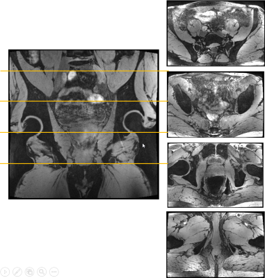 |
80 | A Software-based TIAMO approach to enable high resolution large FOV body imaging at 7T ultra-high field
Jenni Schulz1, Oliver Kraff2, Harald Quick2,3, and Tom Scheenen1,2
1Medical Imaging, Radboud UMC, Nijmegen, Netherlands, 2Erwin L. Hahn Institute for MRI, University Duisburg-Essen, Essen, Germany, 3High-Field and Hybrid MR Imaging, University Hospital Essen, Essen, Germany
Inhomogeneities across large FOVs are problematic at ultra-high field. We propose a software-based TIAMO implementation to enable similar flip angles across the body without hardware interference on the 7T MR scanner. Using an interleaved and complementary CP+/CP2+ excitation scheme, we acquired water-selective and lipid-selective 3D GRE data at high resolution demonstrating homogeneous signal distribution across the pelvis and lower abdomen.
|
||
2091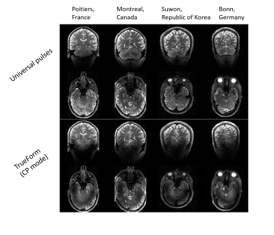 |
81 | Traveling pulses visit 7T Terra sites: Getting ready for Parallel Transmission in routine use
Franck Mauconduit1, Aurélien Massire2, Vincent Gras1, Eberhard Pracht3, Marc Lapert2, Ilana Leppert4, Christine Lucas Tardif4, Sugil Kim5, Kamil Uludag6,7, Tony Stoecker3, Mathieu Naudin8,9,10, Rémy Guillevin8,9,10, Alexandre Vignaud1, and Nicolas Boulant1
1Paris-Saclay University, CEA, CNRS, BAOBAB, NeuroSpin, Gif-sur-yvette, France, 2Siemens Healthcare SAS, Saint-Denis, France, 3German Center for Neurodegenerative Diseases (DZNE), Bonn, Germany, 4Montreal Neurological Institute, McGill University, Montreal, QC, Canada, 5Siemens Healthineers Ltd, Seoul, Korea, Republic of, 6Techna Institute & Koerner Scientist in MR Imaging, University Health Network, Toronto, ON, Canada, 7Center for Neuroscience Imaging Research, Institute for Basic Science & Department of Biomedical Engineering, Sungkyunkwan University, Suwon, Korea, Republic of, 8CHU Poitiers, Poitiers, France, 9LRCOM I3M, University and University Hospital of Poitiers, Poitiers, France, 10Laboratory of Applied Mathematics, UMR CNRS 7348, University of Poitiers, Poitiers, France
Parallel transmission universal pulses (UPs) consist of pre-optimized RF pulse solutions mitigating the RF field inhomogeneity problem for a given RF coil. Optimized offline on a database of representative field maps, they are designed to be robust to intersubject variability and spare the user a cumbersome online calibration. Initially designed for the MAGNETOM 7T Classic system from Siemens Healthineers, with the RF coil being strictly identical, this abstract describes the set of transformations on the pulses necessary to meet the MAGNETOM Terra specifications as well as successful in vivo results already achieved at four Terra sites using the same UPs.
|
||
2092 |
82 | Parallel Transmit DANTE-SPACE for improved black-blood signal suppression at 7 Tesla
Matthijs H.S. de Buck1, James L. Kent1, Aaron T. Hess1, and Peter Jezzard1
1Wellcome Centre for Integrative Neuroimaging, FMRIB, Nuffield Department of Clinical Neurosciences, University of Oxford, Oxford, United Kingdom
DANTE-SPACE can be used for visualizing intracranial vessel walls at 7T, where the flow-sensitive signal attenuation of the DANTE preparation simultaneously suppresses signal from the internal blood and external CSF. However, the efficacy of DANTE is reduced for blood moving upstream through the neck due to limited B1+ coverage at 7T. Here, EPG simulations are used to estimate the effect of this limited B1+ coverage on the achieved contrasts. A pTx-based approach which uses separate RF shims for the DANTE-preparation and the SPACE readout to improve the attenuation of blood moving through the neck is proposed, and validated in vivo.
|
||
2093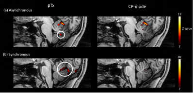 |
83 | Functional MRI of large-scale brain networks involved in motor control using parallel transmission at 7 Tesla
Yidi Lu1, Chia-Yin Wu1,2,3, Shota Hodono1,2, Jin Jin2,4, David Reutens1,2, and Martijn A Cloos1,2
1Centre for Advanced Imaging, The University of Queensland, Brisbane, Australia, 2ARC Training Centre for Innovation in Biomedical Imaging Technology, The University of Queensland, Brisbane, Australia, 3School of Information Technology and Electrical Engineering, The University of Queensland, Brisbane, Australia, 4Siemens Healthcare Pty Ltd, Brisbane, Australia
The standing wave artefact affects SNR and contrast of the images in Ultra-high field (UHF) functional magnetic resonance imaging (fMRI). One way to mitigate this effect is to use parallel transmission (pTx). In this study, we evaluate the benefits of pTx for studies that investigate large-scale brain networks involved in motor control. We show that, compared to the standard circularly polarized (CP) mode, activation patterns in the posterior lobule of the cerebellum, produced by a coordinated finger flexion-extension task in both hands, are better captured using subject-specific pTx pulses.
|
||
2094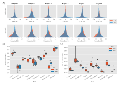 |
84 | Comparison of Nova 1Tx vs 8Tx head coils for routine 7T neuroimaging
Belinda Ding1, Catarina Rua1,2, and Christopher T Rodgers1
1Wolfson Brain Imaging Centre, University of Cambridge, Cambridge, United Kingdom, 2Invicro LLC, A Konica Minolta Company, London, United Kingdom
Most 7T MRI sites have two head coils available for neuroimaging: a single-transmit coil and one capable of parallel transmission (pTx, or 8Tx). The 8Tx coil has a circularly polarised (CP) mode, allowing it to play RF waveforms identical to its 1Tx counterpart. This study compares the performance of a 1Tx coil against an 8Tx coil from the same manufacturer for routine neuroimaging. The 8Tx coil gave better B1+ profiles in posterior brain regions which translates into improvements in other imaging sequences. It also enables the inclusion of pTx-enabled sequences. However, more detailed studies are needed before pooling data.
|
||
2095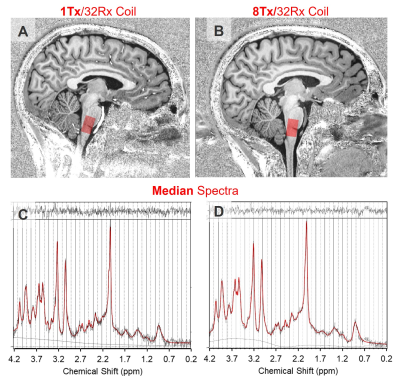 |
85 | Brainstem 7T 1H-MRS: comparison between single-channel (1Tx32Rx) and parallel transmit (8Tx32Rx) coils
Carina Graf1, Belinda Ding1, Catarina Rua1,2, and Christopher T Rodgers1
1Wolfson Brain Imaging Centre, University of Cambridge, Cambridge, United Kingdom, 2Invicro LLC, A Konica Minolta Company, London, United Kingdom 7T MRS offers increased SNR and peak separation. Many Siemens 7T sites have both 1Tx32Rx and 8Tx32Rx head coils. Workflow is similar when using the TrueForm/CP+ mode. We compared MP2RAGE and semiLASER 1H-MRS in brainstem in healthy volunteers to identify a preferred coil for future studies. Results: voxelplacement, linewidth, and concentrations were not significantly different. SNRNAA increased 27% and CRLBs for Gln, GABA & GSH trended to be lower for the 8Tx coil. This encourages regular use of the 8Tx32Rx coil for brainstem 1H-MRS and facilitates method development by allowing full pTx sequences to be added on to scheduled examinations. |
||
2096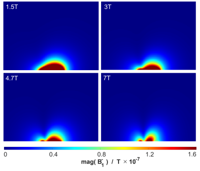 |
86 | Time-Generalized Biot-Savart Law for $$$B_1$$$, $$$B_1^+$$$, and $$$B_1^-$$$ Field Computations up to 7T MRI
Mirsad Mahmutovic1, Sam-Luca JD Hansen1, and Boris Keil1
1Institute of Medical Physics and Radiation Protection, TH Mittelhessen University of Applied Science, Giessen, Germany
Numerous methods for field computations of RF coils have been developed in the past. All these methods are based on Maxwell's equations or their derived wave equation. However, these methods are difficult to implement and not very intuitively to handle. In this work, we have investigated the suitability of the time-generalized Biot-Savart-law for $$$B_1$$$, $$$B_1^+$$$ and $$$B_1^-$$$-field computations. We showed that this method can be easily implemented and that the obtained results are a good approximation when reflections are neglectable.
|
||
2097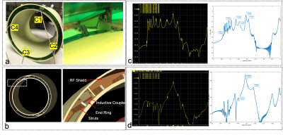 |
87 | Model validation of a 500 MHz Inductive Resonator for 11.7 T Brain MRI
Natalia Gudino1, Jacco A de Zwart2, Peter Van Gelderen2, Stephen J Dodd1, Jeff H Duyn2, and Joe Murphy-Boesch2
1LFMI, NIH, Bethesda, MD, United States, 2NIH, Bethesda, MD, United States
To evaluate safety of a 500 MHz inductive birdcage resonator for human brain MRI at 11.7 T, we compared phantom SAR measurements with predictions based on simulations. On a head-shaped phantom with realistic permittivity and conductivity, good correspondence was achieved in heating distributions when irradiating the phantom with 85 W RF power. The ability to predict SAR at 500MHz with simulation is critical step towards in-vivo MRI at 11.7 T.
|
||
2098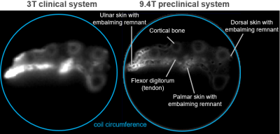 |
88 | Comparison of ultra-high field MRI of ancient remains at 9.4T and 3T
Agazi Samuel Tesfai1, Johannes Fischer1, Ali Caglar Özen1, Sébastien Bär1,2, Patrick Eppenberger3, Lena Öhrström3, Frank Rühli3, Ute Ludwig1, and Michael Bock1
1Dept.of Radiology, Medical Physics, Medical Center University of Freiburg, Faculty of Medicine, University of Freiburg, Freiburg, Germany, 2Department of Neurosurgery, Section for Neuroelectronic Systems, University Medical Center Freiburg, Freiburg, Germany, 3Institute of Evolutionary Medicine, Faculty of Medicine, University of Zurich, Zurich, Switzerland
Ancient mummified samples have ultra-short T2* which decreases with increasing field strength. Here, image quality and T2* relaxation times are compared for different tissues of a mummified hand between a clinical 3T and a preclinical 9.4T system using a 3D UTE sequence. Although T2* is shorter at 9.4T, image quality was superior to 3T, justifying the benefits of ultra-high fields (UHF).
|
||
 Back to Meeting Home
 Back to the Program-at-a-Glance
The International Society for Magnetic Resonance in Medicine is accredited by the Accreditation Council for Continuing Medical Education to provide continuing medical education for physicians.
 Back to Meeting Home
 Back to the Program-at-a-Glance
 View the Presentation