Digital Poster
Low-Field MRI I
Joint Annual Meeting ISMRM-ESMRMB & ISMRT 31st Annual Meeting • 07-12 May 2022 • London, UK

| Computer # | ||||
|---|---|---|---|---|
1806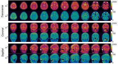 |
76 | The Feasibility of 3D MR Fingerprinting using Cartesian sampling for isotropic 1mm3 resolution at 0.55T
Yun Jiang1, Ruogu Matthew Zhu2, Bo Zhao3,4, and Nicole Seiberlich1
1Department of Radiology, University of Michigan, Ann Arbor, MI, United States, 2Department of Electrical and Computer Engineering, University of Michigan, Ann Arbor, MI, United States, 3Department of Biomedical Engineering, University of Texas at Austin, Austin, TX, United States, 4Oden Institute for Computational Engineering and Sciences, University of Texas at Austin, Austin, TX, United States
The purpose of this work is to assess the feasibility of performing MR Fingperprinting with 3D pseudorandom Cartesian sampling for an isotropic spatial resolution of 1mm3 at 0.55T in the brain.
|
||
1807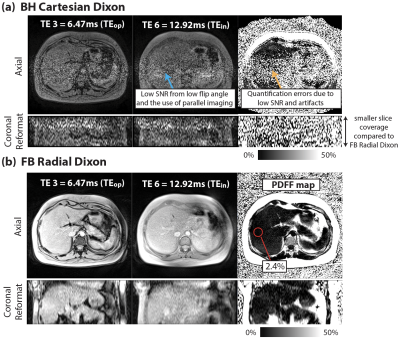 |
77 | Free-Breathing Liver Fat Quantification using Radial Acquisition on a High-Performance 0.55T MRI System
Shu-Fu Shih1,2, Sophia X. Cui3, Xiaodong Zhong3, Bilal Tasdelen4, Ecrin Yagiz4, Krishna S. Nayak4, and Holden H. Wu1,2
1Department of Radiological Sciences, University of California Los Angeles, Los Angeles, CA, United States, 2Department of Bioengineering, University of California Los Angeles, Los Angeles, CA, United States, 3Siemens Medical Solutions USA, Inc., Los Angeles, CA, United States, 4Ming Hsieh Department of Electrical and Computer Engineering, University of Southern California, Los Angeles, CA, United States
MRI proton-density fat fraction (PDFF) is used for non-invasive diagnosis of fatty liver disease, and has been validated at 1.5T and 3T field strengths. There is renewed interest in lower field strengths, such as 0.55T, because of potential advantages such as lower hardware/siting costs and a larger bore diameter. This study investigated PDFF quantification at 0.55T and validated PDFF quantification at 0.55T using Cartesian and Radial Dixon techniques in a reference phantom, and demonstrated the feasibility of in vivo free-breathing radial liver PDFF quantification in healthy subjects at 0.55T.
|
||
1808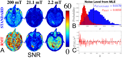 |
78 | Random Matrix Theory reconstruction for T1 quantification on a field-cycling scanner for B0 ranging between 2-200mT
Gregory Lemberskiy1,2, Vasiliki Mallikourti3, Dmitry S Novikov1, and Lionel M Broche3
1Radiology, New York University Grossman School of Medicine, New York, NY, United States, 2Microstructure Imaging INC, New York, NY, United States, 3Aberdeen Biomedical Imaging Centre, University of Aberdeen, Aberdeen, United Kingdom
We showcase high SNR T1 maps over a range of low field strengths on a stroke patient imaged using a field-cycling imaging scanner (FCI), an MR system capable of varying B0 over 2 orders of magnitude. Images from this scanner are denoised using Random Matrix Theory (RMT), leveraging the redundancy over multiple inversion times and field strengths to increase the baseline SNR by 3-fold. Following RMT denoising, we observe the T1 dispersion effect indicating a deviation of R1 frequency dependence from the BPP theory.
|
||
1809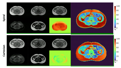 |
79 | Ultra-Fast Water/Fat Imaging of the Abdomen at 0.55T
Krishna S. Nayak1,2, Bilal Tasdelen1, Ecrin Yagiz1, and Sophia X. Cui3
1Ming Hsieh Department of Electrical and Computer Engineering, University of Southern California, Los Angeles, CA, United States, 2Department of Biomedical Engineering, University of Southern California, Los Angeles, CA, United States, 3Siemens Medical Solutions USA, Inc., Los Angeles, CA, United States
We demonstrate single-breath-hold volumetric water/fat imaging of the abdomen using spiral gradient echo imaging at 0.55T. This provides 4x finer spatial resolution with adequate SNR and spatial resolution to measure the visceral adipose tissue compartment.
|
||
1810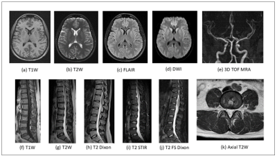 |
80 | Initial Evaluation of Neuroimaging at 0.55T
Anna Lavrova1, Kathleen Ropella-Panagis1, Nancy Dudek1, Joel Morehouse1, Ryo Kurokawa1, Mariko Kurokawa1, Pedro Itriago-Leon2, Toshio Moritani1, and Nicole Seiberlich1
1Department of Radiology, University of Michigan, Ann Arbor, MI, United States, 2Siemens Medical Solutions USA Inc., Houston, TX, United States The purpose of this study was to determine which sequences can be immediately deployed for brain and spine imaging on a low-field 0.55T MRI system, with the goal of generating optimized protocols which can be used at other sites for routine clinical neuroimaging. The image quality of 174 brain and spine images was rated by two neuroradiologists. Almost all brain (T1w, T2w, FLAIR, DWI, MRA) and spine (T1w, T2w, DIXON) sequences received image quality scores corresponding to acceptable diagnostic image quality. These sequences form numerous neurological MRI protocols, which based on these results could be collected at 0.55T. |
||
1811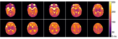 |
81 | T1 and T2 Mapping of the Infant Brain with a Low-Field, Portable MRI System
Nathan S Artz1, Maura S Sien1, Amie S Robinson1, Houchun H Hu2, Rafael O'Halloran2, Megan Poorman2, and Sherwin S Chan1
1Radiology, Children's Mercy Kansas City, Kansas City, MO, United States, 2Hyperfine, Inc., Guilford, CT, United States
Low-field, portable MRI offers great potential to improve diagnostic ability in the neonatal intensive care unit. Brain relaxometry will be critical to tune scan parameters at a low magnetic field for infants, whose brain tissue composition is very different than adults. In this study, ten infants were scanned at bedside on a 64mT system. Whole brain T1 and T2 mapping was performed for five infants each. Regions of interest were drawn in the white matter and thalamus for all subjects. Mean T1 and T2 values were 702ms and 294ms for white matter and 364ms and 139ms for the thalamus.
|
||
1812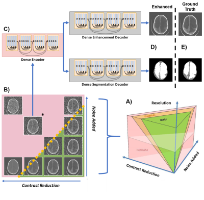 |
82 | Assessing the utility of low-resolution images for Hydrocephalus treatment planning
Joshua Harper1, Venkateswararao Cherukuri2, Tom O'Reilly3, Mingzhao Yu2, Edith Mbabazi-Kabachelor4, Roland Mulando4, Kevin N. Sheth5, Andrew G. Webb3, Benjami C. Warf6, Abhaya V. Kulkarni7, Vishal Monga2, and Steven J. Schiff2
1Engineering Science, Penn State University, State College, PA, United States, 2Penn State University, University Park, PA, United States, 3Leiden University Medical Center, Leiden, Netherlands, 4The CURE Children's Hospital of Uganda, Mbale, Uganda, 5Yale, New Haven, CT, United States, 6Boston Children's Hospital, Harvard Medical School, Boston, MA, United States, 7University of Toronto, Toronto, ON, Canada
Brain images of a quality lower than is conventionally acceptable may still be useful for planning hydrocephalus treatment. Low-field MRI is a technology of growing global interest which has the capability of producing images of sufficient quality for treatment planning and is affordable enough to disseminate to rural regions of the world. Though deep learning enhancement of low-quality images does improve CNR and apparent quality, spatial errors of brain and CSF after enhanced reconstruction add significant risk to treatment management and should be avoided.
|
||
1813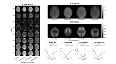 |
83 | In Vivo T1 Mapping of Neonatal Brain Tissue at 64mT
Francesco Padormo1,2, Paul Cawley1, Louise Dillon1, Emer Hughes1, Jennifer Almalbis1, Joanna Robinson1, Alessandra Maggioni1, Daniel Cromb1, Anthony Price1, UNITY Consortium3, Lori Arlinghaus4, Houchun Harry Hu4, Steve Williams3, Shaihan Malik1, Serena Counsell1, Mary Rutherford1, Tomoki Arichi1, A. David Edwards1, and Joseph V. Hajnal1
1Centre for the Developing Brain, King's College London, London, United Kingdom, 2Medical Physics, Guy's & St Thomas' NHS Foundation Trust, London, United Kingdom, 3Centre for Neuroimaging Sciences, King's College London, London, United Kingdom, 4Hyperfine Inc., Guildford, CT, United States
A promising application of ultra-low field portable MRI is neonatal imaging. In this work we show that neonatal brain T1 mapping is viable at 64mT utilising the Hyperfine Swoop portable MRI system.
|
||
1814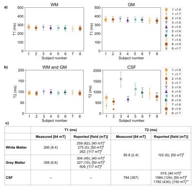 |
84 | Quantitative MRI measurements for T1 and T2 at 0.064T
Kalina V Jordanova1, Michele N Martin1, Karl F Stupic1, Samantha By2, Megan E Poorman2, Rafael O'Halloran2, and Kathryn E Keenan1
1NIST: National Institute of Standards and Technology, Boulder, CO, United States, 2Hyperfine, Inc., Guilford, CT, United States
Increased use of low-field MRI and quantitative MRI suggests a need for accurate quantitative measurements for fields <0.55T. We measure T1 and T2 for normal brain tissues at 0.064T using a Hyperfine scanner. T1 measurements gave a mean of 266±8.4ms for white matter, 358±6.6ms for grey matter; T2 measurements gave a mean of 95.8±2.4ms for combined white matter and grey matter, 794±357ms for CSF. Validation samples are measured on the Hyperfine system and compared to measurements taken on an electromagnet NMR at 0.064T.
|
||
1815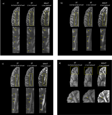 |
85 | Postmortem Multiple Sclerosis Brain Lesion Identification at Low Field
Sharada Balaji1, Adam Dvorak1, Irene M. Vavasour2,3, Megan E. Poorman4, Hanwen Liu1, Emil Ljungberg5,6, Steve Williams6, Sean Deoni7, Cornelia Laule1,2,3,8, David K.B. Li2, G.R. Wayne Moore3,8,9, Alex MacKay1,2, and Shannon H. Kolind1,2,3,9
1Physics and Astronomy, University of British Columbia, Vancouver, BC, Canada, 2Radiology, University of British Columbia, Vancouver, BC, Canada, 3International Collaboration on Repair Discoveries, Vancouver, BC, Canada, 4Hyperfine, Inc., Guilford, CT, United States, 5Medical Radiation Physics, Lund University, Lund, Sweden, 6Neuroimaging, King's College London, London, United Kingdom, 7MNCH D&T, Bill and Melinda Gates Foundation, Seattle, WA, United States, 8Pathology and Laboratory Medicine, University of British Columbia, Vancouver, BC, Canada, 9Medicine, University of British Columbia, Vancouver, BC, Canada
A single hemisphere of postmortem brain tissue from a subject with multiple sclerosis (MS) was scanned at high (Philips Elition 3T) and low (Hyperfine Swoop 64mT) field strength to determine whether lesions could be detected using a portable low field MRI system. T2-weighted scans were acquired at both field strengths with matching resolutions to assess the impact of low field. Comparisons with a high resolution 3T scan showed that of 17 visible lesions, 11 were seen on the lower resolution 3T and 10 were seen on the 64mT scan, demonstrating the feasibility of lesion detection at low field.
|
||
1816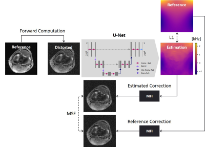 |
86 | Physics-Informed Deep Learning for Image Distortion Correction from B0-inhomogeneities in Low-Field MRI
David Schote1, Lukas Winter1, Christoph Kolbitsch1, Felix Zimmermann1, Thomas O'Reilly2, Andrew Webb2, Frank Seifert1, and Andreas Kofler1
1Physikalisch-Technische Bundesastalt (PTB), Braunschweig and Berlin, Germany, 2C.J. Gorter Center for High Field MRI, Department of Radiology, Leiden University Medical Center, Leiden, Netherlands
A physics-informed deep neural network using a U-Net in combination with a multifrequency interpolation is presented to correct for B0-field image distortions in low-field MRI. Training data is based on 1.5T and 3.0T knee images and realistic measurement-based assumption of SNR and B0-field inhomogeneities from previously constructed Halbach magnets. Significant (p<0.05) improvements are demonstrated applying the suggested methodology to uncorrected images.
|
||
1817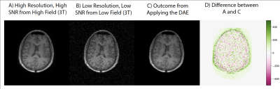 |
87 | Simulation Evidence for use of a Denoising Auto-Encoder (DAE) to Improve Ultra-Low Field (64mT) MRI with a High Field (3T) Prior
M. Ethan MacDonald1,2, Eremiahs Fikre1,2, Fernando Vega1,2, and AbdolJalil Addeh1,2
1Biomedical Engineering, Electrical & Software Engineering, Radiology, University of Calgary, Calgary, AB, Canada, 2Hotchkiss Brain Institute, Foothills Medical Centre, Calgary, AB, Canada
In this work, the denoising autoencoder is applied to simulated low field, low resolution, low signal-to-noise images and used to recover the high field, high resolution, high signal-to-noise paired image. Different types of noise, gaussian and chi-squared, is added in simulation. We found that the denoising autoencoder worked slightly better for normally disturbed noise, but not in all cases. We found a linear trend between the model performance with RMSE and the standard deviation of the added noise. This work demonstrates the use of simple and robust denoising autoencoder to improve low field MRI.
|
||
1818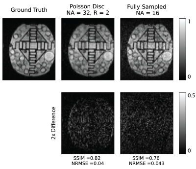 |
88 | Accelerating Ultra-Low Field MRI with Compressed Sensing
David E J Waddington1, Efrat Shimron2, Nicholas Hindley1,3, Neha Koonjoo3, and Matthew S Rosen3,4,5
1ACRF Image X Institute, Faculty of Medicine and Health, The University of Sydney, Sydney, Australia, 22Department of Electrical Engineering and Computer Sciences, UC Berkeley, Berkeley, CA, United States, 3A. A. Martinos Center for Biomedical Imaging, Charlestown, MA, United States, 4Department of Physics, Harvard University, Cambridge, MA, United States, 5Harvard Medical School, Boston, MA, United States Portable MRI scanners that operate at very low magnetic fields are increasingly being deployed in clinical settings. However, the intrinsic low signal-to-noise (SNR) ratio of these low-field MRI scanners often necessitates many signal averages, and therefore excessively long acquisition times. Here we propose to improve SNR through optimized k-space undersampling and Compressed Sensing reconstruction. We demonstrate this approach for 6.5 mT ultra-low-field MRI using: (1) retrospective-subsampling experiments with 2x to 4x acceleration; (2) prospectively-subsampled data acquired from a human brain phantom with a 6.5mT MRI. The results exhibit a higher SNR than the traditional averaging method, without increasing scan time. |
||
1819 |
89 | Scan acceleration by oversampling in a 70 mT MRI scanner
Fernando Galve1,2, José Miguel Algarín1,2, Teresa Guallart3, Rubén Pellicer4, Eduardo Pallás1,2, José Manuel González3, Yolanda Vives3, Rubén Bosch3, Guillermo López1,2, Juan Pablo Rigla3, José Borreguero3, Alfonso Ríos3, José María Benlloch1,2, and Joseba Alonso1,2
1MRILab, Institute for Molecular Imaging and Instrumentation (i3M), Spanish National Research Council (CSIC), Valencia, Spain, 2MRILab, Institute for Molecular Imaging and Instrumentation (i3M), Universitat Politècnica de València (UPV), Valencia, Spain, 3Tesoro Imaging SL, Valencia, Spain, 4Physio MRI SL, Valencia, Spain
Here we present accelerated images by PhasE-Constrained Over Sampled (PECOS) MRI in our home-made Halbach MRI scanner (70 mT). We show projection images of a phantom obtained in our “PhysioMRI” scanner (Fig. 1(a)), a home-made scanner designed for musculoskeletal imaging at low field strengths. We use cartesian turbo spin echo (TSE) sequence to sample the k-space. Image reconstructions are performed by phase conjugate reconstruction and by PECOS.
|
||
1820 |
90 | Evaluation of dynamic 2D-EPI acquisitions for fetal brain tracking with neural networks Video Permission Withheld
Sara Neves Silva1, Irina Grigorescu2, Alena Uus2, Johannes Steinweg2, Maria Deprez2, Jo Hajnal2, Kuberan Pushparajah2, Jana Hutter2, and Enrico De Vita2
1Biomedical Engineering and Imaging Sciences, King's College London, London, United Kingdom, 2King's College London, London, United Kingdom
Fetal MRI and MRS are often compromised due to unpredictable fetal motion and commonly require multiple repetitions for diagnostic studies. To overcome these limitations, we are working towards developing a deep-learning based automatic MRI fetal motion tracking method. Our method uses, as input, rapid dynamic multi-echo 2D-EPI acquisitions and is based on a 3D U-Net for brain localisation and translational motion parameters estimation for brain tracking. The results show rapid low-resolution acquisitions contain sufficient information to allow automated fetal brain localisation. Our technique can be used to assess fetal movements and to build navigation systems for fetal MRI/MRS.
|
||
 Back to Meeting Home
 Back to the Program-at-a-Glance
The International Society for Magnetic Resonance in Medicine is accredited by the Accreditation Council for Continuing Medical Education to provide continuing medical education for physicians.
 Back to Meeting Home
 Back to the Program-at-a-Glance
 View the Presentation