Digital Poster
Low-Field MRI II
Joint Annual Meeting ISMRM-ESMRMB & ISMRT 31st Annual Meeting • 07-12 May 2022 • London, UK

| Computer # | ||||
|---|---|---|---|---|
1895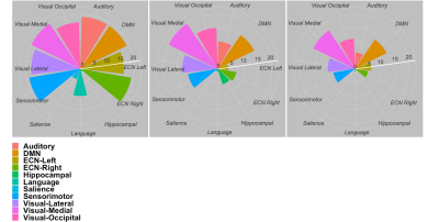 |
75 | Investigating the detection capability of RSNs with fMRI at a low field scanner equipped with a high-performance gradient
Arjama Halder1, Demetrius Riberio de Paula2, William Handler3,4, Andrea Soddu3, and Blaine Chronik1,3,5
1Medical Biophysics, Western University, London, ON, Canada, 2Radbound University, Nijmegen, Netherlands, 3Physics and Astronomy, Western University, London, ON, Canada, 4xMR Labs, London, ON, Canada, 5The xMR Labs, London, ON, Canada
Preliminary evaluation of the feasibility to perform resting state BOLD fMRI at a low field (0.5 T) MR scanners equipped with a high-performance gradient system by investing the detection capability of RSNs at lower field strengths. There was an overall deterioration in the number of detected regions within all RSNs as the tSNR decreased. However, the 0.5 Evry system that will be used for experimental studies offer a better SNR efficiency than the modelled cases considered here suggesting reasonable capability in extracting RSNs that can potentially provide clinical utility.
|
||
1896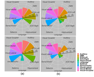 |
76 | Investigating the effect of reduced TR in detection of RSNs at a low field MR scanner
Arjama Halder1, Demetrius Riberio de Paula2, William Handler3,4, Andrea Soddu3, and Blaine A Chronik1,3,4
1Medical Biophysics, Western University, London, ON, Canada, 2Radbound University, Nijmegen, Netherlands, 3Physics and Astronomy, Western University, London, ON, Canada, 4xMR Labs, London, ON, Canada
Initial investigation of effects due to reduced TR on RSNs at a 0.5 T MR scanner. There was a decrease in detected regions within specific networks for the 3 T data as TR was reduced. However, when the tSNR was approximately 17 ignoring the expected decrement of detected regions compared to the 3 T data, there was an increase in positive detection at shorter TR. These result show the possibility of extracting true RSNs for a low field scanner using shorter TRs which can lead to efficient sequence design since T1 is shortened at low fields.
|
||
1897 |
77 | Simulated Impact of Gradient Amplifier Noise on Simple 3D Sequences: Preliminary Results
Mike Twieg1
1Hyperfine, Guilford, CT, United States
It is often stated, without qualification, that MRI gradient systems must be precise and/or accurate in order to ensure MRI image quality. Some imperfections (delay, eddy currents) have been well-studied and documented, while others such as noise, ripple, and quantization have not. Here we simulate the impact of white noise in the gradient fields with simple cartesian imaging sequences for a numerical brain phantom. Preliminary results suggest that high-performance/high-cost components typically marketed for gradient control are excessive, and much cheaper components could be used without significant drawbacks (NRMSE < 1%), especially for low-field/low-cost systems.
|
||
1898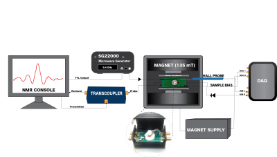 |
78 | Development of a Low Field Tabletop Micro-MRI systemUsing Novel Solid-State Hyperpolarization of Water
Emily Buchanan1,2,3, Karl Stupic1, and Stephen Russek1
1Physical Measurement Laboratory, National Institute of Standards and Technology, Boulder, CO, United States, 2Advanced Imaging Research Center, UT Southwestern Medical Center, Dallas, TX, United States, 3Chemistry and Biochemistry, The University of Texas at Dallas, Richardson, TX, United States
Micro-MRI systems could have transformational applications in pathology and personalized medicine if substantial technological challenges are overcome: resolution needs to be improved from current state-of-the-art to subcellular resolution <10$$$\mu$$$m, contrast needs to be improved to allow delineation of cells and cell structures, and systems need to be miniaturized from the large high-field superconducting-magnet systems to low-cost tabletop systems. A key enabling component is likely to be a room-temperature low-field point-of-use hyperpolarization system. Here we present a tabletop 135mT micro-MRI system that uses a novel solid-state point-of-use water proton hyperpolarization system integrated into the imaging cell that functions as a bioreactor.
|
||
1899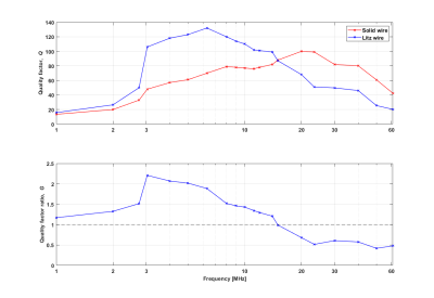 |
79 | Application of Litz Wires in MRI Coil Design up to 15 MHz
Robert Kowal1,2, Ivan Fomin1,2, Marcus Prier1,3, Enrico Pannicke1,2, Georg Rose1,2, and Oliver Speck1,3
1Research Campus STIMULATE, Otto-von-Guericke University, Magdeburg, Germany, 2Institute for Medical Engineering, Otto-von-Guericke University, Magdeburg, Germany, 3Department for Biomedical Magnetic Resonance, Otto-von-Guericke University, Magdeburg, Germany
The use of litz wire in their optimized frequency range can provide increased efficiency of high-frequency coils and lead to higher SNR in MR measurements. This work experimentally explores the added value of such a litz wire using equivalent solenoid coils in the range of 1 to 61 MHz. An improvement could be measured up to a frequency of 15 MHz, whereas the maximum effectiveness of the investigated wire was at 3 MHz. In addition, the SNR gain was validated in agreement on an 0.26 T MR system by comparing 1H FID signals.
|
||
1900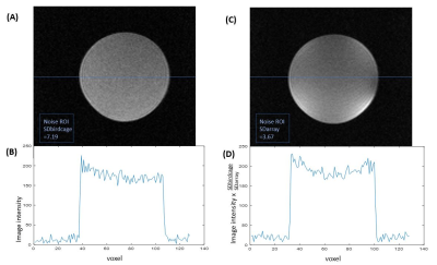 |
80 | A flexible, 4-channel head array for 0.2T readout FCI (Field-Cycling Imaging).
Robert Stormont1,2, James Ross1, Gareth Davies1, Vasiliki Mallikourti1, Amnah Alamri1, Lionel Broche1, and David Lurie1
1Medical Physics, University of Aberdeen, Aberdeen, United Kingdom, 2GE Healthcare, Waukesha, WI, United States A 4-Channel Head Coil RX Array has been created supporting Field-Cycling Imaging studies at an 8.5 MHz readout frequency. Head imaging to date, has been accomplished with a quadrature TX/RX birdcage coil. The array goals include improved SNR, enhanced patient comfort, peripheral compatibility, with fast application to various sized heads. Evaluation with healthy volunteers is now underway. The array appears well tolerated by individuals when compared to the 8-rung birdcage, accommodating a communication headset, and leaving the face quite open. SNR is comparable to the birdcage at the image center and improved as you move towards the array elements. |
||
1901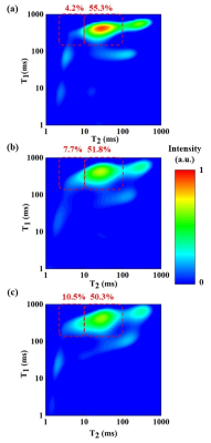 |
81 | Optimized radio frequency coil for low-field magnetic resonance relaxometry in human finger
Junnan Wang1, Rongsheng Lu1, Layale Bazzi2, Xiaowen Jiang1, Yi Chen1, Zhengxiu Wu1, Qing Yang1, Zhonghua Ni1, Hong Yi1, and Dan Xiao2
1School of Mechanical Engineering, Southeast University, Nanjing, China, 2Department of Physics, University of Windsor, Windsor, ON, Canada
The permanent magnet based low-field and low-cost MR systems increase the accessibility and potential of point-of-care diagnosis, where relaxometry measurement is usually preformed due to the low requirement of B0 field homogeneity. However, inhomogeneous B1 field results in errors in the T2 relaxation times, especially in in-vivo localized human tissue measurements. A novel RF coil termed T coil has been developed to provide a better B1 field homogeneity and more accurate in-vivo relaxation measurements than the conventional coils. This may enable the noninvasive diagnosis of diseases through the human finger measurements with portable and affordable MR systems.
|
||
1902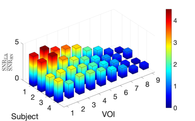 |
82 | Evaluation of a Novel 8-Channel Receive Coil for Speech Production MRI at 0.55 Tesla.
Felix Munoz1, Yongwan Lim2, Sophia X. Cui3, Helmut Stark4, and Krishna Shrinivas Nayak1,2
1Department of Biomedical Engineering, University of Southern California, Los Angeles, CA, United States, 2Ming Hsieh Department of Electrical and Computer Engineering, University of Southern California, Los Angeles, CA, United States, 3Siemens Medical Solutions, Los Angeles, CA, United States, 4Stark Contrast, Erlangen, Germany MRI has enabled significant advances in our understanding of human speech production. This application benefits from a lower magnetic field due to reduced off-resonance effects at air-tissue interfaces. Here, we evaluate the SNR and parallel imaging performance of a dedicated speech coil at 0.55 Tesla, and compare it to that of a head-neck coil. Over the upper airway regions of interest, the dedicated coil shows approximately 1.3- to 4.6-fold improvement in SNR efficiency compared to the head-neck coil. |
||
1903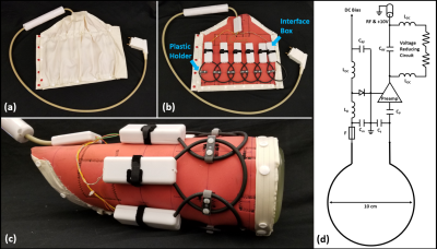 |
83 | Flexible six-channel knee array for MRI at 0.55T
Bili Wang1,2, Syed S. Siddiq1,2, Jerzy Walczyk1,2, Mary Bruno1,2, Jan Fritz1,2, Iman Khodarahmi1, Inge M. Brinkmann3, Robert Rehner4, Karthik Lakshmanan1,2, and Ryan Brown1,2
1Bernard and Irene Schwartz Center for Biomedical Imaging, Department of Radiology, New York University Grossman School of Medicine, New York, NY, United States, 2Center for Advanced Imaging Innovation and Research (CAI2R), Department of Radiology, New York University Grossman School of Medicine, New York, NY, United States, 3Siemens Medical Solutions USA Inc., Malvern, PA, United States, 4Siemens Healthcare GmbH, Erlangen, Germany
New-generation 0.55T systems require a suite of tailored coils to preserve image quality that is expected from higher field systems. We built a six-channel coil array for knee MRI using RG58 coaxial cable loops, which have adequate mechanical flexibility to wrap tightly around the leg and suitably low conductive loss at 23.55MHz. Initial findings show that the dedicated coil outperformed a general-purpose counterpart and suggest that clinical quality knee imaging is feasible.
|
||
1904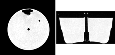 |
84 | Phantom Development for Comparison of Thermal Mapping
Diego Felipe Martinez1, Curtis N. Wiens2, Amgad Louka3, Dereck Gignac1, Will Bradfield Handler1, and Blaine Alexander Chronik1,3
1The xMR Labs, Physics and Astronomy, Western University, London, ON, Canada, 2Research and Development, Synaptive Medical, Toronto, ON, Canada, 3Medical BioPhysics, Western University, London, ON, Canada
Thermal Mapping using MRI is a non-invasive method for probing the body and has both diagnostic and interventional applications. If conducted on more accessible low field strength systems, these can improve the accessibility of the method for wider use. In order to validate and compare thermal mapping, a temperature control phantom was developed with tools for heating the phantom and externally tracking the temperature of the phantom. A heating study was performed with the phantom at two temperatures, and the thermometry methods showed good connection to the change in temperature in the phantom.
|
||
1905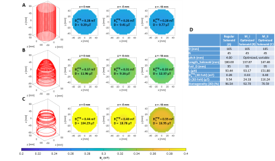 |
85 | Design and Optimization of Asymmetrical Tapered RF Solenoid Tx / Rx Coil for Halbach-Array based Low-field MRI Scanner
Meena Rajendran1 and Shao Ying Huang1
1Singapore University of Technology, Singapore, Singapore The optimization of an asymmetrical tapered solenoid coil using multi-objective Genetic Algorithm is presented to achieve high sensitivity, high homogeneity, low inductance simultaneously at 2.84MHz within a cylindrical volume(95×95х10mm). The asymmetrical solenoid coil has a lower half that consists of turns with variable pitches and a constant radius and an upper half that consist of turns with variable pitches and profile curvature(variable radius). Two coil winding patterns are included: single winding and multiple windings at each groove. The optimal design shows a sensitivity of 138 μT/√W (29.7%increase), 3.8% homogeneity reduction, comparable inductance compared to a traditional solenoid of similar dimensions. |
||
 Back to Meeting Home
 Back to the Program-at-a-Glance
The International Society for Magnetic Resonance in Medicine is accredited by the Accreditation Council for Continuing Medical Education to provide continuing medical education for physicians.
 Back to Meeting Home
 Back to the Program-at-a-Glance
 View the Presentation