Digital Poster
Interventional & Low-Field MRI
Joint Annual Meeting ISMRM-ESMRMB & ISMRT 31st Annual Meeting • 07-12 May 2022 • London, UK

| Computer # | ||||
|---|---|---|---|---|
1193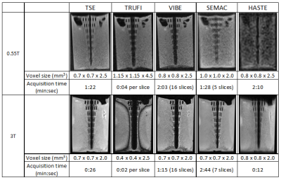 |
98 | Feasibility of MRI-guided Interventions at a Large-bore New-generation 0.55T Low-Field MRI System Video Permission Withheld
Iman Khodarahmi1, Inge M Brinkmann2, Mary Bruno1, Jerzy Walczyk1, Ryan Brown1, and Jan Fritz1
1Department of Radiology, New York University School of Medicine, New York, NY, United States, 2Siemens Medical Solutions USA Inc, Malvern, PA, United States
New generation low-field MRI systems can be ideal for MRI-guided interventions due to wide bore diameters and lower susceptibility artifacts improving patient access and needle visualization, respectively. We evaluated a set of pulse sequences for their suitability for needle visualization using a new-generation 0.55T system and compared the results with those of a clinical 3T system. Interventional needles can be adequately visualized at 0.55T. Using TSE pulse sequences, needle artifacts display more favorable at 0.55T than 3T. SEMAC and HASTE pulse sequences may not facilitate diagnostic needle visualization at 0.55T but are not needed for clinical MR interventions.
|
||
1194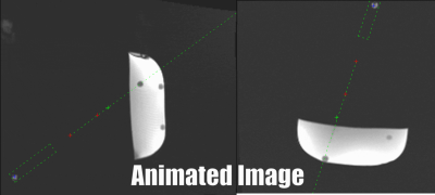 |
99 | Passive Real-Time Needle Tracking and Needle Tip Estimation
Andreas Reichert1, Samantha Hickey1, Johannes Fischer1, Michael Vogele2, Simon Reiss1, and Michael Bock1
1Dept. of Radiology, Medical Physics, Medical Center University of Freiburg, Faculty of Medicine, Freiburg, Germany, 2Interventional Systems GmbH, Kitzbuehel, Austria
A real-time sequence is presented with automatic adjustment of the imaging plane parallel to a needle. The sequence utilizes the phase-only cross correlation (POCC) algorithm to detect the orientation of an end-effector and to visualize the planned needle pathway. Additionally, it detects a passive marker at the distal end of the needle to calculate the position of the needle tip for real-time display during needle insertion. The sequence is evaluated in a phantom experiment, and both lateral and longitudinal needle insertion accuracies are determined.
|
||
1195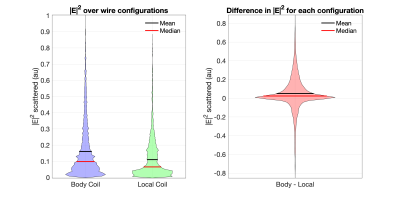 |
100 | A Monte Carlo Analysis of Guidewire Safety Comparing a Body and Local Coil Array
felipe godinez1,2, Jeffrey W Hand2, Nuno J Teixeira3, Bart R Steensma4, Joseph V Hajnal2, alexander J E Raaijmakers5, and shaihan malik2
1Radiology, University of California, Sacramento, CA, United States, 2Biomedical Engineering Department, School of Biomedical Engineering and Imaging Sciences, King's College London, London, United Kingdom, 3Perinatal Imaging, King's College London, London, United Kingdom, 4Center for Image Sciences, University Medical Center Utrecht, Utrecht, Netherlands, 5Biomedical Engineering, Medical Imaging Analysis, Eindhoven University of Technology, Eindhoven, Netherlands
Metallic Endovascular devices can be unsafe when used during MR guided procedures. A potential solution to the heating problem might be to use a local coil instead of a body coil to excite the imaging volume. In this work we present a Monte Carlo analysis using many random guidewire trajectories and the transfer function to estimate the scatter electric field at the guidewire tip imbedded in a heterogenous dielectric.
|
||
1196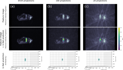 |
101 | Fast 3D Passive Needle Localization for MR-Guided Interventions using Radial White Marker Acquisitions and CNN Postprocessing
Jonas F. Faust1,2, Axel J. Krafft2, Daniel Polak2, Ralf Vogel3, Peter Speier2, Nicolas G. R. Behl2, Mark E. Ladd4, and Florian Maier2
1Ruprecht-Karls-Universität Heidelberg, Heidelberg, Germany, 2Siemens Healthcare GmbH, Erlangen, Germany, 3Pattern Recognition Lab, Friedrich-Alexander-Universität Erlangen-Nürnberg, Erlangen, Germany, 4Medical Physics in Radiology, German Cancer Research Center (DKFZ), Heidelberg, Germany
Accurate 3D localization of biopsy needles during MR-guided interventions is especially challenging due to time restrictions in real-time workflows. While active tracking methods rely on additional RF-sensitive hardware, rapid passive tracking methods often make use of markers or prior knowledge regarding the needle location. In an ex-vivo study, we investigated a novel passive tracking method using heavily undersampled, radial acquisitions in combination with contrast-optimized White Marker imaging and CNN image postprocessing regarding its potential to speed up passive needle artifact localization without the use of additional hardware or prior knowledge on the needle location.
|
||
1197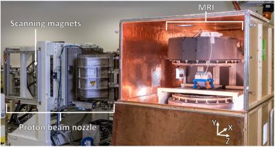 |
102 | Measurement of magnetic fringe fields from a proton pencil beam scanning nozzle causing MR image ghosting artefacts during in-beam MR imaging
Ekaterina Semioshkina1, Bradley M. Oborn2, and Aswin L. Hoffmann1
1Institute of Radiooncology – OncoRay, Helmholtz-Zentrum Dresden – Rossendorf, Dresden, Germany, 2Centre for Medical Radiation Physics (CMRP), University of Wollongong, Wollongong, Australia
Proton therapy (PT) is expected to benefit from real-time MRI guidance. However, the integration of MRI and PT is challenging due to the interaction between the magnetic fringe fields of the PT beamline and the B0-field of the MR scanner. In this study, we measured the magnetic fringe field of the PT beamline. The measurement data will be used to validate a finite element model. With a match to this model, we can proceed to investigate what type of magnetic shielding solution would be needed to decouple the magnetic fields of the PT and MRI systems.
|
||
1198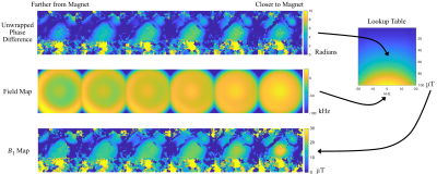 |
103 | Phase-Based B1 Mapping in a 65 mT Single-Sided Prostate MRI Scanner
William A Grissom1, Meredith Sadinski2, Alek Nacev2, and Muller De Matos Gomes2
1Biomedical Engineering, Vanderbilt University, Nashville, TN, United States, 2Promaxo, Oakland, CA, United States
B1 mapping is important to characterize non-uniform coil geometries and to optimize imaging sequences and image reconstructions in low-field MRI, but is made challenging by point-of-care single-sided imaging geometries with large frequency bandwidths and B1 ranges. We propose a high-bandwidth phase-based B1 mapping technique based on a CHORUS pulse sequence with alternated frequency sweep polarities, which has a wide (40 kHz) imaging bandwidth and high B1 dynamic range. The sequence was implemented and validated on a 65 mT single-sided prostate MRI scanner in phantom scans with butterfly coil transmit and local multichannel receive.
|
||
1199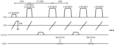 |
104 | Type-III NUFFT Image Reconstruction for a 65 mT Single-Sided Prostate MRI Scanner with Non-Linear Gradient Fields
Meredith Sadinski1, Ram Narayanan1, Alek Nacev1, and William A Grissom2
1Promaxo, Oakland, CA, United States, 2Biomedical Engineering, Vanderbilt University, Nashville, TN, United States
Single-sided low-field MRI scanners can provide image guidance during interventions such as prostate biopsies without restricting surgical access. However, image reconstruction from these scanners is challenging due to the non-linearity of their gradient fields and inhomogeneous B0 fields. At the same time, non-Cartesian sampling is desirable for these scanners to maximize SNR efficiency and provide self-navigation. In this work, we report a fast iterative 3D Type-III NUFFT reconstruction for a 65 mT single-sided prostate imager, with non-linear single-sided gradient fields and a built-in B0 gradient field. The reconstruction was compared with a model-based reconstruction for a high-bandwidth radial RARE acquisition.
|
||
1200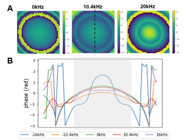 |
105 | Simulating the effects of off-resonance effects from a CHORUS pulse sequence in a single sided, low-field MRI system.
Kartiga Selvaganesan1, Muller Gomes2, Will Grissom3, and Alek Nacev2
1Yale School of Engineering & Applied Science, Yale, New Haven, CT, United States, 2Promaxo, Oakland, CA, United States, 3Biomedical Engineering, Vanderbilt University, Nashville, TN, United States Moving towards single-sided, low-field MRI systems can increase the portability and access to this invaluable clinical tool. However, the B0-fields generated by these systems are highly inhomogeneous, and severe artifacts arise when the RF pulse does not cover the full bandwidth of spin resonances. In this study, we have developed a simulation algorithm that will allow us to better simulate the phase variations induced by B1 and B0 inhomogeneities and help us design RF schemes to remove unwanted phase to produce higher quality images. |
||
1201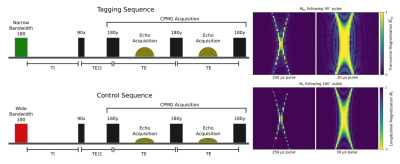 |
106 | Continuous Perfusion Measurement With Singled Sided Low Field MR Video Not Available
Alice Little1,2, Dion Thomas2, Shieak Tzeng3, and Sergei Obruchkov2
1The University of Auckland, Auckland, New Zealand, 2Victoria University of Wellington, Wellington, New Zealand, 3University of Otago, Wellington, New Zealand
In this study we focus on exploring the use of a single sided low-field MR systems to continuously measure perfusion. For this purpose we developed a contrast-free measurement technique - tentatively named Inhomogeneous Flow-sensitive Alternating Inversion Recovery (IFAIR). This new technique, adapted from the FAIR ASL protocol, was designed to be suitable for inhomogeneous field conditions and to function without pulsed gradient fields. The IFAIR signal response displayed a consistent proportional relationship to flow velocity, with potential to allow for quantitative flow-dependent measurements.
|
||
1202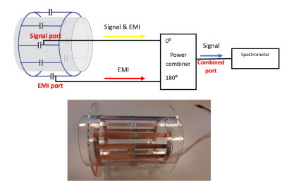 |
107 | A new single channel method for electromagnetic interference reduction on a 50 mT permanent magnet system
Javad Parsa1,2, Thomas O'Reilly2, and Andrew Webb2
1Percuros B.V., Leiden, Netherlands, 2Leiden University Medical Center, Leiden, Netherlands External electromagnetic interference (EMI) in unshielded low field and portable MRI systems can swamp the signal, and various multi-detector methods have been developed to minimize it. A new EMI cancellation method designed for a single-channel receiver-system has been implemented on a 50 mT system by using the MR-inactive channel of a birdcage coil and a 180o power hybrid. This method results in up to a 97% reduction in the standard deviation of external EMI. |
||
1203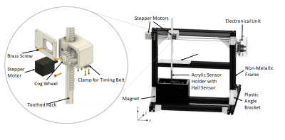 |
108 | Development of a Low-Cost B0 Field Mapping Device Video Permission Withheld
Marcel Eisenmann1,2, Ivan Fomin1,2, Marcus Prier1,3, Georg Rose1,2, and Oliver Speck1,3
1Research Campus STIMULATE, Otto von Guericke University, Magdeburg, Germany, 2Institute for Medical Engineering, Otto von Guericke University, Magdeburg, Germany, 3Department for Biomedical Magnetic Resonance, Otto von Guericke University, Magdeburg, Germany
Field mapping devices are used to determine the spatial distribution of magnetic fields. As magnetic devices are very unique in shape and size, commercially available mapping devices are often complex and customised and therefore too costly for small-format magnetic resonance imaging (MRI) systems and the open-source community. A low-cost field mapping device with a Hall sensor for the OCRA tabletop MRI system1 is developed. Field maps are measured and compared against a commercial Hall probe. The presented measurements show that the developed device can compete with commercially available devices.
|
||
1204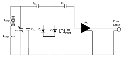 |
109 | Characterising the Magnetic Field Inhomogeneity for Open MRI at 0.5T using a Screened Coil NMR Probe Design
Dipendra Jayantilal Mistry1, Penny Gowland1, Olivier Mougin1, Richard Bowtell1, and Paul Glover1
1Sir Peter Mansfield Imaging Centre, School of Physics and Astronomy, University of Nottingham, Nottingham, United Kingdom
Image distortion caused by field inhomogeneity and instability is a major limitation for open MRI systems. Magnetic Field Monitoring (MFM) is a well-documented approach to characterise and thus compensate for these effects using an array of NMR probes. Here a novel NMR probe with a secondary coil is presented, which is designed to protect the pre-amplifier during transmission of a uniform B1 field. Preliminary data has verified this result and temporal variations of the B0 field have been observed. This approach will be developed with multiple probes to measure and compensate for the spatio-temporal response of the system.
|
||
 Back to Meeting Home
 Back to the Program-at-a-Glance
The International Society for Magnetic Resonance in Medicine is accredited by the Accreditation Council for Continuing Medical Education to provide continuing medical education for physicians.
 Back to Meeting Home
 Back to the Program-at-a-Glance
 View the Presentation