Digital Poster
High-Field RF Coils & Arrays I
Joint Annual Meeting ISMRM-ESMRMB & ISMRT 31st Annual Meeting • 07-12 May 2022 • London, UK

| Computer # | ||||
|---|---|---|---|---|
1444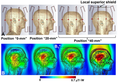 |
68 | Folded-End Dipole Transceiver Array for Human Whole Brain Imaging at 7 T.
Nikolai I. Avdievich1, Georgiy Solomakha2, Loreen Ruhm1, Anton V Nikulin1,3, Arthur Magill4, and Klaus Scheffler1,3
1High-Field MR Center, Max Planck Institute for Biological Cybernetics, Tübingen, Germany, 2Physics and Engineering, ITMO University, St. Petersburg, Russian Federation, 3Biomedical Magnetic Resonance, University of Tübingen, Tübingen, Germany, 4Medical Physics in Radiology, German Cancer Research Center, Heidelberg, Germany
In spite of great benefits of 7T MRI, its further clinical development is associated with difficulties in designing RF coils. Recently, we developed a novel type of dipole antennas, a folded-end dipole. In this work we evaluated an 8-element transceiver folded-end dipole array for 7T human head imaging. The array provided ~20% higher Tx-efficiency and significantly better whole-brain coverage than that of a widely-used commercial array. In addition, we evaluated passive dipoles for decoupling the proposed array. In contrast to the common unfolded dipole array, the passive dipoles produce practically no destructive interference with the RF field of the array.
|
||
1445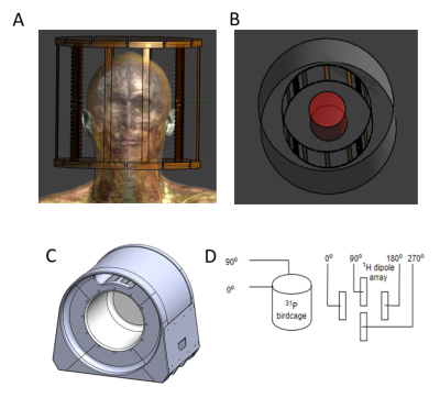 |
69 | A novel 1H-dipole/31P-birdcage transceiver coil for head imaging at 7T Video Permission Withheld
Ria Forner1, Ingmar Voogt2, Bart Steensma3, Kyungmin Nam3, Mark Gosselink3, Debra Rivera4, William T Clarke5,6, Aidin Alihaghnejad2, Arjan Hendriks3, and Dennis Klomp1
1UMC Utrecht, Utrecht, Netherlands, 2Wavetronica B.V., Utrecht, Netherlands, 3University Medical Center Utrecht, Utrecht, Netherlands, 4Eindhoven University of Technology, Eindhoven, Netherlands, 5Wellcome Centre for Integrative Neuroimaging, FMRIB, Nuffield, United Kingdom, 6Department of Clinical Neurosciences, University of Oxford, Oxford, United Kingdom
A novel coil design with a 31P birdcage and a 1H dipole array is presented for the purpose of fMRS applications. This design allows for free line-of-sight for fMRI and fMRS visual stimulation via mirrors and despite a lower fill-factor performs comparably to a smaller dual-tuned birdcage. The matching of both frequencies is adequate for a range of human head circumferences from 52cm through 57cm. The 31P birdcage performance is not significantly degraded by presence of 1H dipoles when compared to a conventional birdcage dual-tuning strategy with Foster networks.
|
||
1446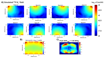 |
70 | Negligible loss in double tuning when stacking 1H to 31P dipoles in an 8-channel dipole array with 24 receivers at 7T
Jabrane Karkouri*1, Ria Forner*2, Martijn Lunenburg3, Ettore Flavio Meliadò3, Catalina Arteaga3, Alexander Raaijmakers4,5, Christopher T. Rodgers†1, and Dennis Klomp†4
1University of Cambridge, Cambridge, United Kingdom, 2Radiology, UMC Utrecht, Utrecht, Netherlands, 3Tesla Dynamic Coils, Zaltbommel, Netherlands, 4UMC Utrecht, Utrecht, Netherlands, 5Biomedical engineering, Eindhoven University of Technology, Utrecht, Netherlands We present a novel dual dipole coil concept for 7T 31P metabolic imaging in the body. We constructed an array of 8x stacked 1H/ 31P dipoles, evaluating their performance by bench measurements, EM modelling, phantom experiments and in vivo scans in human liver. Parameters such as coupling, transmission efficiency and SAR are compared to those of a whole-body 31P birdcage coil. |
||
1447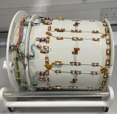 |
71 | An 8-channel transmit 64-channel receive compact head coil for Next Gen 7T scanner with head gradient insert Video Permission Withheld
Shajan Gunamony1,2 and David Feinberg3
1Imaging Centre of Excellence, University of Glasgow, Glasgow, United Kingdom, 2MR CoilTech Limited, Glasgow, United Kingdom, 3Helen Wills Neuroscience Institute, University of California, Berkeley, Berkeley, CA, United States
The NextGen 7T scanner is equipped with a high-performance head gradient insert and hence the radial space available on the patient table for radiofrequency (RF) coils is limited. In addition to a large visual field to support fMRI studies, a split-top receive array on a sliding mechanism is desirable for improved patient comfort. We have developed a novel 8-channel transmit 64-channel receive 7T head coil and implemented a sliding mechanism to operate within the limited space in the head gradient insert. In this abstract, we present the overall coil design, transmit array performance and preliminary phantom results.
|
||
1448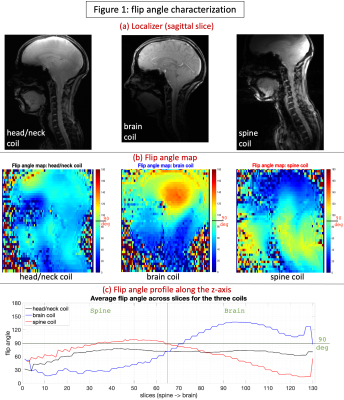 |
72 | Performance characterization of three coils for whole brain and/or cervical spinal cord MRI at 7T
D Rangaprakash1, Bastien Guerin1, Jason P Stockmann1, Markus W May2, Nibardo Lopez Rios3, Kyle M Gilbert4, Yulin Chang5, Lawrence L Wald1,6, Julien Cohen-Adad3,7,8, Boris Keil1,2, and Robert L Barry1,6
1Athinoula A. Martinos Center for Biomedical Imaging, Massachusetts General Hospital, Harvard Medical School, Boston, MA, United States, 2Institute of Medical Physics and Radiation Protection, University of Applied Sciences Mittelhessen, Giessen, Germany, 3NeuroPoly Lab, Institute of Biomedical Engineering, Polytechnique Montreal, Montreal, QC, Canada, 4Centre for Functional and Metabolic Mapping, The University of Western Ontario, London, ON, Canada, 5Siemens Medical Solutions USA Inc., Malvern, PA, United States, 6Harvard-Massachusetts Institute of Technology Division of Health Sciences & Technology, Cambridge, MA, United States, 7Functional Neuroimaging Unit, CRIUGM, Université de Montréal, Montreal, QC, Canada, 8Mila – Quebec AI Institute, Montreal, QC, Canada
Several neurological diseases affect both the brain and spinal cord. 7T MRI enables better imaging of the narrow cord as well as single-subject prediction using fMRI. However, concurrent brain-cord 7T imaging requires a coil with good SNR and acceleration capabilities in both head and neck, which has been unavailable. To overcome this, we performed flip-angle, SNR, g-factor and tSNR characterization of a custom-built 16Tx/64Rx head/neck radiofrequency coil. We also compared it against a custom-built 8Tx/20Rx cervical spine coil as well as a commercial 8Tx/32Rx brain coil. This hardware will pave the way for concurrent brain-cord 7T MRI in the future.
|
||
1449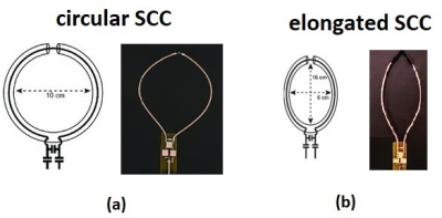 |
73 | Shielded-coaxial-cable (SCC) coils - the array configuration for maximized central SNR at 7T MRI
Sadri Guler1,2, Giovanni Costa3, Vincent Boer2, Maarten Paulides3, Peter Baltus3, Esben Petersen1,2, and Irena Zivkovic3
1Section for Magnetic Resonance, DTU Health Tech, Technical University of Denmark, Copenhagen, Denmark, 2Danish Research Centre for Magnetic Resonance, Centre for Functional and Diagnostic Imaging and Research, Copenhagen University Hospital Amager and Hvidovre, Copenhagen, Denmark, 3Electrical Engineering Department, Technical University of Eindhoven, Eindhoven, Netherlands
The shielded-coaxial-cable (SCC) coils have been proposed recently as highly decoupled elements per se. The SCC coils are not very sensitive to bending, elongation and various degrees of overlapping. We experimentally investigated the central SNR when the SCC coils are placed in three different arrangements on a cylindrical object. The highest central SNR (more than 30% higher than in the other two arrangements) was obtained when the coils were elongated and adjacent to each other. This arrangement is important when the ROI is located deep inside the field of view, such in c-spine spinal cord or deep brain imaging.
|
||
1450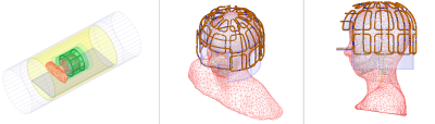 |
74 | SNR Evaluation with High Input Impedance Preamplifier Decoupling Performance for a 2-Layer and 32-Channel Receive Array for Brain Imaging at 7T
Paul-François Gapais1,2, Saadou Almokdad2, Michel Luong3, Eric Giacomini2, Elodie Georget1, and Alexis Amadon2
1Multiwave Imaging SAS, Marseille, France, 2Université Paris-Saclay, CEA, CNRS, BAOBAB, NeuroSpin, Gif-sur-Yvette, France, 3CEA, IRFU, Université Paris-Saclay, Gif-sur-Yvette, France
With the increasing number of elements in receive arrays, the need to accurately predict coil performance is crucial. Decoupling is usually performed with low-input impedance preamplifier decoupling. Recently, new schematics have been proposed that rely on high-input impedance preamplifiers. A simulation method is proposed here to evaluate preamplifier decoupling performance in this context on a 2-layer array at 7T. Results show a strong influence on the thermal SNR according to the impedance presented by the preamplifier to the coil output. The higher this impedance the higher the SNR, but practical implementation need a trade-off between high impedance and noise-matching.
|
||
 Back to Meeting Home
 Back to the Program-at-a-Glance
The International Society for Magnetic Resonance in Medicine is accredited by the Accreditation Council for Continuing Medical Education to provide continuing medical education for physicians.
 Back to Meeting Home
 Back to the Program-at-a-Glance
 View the Presentation