Digital Poster
Hepatocellular Carcinoma & Cirrhosis
Joint Annual Meeting ISMRM-ESMRMB & ISMRT 31st Annual Meeting • 07-12 May 2022 • London, UK

Digital Poster
Hepatocellular Carcinoma & Cirrhosis
| Computer # | ||||
|---|---|---|---|---|
1936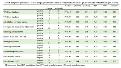 |
31 | Diagnostic performance of histological fibrous capsule and pseudocapsule (a false-positive fibrous capsule) in hepatocellular carcinoma
Atsushi Higaki1, Akira Yamamoto1, Takeshi Fukunaga1, Hirotake Nishimura2, Akihiko Kanki1, and Tsutomu Tamada1
1Radiology, Kawasaki Medical School, Kurashiki, Japan, 2Pathology, Kawasaki Medical School, Kurashiki, Japan
We used contrast-enhanced MRI and CT to evaluate the diagnostic potential of histological fibrous capsule and pseudocapsule in HCC. 51.4% (36/70) of HCCs had capsules, and pseudocapsule was found in 35.3% (18/51) of lesions with enhancing capsule imaging. Among the various imaging features, T1WI low signal rim, arterial phase low signal capsule, enhancing capsule on MRI and CT, and tumor margin enlargement sign on HBP were useful in diagnosing fibrous capsules. Among these, tumor margin enlargement sign on HBP showed the highest accuracy for the diagnosis of capsule, and was the only finding that could diagnose pseudocapsule.
|
||
1937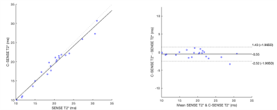 |
32 | SENSE and Compressed SENSE chemical shift encoded multi-gradient echo for T2* quantification in the liver
Elisabeth Sarah Pickles1,2, John Connell2, Stella Nay Kyi Kin2, Alison Telford2, Arina Kazimianec2, Michael Brady2, Daniel Bulte1, Oi Fong Chong3, Han Jun Choo4, Sulaiha Binte Ithnin5, Jason Pik Eu Chang6, Guan Huei Lee7, Marianne Anastasia De Roza8, Kok Kiong Ong9, Sabrina Yi-Mei Wee10, Kee Tung Tan11, Ngiap Chuan Tan12, Han Chong Toh13, Yu Jun Wong14,15, Wei Lyn Yang16,
Xin Yi Yeap17, Cheryl Min En Chua18, Jacelyn Siou Sze Chua18, Jade Shu Qi Goh18, Lynette Soh Han Lai18, Yu Ki Sim18, and Pierce K.H. Chow19,20
1Institute of Biomedical Engineering, University of Oxford, Oxford, United Kingdom, 2Perspectum Ltd, Oxford, United Kingdom, 3SingHealth Polyclinic - Bedok, Singapore, Singapore, 4SingHealth Polyclinic - Sengkang, Singapore, Singapore, 5SingHealth Polyclinic - Tampines, Singapore, Singapore, 6Department of Gastroenterology and Hepatology, Singapore General Hospital, Singapore, Singapore, 7Division of Gastroenterology and Hepatology, National University Hospital, Singapore, Singapore, 8Department of Gastroenterology and Hepatology, Sengkang General Hospital, Singapore, Singapore, 9SingHealth Polyclinic - Outram, Singapore, Singapore, 10SingHealth Polyclinic - Bukit Merah, Singapore, Singapore, 11SingHealth Polyclinic - Marine Parade, Singapore, Singapore, 12SingHealth Polyclinic - Pasir Ris, Singapore, Singapore, 13Division of Medical Oncology, National Cancer Centre Singapore, Singapore, Singapore, 14Department of Gastroenterology & Hepatology, Changi General Hospital, Singapore, Singapore, 15Duke-NUS Medical School, Singapore, Singapore, 16Gastroenterology and Hepatology, Tan Tock Seng Hospital, Singapore, Singapore, 17SingHealth Polyclinic - Punggol, Singapore, Singapore, 18Division of Surgery and Surgical Oncology, National Cancer Centre Singapore, Singapore, Singapore, 19Department of Hepato-pancreato-biliary and Transplant Surgery, Singapore General Hospital and National Cancer Centre Singapore, Singapore, Singapore, 20Surgery Academic Clinical Programme, Duke-NUS Medical School, Singapore, Singapore
T2*, measured using 3D breath-hold chemical shift encoded multi-gradient echo can be used to estimate liver iron concentration. The breath-hold duration (typically 10-20 s) with traditional parallel imaging acceleration techniques such as Sensitivity Encoding (SENSE) can be difficult for patients. Compressed SENSE (C-SENSE) can be used to decrease acquisition time. We compare T2* obtained with SENSE and C-SENSE in 21 patients. We found that C-SENSE acquisition was faster (9 s vs 18 s), with strong correlation and agreement with SENSE. Our findings suggest C-SENSE may potentially improve patient tolerance and turnover speed for T2* acquisitions when compared to SENSE.
|
||
1938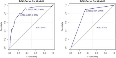 |
33 | Clinical value of MRI-detected extramural vascular invasion in predicting distant metastasis of rectal cancer
Tang Cui1, Jinming Xu2, Moubin Lin3, Shixiong Qiu4, Xiaoming Zuo4, Mengxiao Liu5, and Peijun Wang6
1Yangpu Hospital, School of Medicine, Tongji University,, shang hai, China, 2Department of Radiology, Yangpu Hospital, Tongji University School of Medicine, Shanghai, shang hai, China, 3Department of General Surgery, Yangpu Hospital, Tongji University School of Medicine, Shanghai, Shanghai, China, 4Department of Radiology, Yangpu Hospital, Tongji University School of Medicine, Shanghai, Shanghai, China, 5MR Scientific Marketing, Diagnostic Imaging, Siemens Healthcare Ltd., Shanghai, Shanghai, China, 6Department of Radiology, Tongji Hospital, Tongji University School of Medicine, Shanghai, Shanghai, China
The results of the present study suggest that there was a significant difference in the occurrence of distant metastasis between RC patients with or without mrEMVI, CRM invasion, regional LN metastasis, and MR peritoneal reflex involvement. And mrEMVI and regional LN metastasis were independent risk factors for distant metastasis of rectal cancer. Therefore, MRI can be used to observe the status of EMVI before surgery, which is helpful for the formulation of individualized treatment plans for rectal cancer and assessment of prognosis.
|
||
1939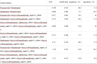 |
34 | The value of diffusion kurtosis imaging texture analysis in predicting treatment response to transcatheter arterial chemoembolization in HCC
Chen Qiao1, Ailian Liu1, Ying Zhao1, Qingwei Song1, Xin Li 1, Yan Guo2, and Tingfan Wu2
1the First Affiliated Hospital of Dalian Medical University, DaLian, China, 2Philips Healthcare, BeiJing, China
This work aimed at texture analysis based on mean apparent diffusion coefficient (MD) of diffusion kurtosis imaging (DKI) to evaluate the treatment efficacy of transcatheter arterial chemoembolization (TACE) in hepatocellular carcinoma (HCC). The results showed that MD texture features can differentiate objective response and non-response groups. When using the single parameter, the GreyLevelNonuniformity_angle135_offset1 achieved the best result (AUC: 0.714, sensitivity: 68.2%, specificity: 81%). In the combined diagnosis of multiple parameters, FrequencySize,MaxIntensity, GreyLevelNonuniformity_angle0_offset1 and GreyLevelNonuniformity_angle135_offset1 were combined to obtain the best combined diagnosis(AUC:0.760,sensitivity: 81.8%, specificity: 66.7%).
|
||
1940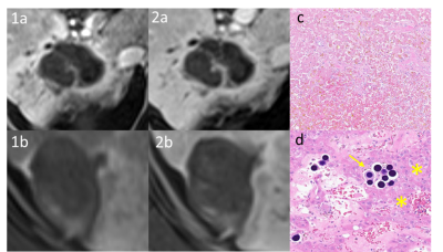 |
35 | MRI Correlation with Liver Explant Pathology: LIRADS Treatment Response Algorithm for Residual HCC After Y-90 Therapy
Matthew Harwood1, Parth Parikh2, Alexander Clinkenbeard1, Sailen Naidu3, Marcela Salomao4, and Alvin Silva1
1Radiology, Mayo Clinic Arizona, Phoenix, AZ, United States, 2Mayo Clinic Alix School of Medicine, Arizona, Scottsdale, AZ, United States, 3Interventional Radiology, Mayo Clinic Arizona, Phoenix, AZ, United States, 4Pathology, Mayo Clinic Arizona, Phoenix, AZ, United States
The liver imaging and reporting data system treatment response algorithm (LIRADS TRA) for CT and MRI has been used to predict residual viable tumor after treatment with yttrium-90 (Y-90). This single-center cohort of 20 patients were reviewed using the LIRADS TRA assessing residual viable tumor following Y-90 therapy. Pathology of subsequent liver explants was the reference standard. The LIRADS TRA predicted positive treatment effect (nonviable): NPV 82.4% (95% CI 0.47-1.55), accuracy 75% (72.0-89.4). The results of this preliminary study will need further validation in a larger, similar, pathologic-confirmed cohort.
|
||
1941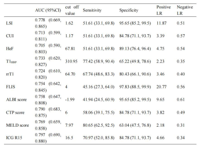 |
36 | Does Functional Parameters from Gadoxetic Acid–enhanced MRI Predict Decompensation in Cirrhosis? Video Not Available
Xueqin ZHANG1, Tao ZHANG1, Lei XU1, and Xiance ZHAO2
1Affiliated Nantong Hospital 3 of Nantong University, Nantong, China, 2Philips Healthcare, Shanghai, China
We used Look-Locker sequences to acquire T1 mapping images pre and post-contrast at 20 minutes after gadoxetic acid administration. Liver-to-spleen contrast index (LSI), contrast uptake index (CUI), relative liver enhancement (RLE), hepatocyte fraction (HeF), T1 relaxation times on unenhanced phase (T1unenh), T1 relaxation times on hepatobiliary phase (T1HBP), reduction rates of T1 relaxation times (rrT1), and the functional liver imaging score (FLIS) of the liver were measured and calculated by two radiologists independently. Our study showed that LSI, CUI, HeF, T1HBP, rrT1 and FLIS based on gadoxetic acid–enhanced MRI are help for the evaluation of liver function.
|
||
1942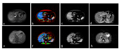 |
37 | Radiomics assessment of liver fibrosis in patients with chronic liver disease using MRI: a machine learning approach
Farzin Mobayyyen1,2, Behrooz Taghvainia3, Hassan Homayoun2, Ali Abbasian Ardakani4, Anahita Fathi Kazerooni2, Hanieh Mobarak Salari2, Nasser Rakhshani5, Hamidreza Salighehrad1,2, and Hamid Reza haghighatkhah6
1Department of Medical Physics and Biomedical Engineering, School of Medicine, Tehran University of Medical Sciences, Tehran, Iran (Islamic Republic of), Tehran, Iran (Islamic Republic of), 2Quantitative MR Imaging and Spectroscopy Group, Research Center for Molecular and Cellular Imaging, Tehran University of Medical Sciences, Tehran, Iran (Islamic Republic of), 3Shahid Beheshti University of Medical Science, Tehran, Iran (Islamic Republic of), 4Department of Radiology Technology, School of Allied Medical Sciences, Shahid Beheshti University of Medical Sciences, Tehran, Iran (Islamic Republic of), 5Gastrointestinal and Liver Disease Research Center, Firoozgar Hospital, Iran University of Medical Sciences, Tehran, Iran (Islamic Republic of), 6Department of Diagnostic Imaging, Shohada-e-Tajrish Hospital, Shahid Beheshti University of Medical Science, Tehran, Iran (Islamic Republic of)
Neither biochemical markers nor a qualitative assessment of medical images are reliable to differentiate mild from moderate stages of liver fibrosis. The main purpose of this study is to develop a machine learning model to classify mild and moderate liver fibrosis based on radiomic features extracted from MRI images (T1-w, T2*-map & ADC-map). Nu-SVC classifier was employed as the classification technique, trained by the extracted data from image series of 29 patients with histopathology-confirmed mild and moderate liver fibrosis. Results demonstrate that radiomic analysis of T2*-map and ADC-map has high potential in classifying different stages of liver fibrosis.
|
||
 Back to Meeting Home
 Back to the Program-at-a-Glance
The International Society for Magnetic Resonance in Medicine is accredited by the Accreditation Council for Continuing Medical Education to provide continuing medical education for physicians.
 Back to Meeting Home
 Back to the Program-at-a-Glance
 View the Presentation