Digital Poster
Kidney
Joint Annual Meeting ISMRM-ESMRMB & ISMRT 31st Annual Meeting • 07-12 May 2022 • London, UK

| Computer # | ||||
|---|---|---|---|---|
2482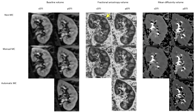 |
14 | Performance of small field of view rotated 2D selective RF excitation Diffusion Tensor (DTI) Imaging of kidneys with automatic motion correction
Dee Zhen Lim1, Julia Williams1, Jason Wong1, Lucy McKenna1, Julie Smith1, Emma Hornsey1, Dominic Italiano2, Leonid Churilov2, Daniel Staeb3, Thomas Benkert4, Elif Ekinci5,6, and Ruth P Lim1,7
1Department of Radiology, Austin Health, Heidelberg, Australia, 2Melbourne Medical School, University of Melbourne, Melbourne, Australia, 3MR Research Collaborations, Siemens Healthcare Pty Ltd, Melbourne, Australia, 4MR Application Predevelopment, Siemens Healthcare GmbH, Erlangen, Germany, 5Department of Endocrinology, Austin Health, Heidelberg, Australia, 6Department of Medicine, University of Melbourne, Melbourne, Australia, 7Departments of Radiology and Surgery, University of Melbourne, Melbourne, Australia Renal diffusion tensor imaging (DTI) is a promising, non-invasive biomarker for kidney function. Cortical and medullary fractional anisotropy (FA) and mean diffusivity (MD) correlates with early microstructural and functional changes in kidney diseases. However, reliability of DTI metrics is affected by artifacts. The ideal renal DTI sequence for robust and reproducible measurements remains under investigation. We report on a prototype for small field-of-view (FOV) DTI incorporating 2D selective and rotated RF excitation and automatic motion correction to minimize artifacts in 9 diabetic patients and 9 controls, demonstrating high inter-reader agreement for measured FA and MD and high image quality. |
||
2483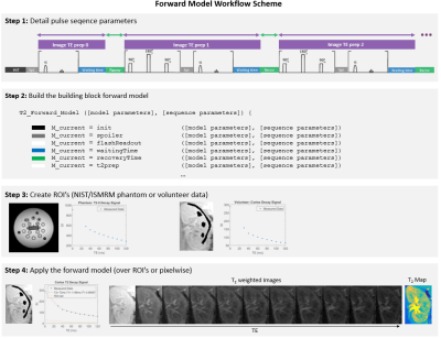 |
15 | Renal T2-mapping with a T2-preparation sequence: comparison of signal models
Joao Periquito1, Kanishka Sharma1, Bashair Alhummiany2, Joao Sousa1, and Steven Sourbron1
1The University of Sheffield, Sheffield, United Kingdom, 2University of Leeds, Leeds, United Kingdom
A recent consensus paper recommends a mono-exponential signal model to determine T2-values from a T2-preparation sequence. However, this assumes complete signal recovery after each readout, and therefore necessitates long acquisition times. In this study, we compare the mono-exponential model against a forward modelling approach which is also accurate with incomplete recovery. Simulations, phantom data and repeatability data in healthy volunteers show the forward model is significantly more accurate and allows for a 7-fold reduction in acquisition time with a negligible cost in T2 precision.
|
||
2484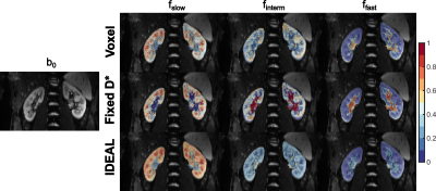 |
16 | Image Downsampling Expedited Adaptive Least-squares (IDEAL) fitting improves IVIM analysis in the human kidney
Julia Stabinska1,2,3, Helge Jörn Zöllner1,2, Hans-Jörg Wittsack3, and Alexandra Ljimani3
1F.M. Kirby Research Center for Functional Brain Imaging, Kennedy Krieger Institute, Baltimore, MD, United States, 2The Russell H. Morgan Department of Radiology and Radiological Science, The Johns Hopkins University School of Medicine, Baltimore, MD, United States, 3Department of Diagnostic and Interventional Radiology, Heinrich Heine University Dusseldorf, Dusseldorf, Germany
Recent studies have shown that a three-compartment model is preferable when analyzing renal DWI signal, as the conventional bi-exponential IVIM fitting does not differentiate between pure water diffusion and incoherent intra-tubular fluid motion. However, triexponential IVIM modelling in the kidney is challenging due to suboptimal SNR of diffusion-weighted images. In the present study, we applied Image Downsampling Expedited Adaptive Least-squared (IDEAL) fitting, which utilizes image downsampling to generate high SNR images to iteratively update the initial model parameters until the final image resolution is reached, and achieved more reliable estimates of the IVIM-related parameters compared with the conventional fitting methods.
|
||
2485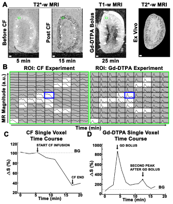 |
17 | Mapping spatially heterogeneous single nephron function in the kidney using MRI.
Edwin J. Baldelomar1, Jennifer Charlton2, and Kevin M Bennett3
1Radiology, Washington University in St. Louis School of Medicine, St. Louis, MO, United States, 2Pediatric Nephrology, University of Virginia, Charlottesville, VA, United States, 3Radiology, Washington University in St. Louis School of Medicine, Saint Louis, MO, United States
Nephrons are the functional units of the kidney, coordinated with surrounding nephrons through the vasculature to filter blood plasma. We used in vitro CFE-MRI combined with Gd-DTPA contrast enhanced MRI to dynamically map single nephron physiology throughout the isolated, perfused rat kidney. We were able to map the heterogeneity and spatial variability of macromolecular binding and single nephron filtration. MRI appears to measure similar dynamics in single nephron filtration as intra-vital optical microscopy, with the advantage of measuring all nephrons in the kidney.
|
||
2486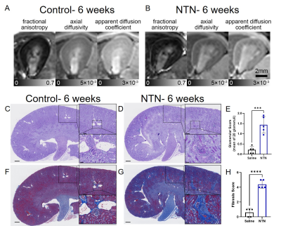 |
18 | Evaluating renal fibrosis in a nephrotoxic nephritis model using DTI-MRI
Shimrit Avraham1, Rohan Virgincar2, Joshua Webster3, Andrey Shaw1, and Luke Xie2
1Research Biology, Genentech, South San Francisco, CA, United States, 2Biomedical Imaging, Genentech, South San Francisco, CA, United States, 3Research Pathology, Genentech, South San Francisco, CA, United States
Accurate estimation of kidney function is crucial to the detection, evaluation, and treatment of chronic kidney disease (CKD). The traditional methods are inaccurate or invasive. Here, we used DTI-MRI to evaluate kidney function with disease progression in nephrotoxic serum- induced CKD model (NTN). We detect changes associated with renal inflammation and fibrosis prior to functional decline. Moreover, AD and ADC of the cortex-outer medulla border were highly correlated with histologic evidence of fibrosis. These results suggest that DTI-MRI can detect early stages of CKD non-invasively in-vivo and can be used for fibrosis mapping in a variety of renal diseases.
|
||
| 2487 | 19 | Spatial Heterogeneity in Quantitative Renal ADC, ASL perfusion and R2* Maps Using Texture Analysis Video Permission Withheld
Lu-Ping Li1,2, Emily Wilt1, Artem Mikheev3, Henry Rusinek3, Stuart Sprague1,2, Orly Kohn2, and Pottumarthi Prasad1,2
1NorthShore University HealthSystem, Evanston, IL, United States, 2Pritzker School of Medicine, University of Chicago, Chicago, IL, United States, 3Langone School of Medicine, New York University School of Medicine, New York, NY, United States
While regions of interest analysis is widely used in quantitative MRI, emphasis usually is placed only on the spatial average and information of spatial heterogeneity is ignored. Texture analysis has gained increasing interest in the context of applying artificial intelligence. These Radiomic tools are now readily available in image analysis tool boxes for more widespread adoption. We illustrate an application of such analysis on quantitative renal MRI, including ADC, ASL and R2* maps. Our results show that several measures of heterogeneity of cortical voxel-wise maps discriminate between healthy and individuals with chronic kidney disease.
|
||
2488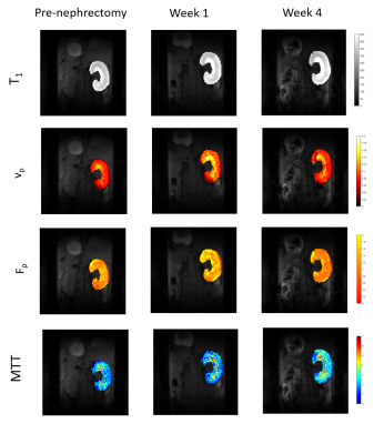 |
20 | Preclinical Investigation of Kidney Volume and Perfusion after Nephrectomy using Dynamic Contrast-Enhanced MRI.
Penny L Hubbard Cristinacce1, Franziska Lausecker2, Shanshan Li2, Ben R Dickie3,4, Michael Berks1, Ross A Little1, Alastair Hutchison5,6, James PB O'Connor1,7, and Rachel Lennon2
1Division of Cancer Sciences, The University of Manchester, Manchester, United Kingdom, 2Division of Cell-Matrix Biology and Regenerative Medicine, The University of Manchester, Manchester, United Kingdom, 3Division of Informatics, Imaging and Data Science, The University of Manchester, Manchester, United Kingdom, 4Geoffrey Jefferson Brain Research Centre , Manchester Academic Health Science Centre, Northern Care Alliance & University of Manchester, Manchester, United Kingdom, 5Division of Cardiovascular Sciences, The University of Manchester, Manchester, United Kingdom, 6Dorset Count Hospital NHS Foundation Trust, Dorset, United Kingdom, 7Division of Radiotherapy and Imaging, Institute of Cancer Research, London, United Kingdom
Compensatory hypertrophy of the remaining kidney is observed in mice after nephrectomy using structural MRI. By fitting the two-compartment filtration model to dynamic contrast-enhanced MRI data we examined the effects of nephrectomy on the passage of blood through the remaining kidney over time. Total kidney plasma volume (vp) increases at 1 week post-nephrectomy, along with T1. This is followed by a decrease at week 4, which is coupled to a decrease in total and per unit volume plasma flow and an increase in plasma mean transit time.
|
||
2489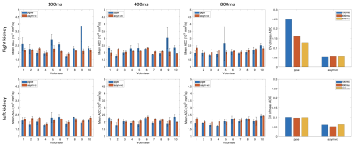 |
21 | Velocity-compensated optimized diffusion encoding in respiratory-triggered renal ADC mapping
Sean McTavish1, Anh T. Van1, Johannes M. Peeters2, Kilian Weiss3, Marcus R. Makowski1, Rickmer F. Braren1, and Dimitrios C. Karampinos1
1Department of Diagnostic and Interventional Radiology, Technical University of Munich, Munich, Germany, 2Philips Healthcare, Best, Netherlands, 3Philips GmbH Market DACH, Hamburg, Germany
Diffusion-weighted imaging (DWI) and ADC mapping remains a potential biomarker for the assessment of renal fibrosis in chronic kidney disease and characterization of renal carcinoma. However, for body DWI applications including renal DWI, motion effects can confound the robustness of abdominal DWI. Motion-compensated diffusion encoding designs have been proposed as alternatives in overcoming the effect motion in other organs such as the liver and the pancreas. The purpose of this work is to assess the effect of respiratory motion and to apply motion compensated diffusion encoding waveforms in the context of coronal respiratory-triggered ADC mapping in the kidney.
|
||
2490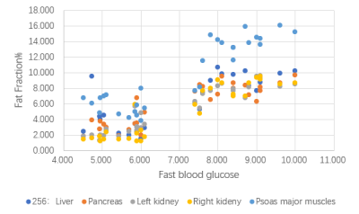 |
22 | Dixon-MRI to quantitatively evaluate the correlation between abnormal fat deposition and chronic kidney disease with diabetic complications Video Not Available
Fangjie Xia1,2, Wen Shen3, Lihua Chen3, and Jinxia Zhu4
1Tianjin First Center Hospital, Tianjin medical university, Tianjin, China, 2Tianjin medical university, Tianjin, China, 3Tianjin First Center Hospital, Tianjin, China, 4Siemens Healthcare, Beijing, China
The study use Dixon and DKI sequence to quantitative analysis the ectopic fat deposition in chronic kidney disease and diabetic nephropathy. Compare the degree of fat infiltration in different organs and tissues including Liver, pancreas, kidney and muscles, combined with clinical laboratory experiments, to study the relationship between fat deposition and the development of CKD or diabetic nephropathy. Aim to point out the significance of treatment to fat deposition for CKD or diabetic nephropathy patient.
|
||
2491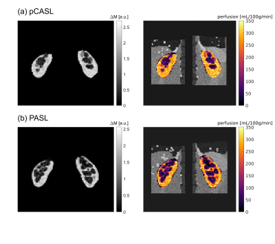 |
23 | Synthetic arterial spin labelling datasets of the kidneys for pipeline evaluation and comparison
Irène Brumer1, Dominik F. Bauer1, Lothar R. Schad1, and Frank G. Zöllner1
1Computer Assisted Clinicial Medicine, Mannheim Institute for Intelligent Systems in Medicine, Medical Faculty Mannheim, Heidelberg University, Mannheim, Germany
Synthetic kidney ASL data with respiratory motion was generated using models from the XCAT phantom and matching recommendations for in-vivo acquisitions. Both pCASL and PASL datasets with 1 M0 and 25 control-label pairs were created and analysed using an in-house developed processing pipeline including registration, manual segmentation, calculation of mean perfusion-weighted image and perfusion map. The registration performed well on the synthetic data and the perfusion maps yielded good cortex/medulla contrast. The presented method allows a wide range of parameter choices for creating synthetic ASL datasets valuable for testing processing pipelines and comparing them across research and clinical imaging centres.
|
||
2492 |
24 | Parametric T1 MR Microscopy Detects Gadolinium Residuals in Ex Vivo Rat Kidneys
Ehsan Tasbihi1, Luis Hummel2, Thomas Gladytz1, Ludger Starke1, Jason M. Millward1, Erdmann Seeliger2, and Thoralf Niendorf1,3
1Berlin Ultrahigh Field Facility (B.U.F.F.), Max Delbrueck Center for Molecular Medicine in the Helmholtz Association, Berlin, Germany, Berlin, Germany, 2Institute of Vegetative Physiology, Charité – Universitätsmedizin Berlin, Berlin, Germany, Berlin, Germany, 3Experimental and Clinical Research Center, a joint cooperation between the Charité Medical Faculty and the MAX Delbrück Center for Molecular Medicine in the Helmholtz Association, Berlin, Germany, Berlin, Germany
Accumulation of gadolinium (Gd) following use of Gd-based contrast agents (GBCA) is a concern. To study the potential of T1 microscopy for assessing Gd residues in the kidney, rats were administered 8 intravenous doses of various GBCAs over a period of two weeks. Five days following the last administration, the kidneys were collected and fixed in formalin. High resolution T1 maps obtained from ex vivo scans showed significantly reduced T1 levels for both linear and macrocyclic GBCAs. This demonstrates to potential for quantitative T1 mapping to detect Gd residues non-invasively in renal tissue.
|
||
2493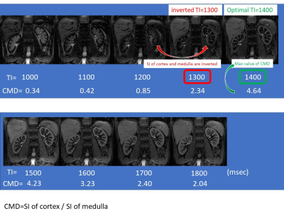 |
25 | Multiparametric MR imaging in diabetic nephropathy: New insights to evaluate early diabetic nephropathy noninvasively
Akira Yamamoto1, Tsutomu Tamada2, Yu Ueda3, Mitsuru Takeuchi4, Ayumu Kido2, Atsushi Higaki2, and Akihiko Kanki2
1Radiology, Kawasaki Medical School, Kurashiki, Japan, 2Kawasaki Medical School, Kurashiki, Japan, 3Phillips Japan, Tokyo, Japan, 4Radiolonet Tokai, Nagoya, Japan
The purpose of this study was to identify the changes in multiparametric magnetic resonance imaging (MRI) findings in early diabetic nephropathy (DN). Multiparametric measurements were made of the renal cortex and medulla. Significant differences were seen between the healthy and early DN (grade1) in optimal TI and inverted TI value. T2 value of cortex is in marginal significant (p=0.064). This study suggests the possibility that MRI using the values of optimal TI, inverted TI value and T2 value of cortex can be used to evaluate early diabetic nephropathy non-invasively and in a short period of time.
|
||
2494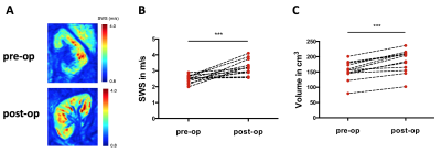 |
26 | Stiffening of renal transplants after donation assessed by MR elastography – physiological adaption from donor to recipient Video Permission Withheld
Stephan Rodrigo Marticorena Garcia1, Frank Friederdorff2, Josef Mang2, Alexandra Webster1, Bernd Hamm1, Jürgen Braun3, and Ingolf Sack1
1Radiology, Charité – Universitätsmedizin Berlin, Berlin, Germany, 2Urology, Charité – Universitätsmedizin Berlin, Berlin, Germany, 3Medical Informatics, Charité – Universitätsmedizin Berlin, Berlin, Germany
Renal stiffness was investigated by using magnetic resonance elastography (MRE) with tomoelastography-postprocessing in 24 participants (12 renal donor-recipient couples) to explore the influence of kidney transplantation on renal viscoelasticity for the first time. Renal stiffness increases immediately after transplantation. These early changes could be due to changes in renal perfusion and increase in renal metabolism, which is also reflected by increase in renal volume. Cold ischemia time was found in being a contributor to changes of solid renal structures. Inccrease in renal stiffness after transplantation could be a novel predictive biomarker for better renal outcome.
|
||
2495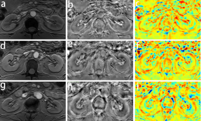 |
27 | Quantitative evaluation of renal injury in the early chronic kidney disease with quantitative susceptibility mapping
Shan Jiayuan1, Zhang Jinggang1, Chen Jie1, Xing Wei1, Wang Yishi2, and Zhang Jilei3
1Third Affiliated Hospital of Soochow University, Changzhou, China, China, 2Philips Healthcare, Beijing, China, 3Philips Healthcare, Shanghai, China
To explore the value of quantitative susceptibility mapping (QSM) in evaluating renal injury in patients with chronic kidney disease (CKD). 40 CKD patients were included in the study to evaluate the potential clinical value of QSM. We found that with the progress of CKD staging, renal medulla susceptibility values decreased significantly. Susceptibility value of the medulla was highly correlated with estimated glomerular filtration rate (eGFR). QSM could serve as a quantitative biomarker to assess the renal injury of early CKD.
|
||
2496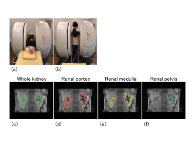 |
28 | Analysis of postural effect on kidney volume using multiposture MRI
Seiya Nakagawa1, Tosiaki Miyati1, Naoki Ohno1, Koga Kawano1, Yuki Oda1, and Satoshi Kobayashi1
1Division of Health Sciences, Graduate School of Medical Sciences, Kanazawa University, Kanazawa, Ishikawa, Japan
We assessed the effect of posture on kidney volume measured in the supine and upright positions using an original MRI system that can image in any posture (multiposture MRI). We compared the kidney volume (whole kidney, renal cortex, medulla, and pelvis) between the supine and upright positions. Upright position reduced whole kidney, renal cortex, and medulla volumes, and these differences between postures potentially provide new diagnostic information.
|
||
 Back to Meeting Home
 Back to the Program-at-a-Glance
The International Society for Magnetic Resonance in Medicine is accredited by the Accreditation Council for Continuing Medical Education to provide continuing medical education for physicians.
 Back to Meeting Home
 Back to the Program-at-a-Glance
 View the Presentation