Digital Poster
Breast Imaging: Beyond Morphology
Joint Annual Meeting ISMRM-ESMRMB & ISMRT 31st Annual Meeting • 07-12 May 2022 • London, UK

Digital Poster
Breast Imaging: Beyond Morphology
| Computer # | ||||
|---|---|---|---|---|
1679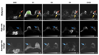 |
48 | Discrimination Between Benign and Malignant Lesions in the Breast with Restriction Spectrum Imaging on a Screening Breast MRI Cohort
Stephane Loubrie1, Ana Rodriguez-Soto1, Lauren Fang1, Christopher Conlin1, Maren MS Andreassen2, Tyler Seibert1,3,4, Michael Hahn1, Vandana Dialani5,6, Catherine J Wei7, Zahra Karimi5, Joshua Kuperman1, Anders Dale1,8, Etta Pisano5,9, and Rebecca Rakow-Penner1,4
1Radiology, UCSD, San Diego, CA, United States, 2Department of Circulation and Medical Imaging, Norwegian University of Science and Technology, Trondheim, Norway, 3Radiation medecine, UCSD, San Diego, CA, United States, 4Bioengineering, UCSD, San Diego, CA, United States, 5Beth Israel Hospital, Boston, MA, United States, 6Harvard Medical School, Bosston, MA, United States, 7Commonwealth Radiology Associates, Boston, MA, United States, 8Neurosciences, UCSD, San Diego, CA, United States, 9American college of radiology, Reston, VA, United States
Diffusion weighted imaging (DWI) holds great potential in improving specificity of findings detected on contrast enhanced breast MRI. Restriction spectrum imaging (RSI), an advanced diffusion imaging model, has potential in discriminating between malignant and fibroglandular breast tissue. In this abstract, we evaluate RSI’s performance in differentiating malignant from benign lesions in a prospective study performed on a breast screening population. All lesions were biopsy proven. The breast RSI model allowed discrimination between malignant, high-risk and low-risk benign lesions and healthy fibroglandular tissue.
|
||
1680 |
49 | In vivo mapping of sodium relaxation times in breast fibroadenoma at 7T
Carlotta Ianniello1,2, Ryan Brown1,2, Linda Moy1,2,3, Justin Fogarty1, Freya Schnabel3,4, Deborah Axelrod3,4, and Guillaume Madelin1,2
1Center for Advanced Imaging Innovation and Research (CAI2R), Department of Radiology, New York University Grossman School of Medicine, New York, NY, United States, 2Center for Biomedical Imaging, Department of Radiology, New York University Grossman School of Medicine, New York, NY, United States, 3Perlmutter Cancer Center, New York University Grossman School of Medicine, New York, NY, United States, 4Department of Surgery, New York University Grossman School of Medicine, New York, NY, United States Sodium (23Na) MRI has been employed to measure the total sodium concentration (TSC) in breast lesions, which is often elevated compared to healthy breast. The knowledge of the 23Na relaxation times of the breast, which could be subject to variations in lesions, could increase the accuracy of TSC estimation. In this work we measured the 23Na relaxation times in six fibroadenoma patients and found a 71% increase in T2,s and a 28% decrease in T1 in the lesions compared to healthy tissue. Therefore, using healthy breast relaxation times in TSC quantification could result in errors in lesions. |
||
1681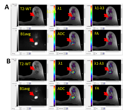 |
50 | Breast DTI: A Prospective Study in Individuals Referred for Biopsy of Indeterminate Breast Lesions
Jacob S Ecanow1, David B Ecanow2, Nondas Leloudas1, Bradley Hack1, and Pottumarthi V Prasad2
1NorthShore University HealthSystem, Evanston, IL, United States, 2Radiology, NorthShore University HealthSystem, Evanston, IL, United States
We present preliminary data from an ongoing study in individuals referred for biopsy of BIRADS 3, 4, and 5 breast lesions based on mammography, ultrasound, and/or screening MRI. DTI data was acquired using single shot EPI with 30 different directions and 2 mm isotropic resolution. MRI data was analyzed using BIT-Motion software. All cancers showed low ADC and λ1 values compared to control regions. FA, consistent with prior reports showed minimal difference and so did (λ1-λ3).
|
||
1682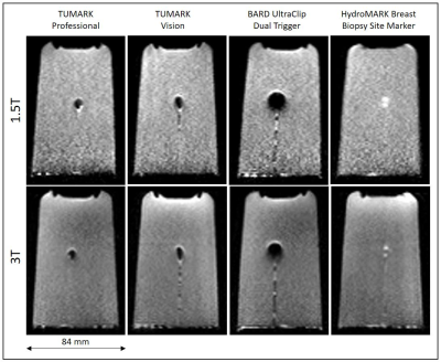 |
51 | Influence of breast biopsy clips on diffusion weighted MRI of the breast
Christian Kremser1, Birgit Amort1, and Martin Daniaux1
1Dept. of Radiology, Medical University of Innsbruck, Innsbruck, Austria
Patients undergoing breast MRI frequently have metallic biopsy clips placed within or adjacent to the lesion, producing metallic artifacts, which could lead to misinterpretation. The aim of our study was to investigate the extent of the artifact of four different clips commonly used in our hospital on diffusion-weighted images obtained with a readout-segmented sequence (RESOLVE) and their influence on ADC maps. It is found that artifacts vary in shape and size between different clips, DWI images and ADC-maps. For the marker with the smallest artifact the minimum size of detectable lesion seems to be approximately 10mm.
|
||
1683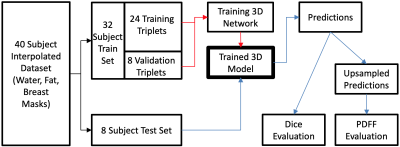 |
52 | Automated breast segmentation using deep-learning in water-fat breast MRI: application to breast density assessment
Arun Somasundaram1, Mingming Wu1, Tabea Borde1, Marcus R. Makowski1, Eva M. Fallenberg1, Yu Zhao2, and Dimitrios C. Karampinos1,3
1Department of Diagnostic and Interventional Radiology, School of Medicine, Technical University of Munich, Munich, Germany, 2AI Lab, Tencent, Shenzhen, China, 3Munich Institute of Biomedical Engineering, Technical University of Munich, Munich, Germany
Conventional methods for semi-automatic and rule-based breast segmentation on MR images have a tradeoff between accuracy and segmentation speed. To overcome this, several 2D-deep learning approaches have been proposed, which usually focus on using T1-weighted images to train their models. Our aim is to train a 3D-network for breast segmentation, trained on water-fat MR images, which can be used to measure the breast density based on the proton density fat fraction (PDFF). We show that our model segments both fast and accurately, and can visually outperform our ground truth segmentations while requiring only a few seconds to generate labels.
|
||
1684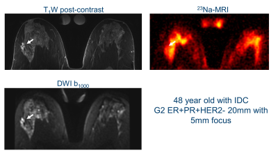 |
53 | Sodium Breast Imaging of Ductal Carcinomas at 3 T Video Not Available
Joshua D Kaggie1, Gabrielle C Baxter1, Mary A McLean1,2, Ramona Woitek1, Ferdia A Gallagher1,2, Andrew D James3,4, Aneurin J Kennerley4,5, Rolf F Schulte6, William J Brackenbury3,5, and Fiona J Gilbert1
1Radiology, University of Cambridge, Cambridge, United Kingdom, 2Cambridge Institute, Cancer Research UK, Cambridge, United Kingdom, 3Biology, University of York, York, United Kingdom, 4York Biomedical Research Institute, University of York, York, United Kingdom, 5Chemistry, University of York, York, United Kingdom, 6GE Healthcare, Munich, Germany
Sodium (23Na)-MRI was performed using a dual-tuned bilateral 23Na/1H breast coil on seven patients with ductal carcinoma, one patient with lobular carcinoma, and ten normal volunteers. Several patients had [23Na] measurements that were 2-3 times higher than the mean normal fibroglandular [23Na] measurements. There were no strong correlations between 23Na-MRI and either DCE or DWI measurements in the patients. These measurements suggest that [23Na] may provide an additional physiological imaging mechanism of breast cancer.
|
||
1685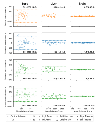 |
54 | Variability of ADC Estimates Between-Scanners from Whole Body Imaging is Dominated by Within-Scanner Variance
Alistair Lamb1, Alan Bainbridge2, Tom Parry3, Harriet Rogers4, Stuart A Taylor3, Hui Zhang5, and Anna Barnes6
1Department of Medical Physics & Biomedical Engineering, University College London, London, United Kingdom, 2Department of Medical Physics and Biomedical Engineering, University College London Hospitals NHS Foundation Trust, London, United Kingdom, 3Centre for Medical Imaging, University College London, London, United Kingdom, 4Institute of Nuclear Medicine, University College London Hospitals NHS Foundation Trust, London, United Kingdom, 5Centre for Medical Image Computing, University College London, London, United Kingdom, 6King's Technology Evaluation Centre, King’s College London, London, United Kingdom
We investigate the reliability of Whole-Body Imaging ADC estimates from subjects tested and retested within- and between-scanners from different vendors with minimal differences in acquisition protocol and post-acquisition analysis. We show substantial within-subject variation in extracranial ADC estimates within- and between-scanners as measured by Limits of Agreement. We additionally show between-scan variability between scanners is dominated by between-scan variability within a scanner. Furthermore, averaging across subsequent within-scanner examinations does not substantially improve reliability of ADC estimates. We therefore conclude a post-acquisition method for reducing within-scanner variation is required to improve the reliability of ADC estimates.
|
||
 Back to Meeting Home
 Back to the Program-at-a-Glance
The International Society for Magnetic Resonance in Medicine is accredited by the Accreditation Council for Continuing Medical Education to provide continuing medical education for physicians.
 Back to Meeting Home
 Back to the Program-at-a-Glance
 View the Presentation