Onsite Tutorial
MSK MRI 101: Practical Considerations for Anatomy & Techniques
Joint Annual Meeting ISMRM-ESMRMB & ISMRT 31st Annual Meeting • 07-12 May 2022 • London, UK

Onsite Tutorial
MSK MRI 101: Practical Considerations for Anatomy & Techniques
| MSK MR Tutorial I | |||
| 13:00 |  |
MSK MR Protocols
Feliks Kogan
MRI has become the hallmark for imaging of the musculoskeletal system due to its ability to image MSK tissues at high resolutions with multiple contrasts. Tailoring MRI sequences allows for optimization of anatomy or pathology of interest, removal of fat, imaging at higher resolutions or with more signal to noise, among other advantages. In this educational talk, we will discuss the MRI image contrast mechanisms, methods for suppression of fat signal, common MSK MRI pulse sequences and image considerations. Further, we will briefly cover novel MSK imaging approaches including accelerated imaging and reconstruction, non-cartesian MRI, bilateral scanning, and contrast/non-contrast imaging.
|
|
| 13:30 | 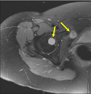 |
Artifacts and Pitfalls in MSK MR Imaging
Andrew Fagan
Image artifacts are commonly encountered in musculoskeletal MRI, and can lead to difficulties in correct image interpretation. While some artifacts are very obvious in an image, and the challenge then becomes one of ‘reading around’ the artifact, others can be more subtle and could be easily misinterpreted as a pathology. Understanding the source of an artifact can help to minimize its prevalence during set-up/scanning, or to differentiate artifact from genuine pathology when present in an image. This interactive talk will present a range of common (and some not-so-common) image artifacts, and discuss their source and strategies to minimize their impact.
|
|
| 14:00 | MRI of the Hand & Wrist
James Teh
|
||
| MSK MR Tutorial II | |||
| 14:30 | MR Anatomy of the Knee
Hakan Ilaslan
|
||
| 15:00 | Break & Meet the Teachers |
||
| 15:30 | MR Anatomy of the Foot and Ankle Video Unavailable
Currently available MR pulse sequences for daily work evaluating foot and ankle pathology will be presented. Anatomy and specific foot and ankle disorders will be reviewed with an emphasis on progressive collapsing foot deformity, the spring and deltoid ligaments and articular cartilage injuries and repair and their MR appearances. A discussion of protocol considerations in the setting of metal will also be included, specifically with regards to imaging the painful total ankle replacement.
|
||
| 16:00 | MR Anatomy of the Hip
Benjamin Fritz
|
||
| 16:30 | MR Anatomy of the Shoulder
Francis Baffour
This presentation will review the anatomy of the shoulder with specific attention to structures identified on magnetic resonance imaging. The anatomy of the shoulder will be discussed in the context of clinical cases.
|
||
 Back to Meeting Home
 Back to the Program-at-a-Glance
The International Society for Magnetic Resonance in Medicine is accredited by the Accreditation Council for Continuing Medical Education to provide continuing medical education for physicians.
 Back to Meeting Home
 Back to the Program-at-a-Glance
 View the Presentation