Online Gather.town Pitches
Processing & Analysis II
Joint Annual Meeting ISMRM-ESMRMB & ISMRT 31st Annual Meeting • 07-12 May 2022 • London, UK

Online Gather.town Pitches
Processing & Analysis II
| Booth # | ||||
|---|---|---|---|---|
3157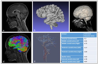 |
1 | Developing a comprehensive whole body MR protocol and model generation pipeline for children Video Not Available
Haribalan Kumar*1,2,3, Robby Green*1, Samantha Holdsworth1,4, Daniel Cornfeld1, Paul Condron1, Eryn Kwon1, Taylor Emsden1, Julie Choisne3, Thor Besier3, Justin Fernandez3, Kat Gilbert3, Martyn Nash3, Soroush Safaei3, Gonzalo Maso Talou3, Davidson Taylor1,5,6, Luke Rolfe1,7, Lucy McHugh1,8,9, Graham Hingangaroa Smith10,11,12,13,14,15, Leigh Potter11,16,17,18, Merryn Tawhai3,
Kelly Burrowes3, Alys Clark3, Jiantao Shan3, Elias Soltani3, Erana Hogarth1,17, Rinki Murphy4, Josh McGeown1, Laura Carman3, Alan Wang1,3, Maryam Tayebi1,3, Ali Mirjalili4, Grace Cleland-Pottie1,19,20, Mahyar Osanlouy3, Vickie Shim1,3, Jerome Maller2, Reweti Ropiha21, and Graham Wilson1,8
1Mātai Medical Research Institute, Gisborne, New Zealand, 2General Electric Healthcare AUS/NZ, Melbourne, Australia, 3Auckland Bioengineering Institute, Auckland, New Zealand, 4Faculty of Medical and Health Sciences, University of Auckland, Auckland, New Zealand, 5Ngāi Tāmanuhiri, Tairāwhiti, New Zealand, 6Rongowhakaata, Tairāwhiti, New Zealand, 7Ngāti Pāhauwera, Hawke's Bay, New Zealand, 8University of Otago, Dunedin, New Zealand, 9Ngāi Tahu, Te Waipounamu, New Zealand, 10Kāti Māmoe, North Otago, New Zealand, 11Mātai Ngā Māngai Māori, Gisborne, New Zealand, 12Te Aitanga a Hauiti, Tairāwhiti, New Zealand, 13Ngāti Apa, Rangitīkei, New Zealand, 14Massey University, Palmerston North, New Zealand, 15Ngāti Kahungunu, Wairarapa, New Zealand, 16Rongomaiwahine, Mahia, New Zealand, 17Ngāti Porou, Tairāwhiti, New Zealand, 18Ngāti Kahungunu, Wairoa, New Zealand, 19University of Cantebury, Christchurch, New Zealand, 20Ngāpuhi, Te Tai Tokerau, New Zealand, 21Turanga Health, Tairāwhiti, New Zealand Approaches to magnetic resonance (MR) image acquisition and computational modelling often exist in silo. The application of computational modelling to paediatric data is in its infancy. This collaborative study addresses this gap for multiple organ systems. State-of-the art MR imaging sequences were selected and refined to provide imaging data for the creation of subject-specific geometric models. The MR protocol and modelling outputs will underpin a longitudinal paediatric study set in Aotearoa New Zealand, generating normative data in healthy children, and identifying early biomarkers of pathological processes. This will enable the development of new personalised, predictive, and preventative models of care. |
||
3158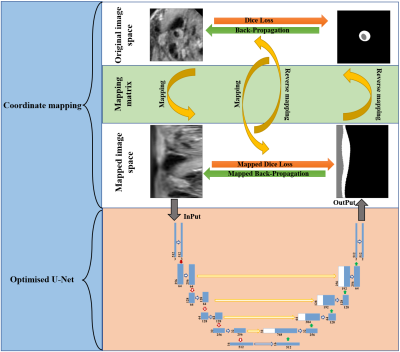 |
2 | Automatic analysis of carotid vessel wall in MR black blood images using custom convolutional trajectories
Shuai Shen1,2, Wenjing Xu1, Hongbing Ma3, Xiaoyi Lv2, Guanxun Cheng4, Liwen Wan1, Lei Zhang1, Ye Li1, Dong Liang1, Xin Liu1, Hairong Zheng1, and Na Zhang1
1Paul C. Lauterbur Research Center for Biomedical Imaging, Shenzhen Institute of Advanced Technology, Chinese Academy of Sciences, Shenzhen, China, 2College of Software, xinjiang University, Xinjiang, China, 3Department of Electronic Engineering, Tsinghua University, Beijing, China, 4Department of Radiology, Peking university shenzhen hospital, Beijing, China
Atherosclerotic plaque is a major cause of ischemic stroke. Some arterial morphological features obtained from MR vessel wall images show great potential for identifying high-risk plaques. Deep learning has now been applied to the automatic segmentation of vessel walls to accurately and efficiently measure arterial morphological features. However, the accuracy of the existing segmentation methods is not yet high enough for clinical practical applications. This study proposed a new segmentation framework with custom convolutional trajectories for automatic segmentation of arterial vessel wall and the framework improved the accuracy of vessel wall segmentation.
|
||
3159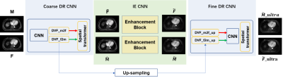 |
3 | A unified deep learning method for 4D-MRI motion deformation estimation and image enhancement
Shaohua Zhi1, Haonan Xiao1, Yinghui Wang1, Wen Li1, Tian Li1, and Jing Cai1
1Department of Health Technology and Informatics, Hong Kong, China
We proposed a unified deep learning framework for predicting the motion deformation between different phases of 4D-MRI with simultaneous image quality enhancement. The network combines a coarse-to-fine unsupervised registration model to estimate the deformation vector fields (DVFs) in different image scales and a GAN-based enhancement network to restore anatomic features. Particularly, a prior knowledge of 4D-MRI is incorporated into the unified model, guiding an accurate DVF prediction and maintaining image topology. Both qualitative and quantitative results showed that the predicted DVFs and resultant 4D-MRI images achieved improved performance compared with the traditional method without modifications.
|
||
3160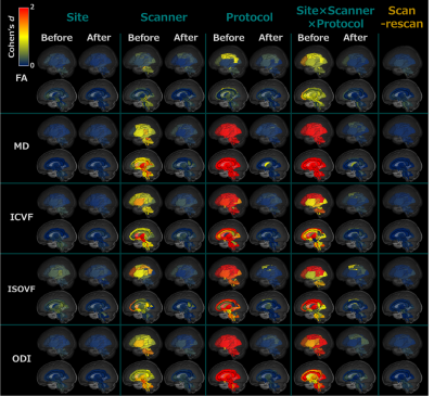 |
4 | Harmonization of multi-site DTI and NODDI data using the combined association test Video Permission Withheld
Yuya Saito1, Koji Kamagata1, Norihide Maikusa2, Christina Andica1, Wataru Uchida1, Hayato Nozaki1,3, Mana Owaki1,3, Akifumi Hagiwara1, Shohei Fujita1,4, Toshiaki Akashi1, Akihiko Wada1, Shinsuke Koike2, Masaaki Hori5, and Shigeki Aoki1
1Department of Radiology, Graduate School of Medicine, Juntendo University, Tokyo, Japan, 2Center for Evolutionary Cognitive Sciences, Graduate School of Art and Sciences, The University of Tokyo, Tokyo, Japan, 3Department of Radiological Sciences, Graduate School of Human Health Sciences, Tokyo Metropolitan University, Tokyo, Japan, 4Department of Radiology, University of Tokyo, Tokyo, Japan, 5Department of Radiology, Toho University Omori Medical Center, Tokyo, Japan When analyzing multi-site diffusion MRI (dMRI) data, metrics should be harmonized to remove the site effect. In this study, we applied the combined association test (ComBat), which uses regression of covariates with an empirical Bayes framework, for diffusion metrics based on diffusion tensor imaging (DTI) and neurite orientation dispersion and density imaging (NODDI). The results showed that ComBat can harmonize site-related effects in DTI and NODDI metrics based on multi-site dMRI while preserving subject biological information, such as sex differences and correlation with age. Thus, ComBat could be applied in large multi-site studies to identify subtle white matter changes. |
||
3161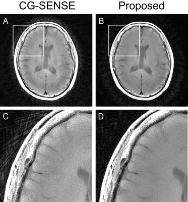 |
5 | Accelerated T1 weighted PROPELLER imaging of the brain with deep-learning based parallel imaging reconstruction
Motohide Kawamura1, Daiki Tamada1, Kazuyuki Sato2, Masahiro Hamasaki2, Satoshi Funayama1, Tetsuya Wakayama3, Utaroh Motosugi4, Hiroyuki Morisaka1, and Hiroshi Onishi1
1Department of Radiology, University of Yamanashi, Chuo, Japan, 2Division of Radiology, University of Yamanashi Hospital, Chuo, Japan, 3MR Collaboration and Development, GE Healthcare, Hino, Japan, 4Department of Radiology, Kofu-Kyoritsu Hospital, Kofu, Japan
PROPELLER sequence is useful because of its robustness to patient motion. Longer acquisition than FSE is a major drawback limiting its wider application in clinical practice. Here, we propose an accelerated T1 weighted PROPELLER of the brain using deep learning based parallel imaging (PI) reconstruction. Our method can unfold highly undersampled aliased images (PI factor = 7), enabling 2.3 times faster acquisition than full-sampling. A preliminary reader study with prospectively undersampled data showed that the proposed method significantly outperformed a conventional SENSE reconstruction in terms of streak artifact.
|
||
3162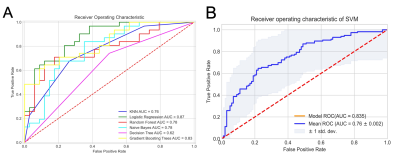 |
6 | CE-MRI Based Radiomics Model to identify Crohn’s disease and Ulcerative colitis Video Permission Withheld
Jun Wang1, Guangyao Liu1, Pengfei Zhang1, Kai Ai2, Laiyang Ma1, Wanjun Hu1, and Jing Zhang1
1Magnetic Resonance, Lanzhou University Second Hospital, Lanzhou, China, 2Philips Healthcare, Xi’an, China
Diagnosis and differential diagnosis of Inflammatory Bowel Disease (IBD) remains challenge since the particularity of small bowel. The objective of this study was to use the contrast enhanced MRI (CE-MRI) radiomics to distinguish subtypes of IBD patients. A total of 216 patients confirmed by pathology underwent small bowel CE-MRI. After the pipeline of radiomics, it was found that wavelet transformed texture features can effectively identify ulcerative colitis (UC) and Crohn’s disease (CD) with high performance. This work provides more detailed and microscopic details for the differential diagnosis of IBD subtypes based on the preliminary results.
|
||
3163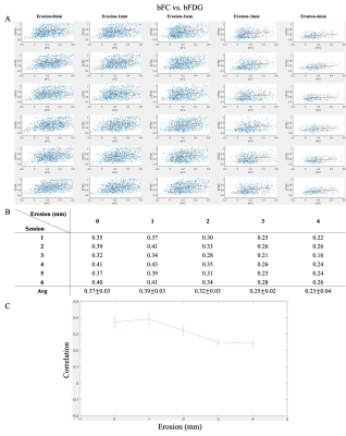 |
7 | Correlations between functional connectivity and glucose uptake in white matter
Bin Guo1,2, Fugen Zhou1, Muwei Li2,3, Zhaohua Ding2,4,5, and John C. Gore2,3,5
1Image Processing Center, School of Astronautics, Beihang University, Beijing, China, 2Vanderbilt University Institute of Imaging Science, Nashville, TN, United States, 3Department of Radiology and Radiological Sciences, Vanderbilt University Medical Center, Nashville, TN, United States, 4Department of Electrical Engineering and Computer Science, Vanderbilt University, Nashville, TN, United States, 5Department of Biomedical Engineering, Vanderbilt University, Nashville, TN, United States
Blood oxygenation-level dependent (BOLD) MRI signals have been reliably detected in white matter (WM) in both task and resting states in numerous studies. However, the relationship between WM BOLD signals and regional metabolism remains to be elucidated. In the present study, we investigated the relationship between resting state functional connectivity and glucose uptake in WM using simultaneous MRI and PET studies of human subjects. We find a significant correlation between these two measurements, suggesting that functional involvement of WM in neural activities was accompanied by an increase in glucose metabolism.
|
||
3164 |
8 | Robust MR image quality assessment algorithm irrespective of the quality of reference images
Yoichiro Ikushima1,2, Shogo Tokurei1, Hiroyuki Tarewaki3, Junji Morishita4, and Hidetake Yabuuchi4
1Department of Radiological Science, Faculty of Health Sciences, Junshin Gakuen University, Fukuoka, Japan, 2Department of Health Sciences, Graduate School of Medical Sciences, Kyushu University, Fukuoka, Japan, 3Division of Radiology, Department of Medical Technology, Osaka University Hospital, Suita, Japan, 4Department of Health Sciences, Faculty of Medical Sciences, Kyushu University, Fukuoka, Japan
The QEMDIM, a no-reference image quality assessment (IQA) algorithm, calculates the quality scores based on the quality difference between an image and reference images. Therefore, scores can be affected by the quality of the reference images. We modified the QEMDIM by changing the scoring method to compensate for this drawback. Brain MR images of various qualities were scored by subjective IQA (our gold standard), QEMDIM, and the modified algorithm. Modified scores showed a higher correlation with subjective IQA scores than the QEMDIM scores, using reference images of varied qualities. Our modified algorithm would be more clinically advantageous than the QEMDIM.
|
||
3165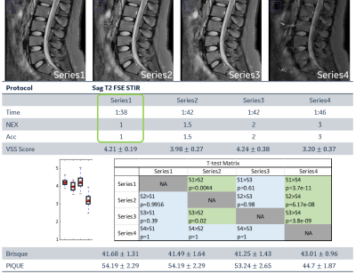 |
9 | Automatic no-reference image quality rating metrics in DL-reconstructed image quality assessment and protocol optimization
Yaan Ge1, Xiaolan Liu1, Qingyu Dai1, and Kun Wang1
1GE Healthcare, Beijing, China
This study proposed an automatic no-reference image quality rating metrics (VSS score) based on SVR model, that not requiring clinical expert labeled data, simulating human visual sense and applicable to all anatomies and contrast. The feasibility of applying this rating metrics in DL-recon integrated rapid scan protocol automatic evaluation is demonstrated. The result shows VSS score is in good correlation with visual sense to image quality and outperformed BRISQUE and PIQUE rating algorithms.
|
||
3166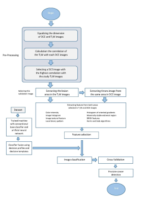 |
10 | Classifier fusion improves prostate cancer detection using MP-MRI Video Permission Withheld
Ghazaleh Jamshidi1, Ali Abbasian Ardakani2, Farshid Babapour Mofrad1, Hamidreza Saligheh Rad3,4, and Mahyar Ghafoori5
11- Department of Medical Radiation Engineering, Science and Research Branch, Islamic Azad University, Tehran, Iran (Islamic Republic of), 22- Department of Radiology Technology, School of Allied Medical Sciences, Shahid Beheshti University of Medical Sciences, Tehran, Iran (Islamic Republic of), 3Tehran University of Medical Science, Tehran, Iran (Islamic Republic of), 4Quantitative MR Imaging and Spectroscopy Group, Research Center for Molecular and Cellular Imaging, Tehran University of Medical Sciences, Tehran, Iran (Islamic Republic of), 53- Department of Radiology, Hazrat Rasoul Akram Hospital, School of Medicine, Iran University of Medical Sciences, Tehran, Iran (Islamic Republic of)
Multiparametric MRI (MP-MRI) has been widely used for detection of Prostate Cancer (PCa). In this study, we propose a new method using MP-MRI including T2-weighted (T2W) and dynamic contrast enhanced (DCE-) MRI for detection of PCa. 32 patients who had high prostate specific antigen (PSA) level recruited. We generated predictive models by extracting radiomics features and classifying benign and malignant lesions. The feature scores are evaluated with Relieff feature selection for each of the modalities. The fused classifier using decision template method showed the highest performance with accuracy, specificity, and sensitivity of 100.
|
||
3167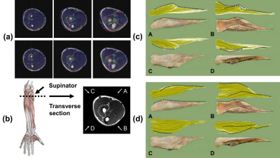 |
11 | Tracking forearm muscle fibers from Diffusion MRI during dynamic contractions
Yang Li1, Shihan Ma1, Qing Li2, Bo Xiong3, Weiwei Wang4, Xinjun Sheng1, and Xiangyang Zhu1
1Shanghai Jiao Tong University, Shanghai, China, 2MR Collaborations, Siemens Healthineers Ltd., Shanghai, China, 3Siemens Digital Medical Technology (Shanghai) Co., Ltd., Shanghai, China, 4Huashan Hospital affiliated to Fudan University, Shanghai, China
To reconstruct the trajectories of forearm muscle fibers during dynamic contractions, we designed a customized MRI-compatible rig to help acquire MRI data of the forearm muscles under three wrist postures. A piecewise registration method was proposed to correct the misregistration between DTI and anatomical images. The reconstructed supinator was highly consistent with the cadaveric dissection reference. Diffusion and architectural parameters were calculated to characterize voluntary contractions. During the wrist flexion and extension, the fascicle length of the supinator was inversely related to the pennation angle. The diffusion parameters increased slightly from neutral position to the other two postures.
|
||
3168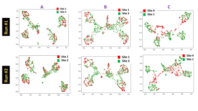 |
12 | Data Selection for Deep Learning via diversity visualization and scoring Video Permission Withheld
Deepa Anand1, Dattesh Dayanand Shanbhag1, and Rakesh Mullick1
1Advanced Technology Group, GE Healthcare, Bangalore, India
Data diversity is a key ingredient for robust deep learning models, especially in the medical domain. We present a diversity visualization and quantification scheme which enables decisions on data selection different enough from already existing data. Out experiments amply validate the usefulness of the proposed diversity metric in terms of enhancement in accuracy of models resulting from using them in data selection decision process with accuracy improvement from 3%->10% across different sites.
|
||
3169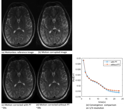 |
13 | Motion correction using Pilot Tone: a general model-based approach
YAN TU HUANG1, Peter Speier2, Tom Hilbert3,4,5, and Tobias Kober3,4,5
1Magnetic Resonance, Siemens Shenzhen Magnetic Resonance Ltd., Shenzhen, China, 2Magnetic Resonance, Siemens Healthcare, Erlangen, Germany, 3Advanced Clinical Imaging Technology, Siemens Healthcare, Lausanne, Switzerland, 4Department of Radiology, Lausanne University Hospital and University of Lausanne, Lausanne, Switzerland, 5LTS5, École Polytechnique Fédérale de Lausanne (EPFL), Lausanne, Switzerland
Motion continues to be a major impediment in clinical MRI. Using Pilot Tone to correct for motion is highly attractive as it uses inexpensive hardware and is independent from sequence. Here, we propose a model-based approach to calculate motion parameters from Pilot Tone signal. To avoid non-convergence those models are prone to, we introduce predefined displacement fields with the Pilot Tone as auxiliary, resulting in a linear motion model with fewer unknowns. Validation is performed on numerically simulated data and in-vivo measurements, comparing different motion models. Image reconstructions with the Pilot Tone showed fewer residual motion artefacts and converged faster.
|
||
3170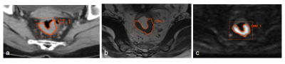 |
14 | Preoperative Prediction of Lymph Node Metastasis in Patients With Rectal Cancer Using A Multi-Modality Radiomics Model Video Not Available
Rui Wu1, Weiqiang Dou2, and Aiyin Li3
1Department of radiology, Shandong Provincial Qianfoshan Hospital, Cheeloo college of Medicine, Shandong University, Jinan, China, 2MR Research, GE Healthcare, Beijing, China, 3Department of radiology, The First Affiliated Hospital of Shandong First Medical University & Shandong Provincial Qianfoshan Hospital, Jinan, China
This study mainly aimed to build a multi-modality radiomics model for predicting lymph node metastasis (LNM) of rectal cancer (RC) preoperatively. 82 RC patients were enrolled in this study. Two, six, four and eleven optimal features were separately selected for venous phase CT, HR-T2WI, DWI and multi-modality by incorporating CT, HR-T2WI and DWI together. Together with MRI-reported LN status feature, clinical features, and the combination of multi-modality and clinical features, in total seven support vector machine (SVM) classification models were respectively built. Incorporated with clinical features, multi-modality radiomics model provided the most robust predictive performance in predicting LNM.
|
||
3171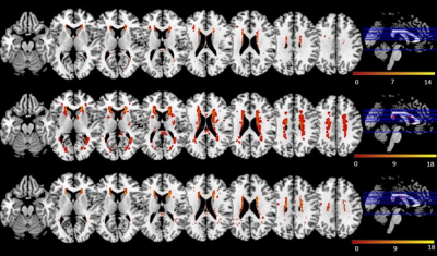 |
15 | Association between white matter hyperintensity and diabetes-related mild cognitive impairment Video Permission Withheld
Yashi Nan1, Jian Zhang1, Jianming Ni2, and Silun Wang1
1Yiwei Medical Technology, Shenzhen, China, 2Department of Radiology, Wuxi No2. People’s Hospital, Affiliated Wuxi Clinical College of Nantong University, Wuxi, China
White matter hyperintensities (WMH) features may assist detecting diabetes-related MCI. We aimed to identify the correlations between WMH and MCI in T2D using brain magnetic resonance imaging. Fifty participants, matched for age, were included. Total and regional WMH volumes were calculated using automated segmentation approach. WMH patterns were compared between groups using a voxel-wise analysis. Results show that cognitive impairment was related to a higher prevalence of regional WMH. Subcortical WMH volumes in specific brain regions. The findings suggested that the presence of WMH in specific regions rather the WMH volumes appears to be correlated with the MCI in T2D.
|
||
 Back to Meeting Home
 Back to the Program-at-a-Glance
The International Society for Magnetic Resonance in Medicine is accredited by the Accreditation Council for Continuing Medical Education to provide continuing medical education for physicians.
 Back to Meeting Home
 Back to the Program-at-a-Glance
 View the Presentation