Online Gather.town Pitches
Processing & Analysis I
Joint Annual Meeting ISMRM-ESMRMB & ISMRT 31st Annual Meeting • 07-12 May 2022 • London, UK

Online Gather.town Pitches
Processing & Analysis I
| Booth # | ||||
|---|---|---|---|---|
3791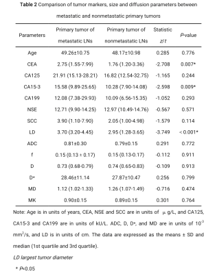 |
1 | Differentiating metastatic from nonmetastatic lymph nodes in cervical cancer patients by IVIM-DWI and DKI
Suixing Zhong1, Ya Zhang1, Yingying Ding1, Xiaoyong Zhang2, Jing Tan1, Conghui Ai1, Yan Jin1, Hongbo Wang1, Huimei Zhang1, Miaomiao Li1, Rong Zhu1, and Shangwei Gu1
1Department of Radiology, Yunnan Cancer Hospital, Third Affiliated Hospital of Kunming Medical University, Kunming, China, 2Clinical Science, Philips Healthcare, Chengdu, China
This study aimed to investigate the diagnostic value of intravoxel incoherent motion (IVIM) diffusion-weighted imaging and diffusion kurtosis imaging (DKI) in distinguishing metastatic from nonmetastatic lymph nodes (LNs) in cervical cancer patients. The clinical and imaging data of 102 patients with cervical cancer were collected prospectively, including 38 patients with LN metastasis and 64 patients without LN metastasis. The morphological parameters, diffusion parameters and tumor markers of primary tumors and LNs were measured and compared between the two groups. The results showed that the diagnostic efficiency of diffusion parameters were not as good as morphological parameters.
|
||
3792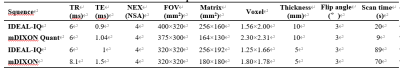 |
2 | Comparison of fat quantification in abdomen and vertebrae by IDEAL-IQ and mDIXON Quant Video Not Available
Na Liu1, Wei qing Song1, Wei yan Miao1, Lian ai Liu1, Jie Liang Lin2, and Fang Xiao Xu3
1the First Affiliated Hospital of Dalian Medical University, Da Lian, China, 2Philips Healthcare, Da Lian, China, 3Philips Healthcare, Bei Jing, China
IDEAL-IQ by GE and mDixon Quant by Philips are two MR fat quantification techniques that have been widely used for clinical disease evaluation and diagnosis in recent years. This study aims to compare performance of the two similar methods for fat quantification in the liver, pancreatic and lumbar vertebral. Results showed no significant difference between IDEAL-IQ and mDixon Quant on the liver, pancreas, and lumbar vertebral fat quantification in the same cohort of heathy volunteers
|
||
3793 |
3 | Acceleration of T2-weighted head Multivane imaging by compressed sensing Video Not Available
Na Liu1, Wei Qing Song1, Jie Liang Lin2, Nan Wang1, Lian ai Liu1, and Wei Yan Miao1
1the First Affiliated Hospital of Dalian Medical University, Da Lian, China, 2Philips Healthcare, BeiJing, China
Multivane (MV) imaging, based on the blade rotating acquisition in k space, is a novel sequence for motion reduction imaging. The MV acquisition percentage is critical parameter in MV imaging for balanced image quality and scan time. This study aims to assess the performance of compressed sensing for accelerated T2-weighted head MV imaging under altered MV acquisition percentages. Results indicated that scan time of head Multivane (MV) T2WI was significantly reduced by compressed sensing (CS) without reduced image quality.
|
||
3794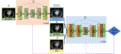 |
4 | 4D-MRI image enhancement via a deep learning-based adversarial network
Yinghui Wang1, Shaohua Zhi1, Haonan Xiao1, Tian Li1, and Jing Cai1
1Department of Health Technology and Informatics, The Hong Kong Polytechnic University, Hong Kong, China
We developed and evaluated a deep learning technique for enhancing four-dimensional MRI (4D-MRI) image quality based on conditional adversarial networks. The quantitative and qualitative evaluative results demonstrated that the proposed model was able to reduce artifacts in low-quality 4D-MRI images and recover the details obtained from high-quality MR images, and performed better as compared with a state-of-the-art method.
|
||
3795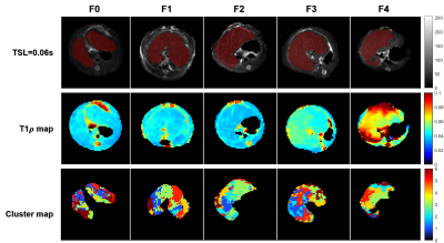 |
5 | Multiexponential T1ρ spectroscopy in liver fibrosis: A prospective animal experiment at 11.7T MRI
Junqi Xu1, Qianfeng Wang1, Xuchen Yu1, Jiawei Han1,2, Yimei Lu3, and He Wang1,2
1Institute of Science and Technology for Brain-Inspired Intelligence, Fudan University, ShangHai, China, 2Human Phenome Institute, Fudan University, ShangHai, China, 3Department of Radiology, Xinhua Hospital, Shanghai Jiao Tong University, ShangHai, China
This study proposed a new method to assess the stage of liver fibrosis by signal curves clustering and multiexponential decay T1ρ spectral analysis. Compared to single component in normal tissue, liver fibrosis tissue has two T1ρ components, in carbon tetrachloride (CCI4) rat model. Meanwhile, T1ρ tends to increase with fibrosis score. Due to the spatial distribution of clusters reveal the fibrosis region, this method might contribute to diagnosis and staging of liver fibrosis.
|
||
3796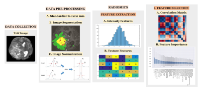 |
6 | Effect of varying intensity discretization and normalization parameters on first order radiomics features.
Abhilasha Indoria1, Sachin Patalasingh1, subhas Konar1, and Jitender Saini1
1NIMHANS, Bengaluru, India
This study assessed the impact of normalization scale and intensity discretization on first order radiomics features. Features were extracted from T2W images by varying the normalization parameter and bin width parameter of Pyradiomics library; Un-normalized (Without_normalization), normalized to scale1, normalized to scale 100 while keeping the bin width constant; and Bin width set to 20, bin width set to 40, bin width set to 60 while keeping the normalization scale set to 1. We found that the radiomics features were highly dependent on normalization scale and independent of bin width parameter.
|
||
3797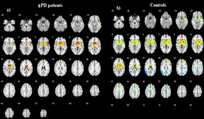 |
7 | Basal Ganglia Connectivity as Assessed by Resting-State fMRI in Genetic Parkinson’s Disease
hacer dasgin1, basak soydas2, Gul Yalcin Cakmakli3, Bilge Volkan Salanci2, Eser Lay Ergun2, Bulent Elibol3, and Kader K. Oguz4
1Aysel Sabuncu Brain Research Center-UMRAM, Ankara, Turkey, 2Department of Nuclear Medicine, Hacettepe University, Ankara, Turkey, 3Department of Neurology, Hacettepe University, Ankara, Turkey, 4Department of Radiology, Hacettepe University, Ankara, Turkey
Genetic Parkinson’s Disease (gPD) displays distinct clinical, demographic and pathological features from idiopathic Parkinson’s Disease (PD). The present study aims to search for alterations in the basal ganglia network (BGN) including substantia nigra (SN) and Subthalamic nucleus (STN) in a peculiar group of gPD using resting state (RS) fMRI. Genetic PD patients revealed less activation in the basal ganglia, SN+STN pathway and reduced connectivity with ACC, MCC and insula, which may reflect frontal lobe and autonomic dysfunction in these patients.
|
||
3798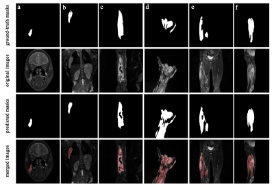 |
8 | Lesion Segmentation for Venous Malformations Based on Unet++ Architecture Video Permission Withheld
Jian Dong1, Yaping Wu1, Yan Bai1, and Meiyun Wang1
1Department of Medical Imaging, Henan Provincial People's Hospital, Zhengzhou, China In this study, we trained a fully automatic lesion segmentation model of venous malformations based on Unet++ structure and fat-saturated T2-weighted images. The results of automatic segmentation of lesions in different sequence directions and locations of the lesions are consistent with the results of manual segmentation by radiologists. The Dice coefficient on the test set reached 0.847. This fully automatic lesion segmentation model can provide support for subsequent automatic diagnosis studies related to venous malformations. |
||
3799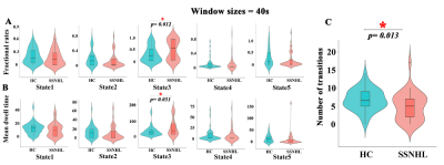 |
9 | What happens “when one door is half closed, another does not open yet” for the unilateral sudden sensorineural hearing loss patients
Yuting Li1, Yuxuan Shang1, Zhuhong Chen1, Jingting Sun1, and Xiaochen Wei2
1Tangdu Hospital, Fourth Military Medical University, Xi'an, China, 2GE Healthcare, MR Research China, Beijing, Beijing, China
Featured alterations in dynamic brain functions, neurovascular coupling, or brain structure occurs at early-, intermediate- or late-stage of unilateral sudden sensorineural hearing loss (SSNHL) “when one door is half closed, another does not open yet”, respectively, and may serve as the neuroimaging markers for SSNHL.
|
||
3800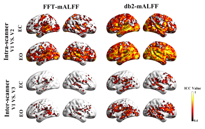 |
10 | Higher reliability and validity of Wavelet-based ALFF of resting-state fMRI: evidence from multicenter eyes open and eyes closed databases Video Permission Withheld
Juan Yue1,2,3, Na Zhao1,2,3,4,5, Yang Qiao1,2,3,5,6, Zi-Jian Feng1,2,3, Yun-Song Hu1,2,3, Yong Zhang 7, Yu-Feng Zang1,2,3, and Jue Wang8
1Center for Cognition and Brain Disorders, The Affiliated Hospital of Hangzhou Normal University, Hangzhou, China, 2Institute of Psychological Sciences, Hangzhou Normal University, Hangzhou, China, 3Zhejiang Key Laboratory for Research in Assessment of Cognitive Impairments, Hangzhou, China, 4Unit of Psychiatry, Department of Public Health and Medicinal Administration, & Institute of Translational Medicine, Faculty of Health Sciences, University of Macau, Macao SAR, China, 5Centre for Cognitive and Brain Sciences, University of Macau, Macao SAR, China, 6Faculty of Health Sciences, University of Macau, Macao SAR, China, 7MR Research, GE Healthcare, Shanghai, China, 8Institute of Sports Medicine and Health, Chengdu Sport University, Chengdu, China
Using multi-center resting-state fMRI databases under eyes closed (EC) and eyes open (EO), this study systematically investigated the intra- and inter-scanner reliability and validity of Wavelet-mALFF across multiple frequency bands. We found that Wavelet-mALFF outperformed FFT-mALFF in terms of intra- and inter-reliability and validity, particularly in the higher frequency band slow-2 (0.1992-0.25 Hz), where db2-mALFF is the top performer in Wavelet-mALFF. We propose that instead of FFT-ALFF, db2-ALFF can be employed to measure the spontaneous oscillations of local brain activity in future studies.
|
||
3801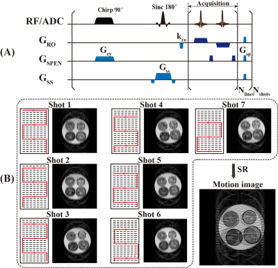 |
11 | Motion correction of high-resolution spatiotemporally encoded MRI based on deep learning
Wei Wang1, Shuhui Cai1, Lin Chen1, Zhigang Wu2, Congbo Cai1, and Zhong Chen1
1Xiamen University, Xiamen, China, 2MSC Clinical & Technical Solutions, Philips Healthcare, Xiamen, China
In multi-shot high-resolution MRI, the motion of patients often leads to serious degradation of imaging quality. In this study, a novel motion correction method based on deep learning and spatiotemporally encoded MRI was proposed to address this problem. The proposed method is robust to motion without utilizing extra scan or parallel reconstruction. The results of simulation and in vivo rat brain experiments demonstrate its efficacy in reducing image motion artifacts when subject movement exists.
|
||
3802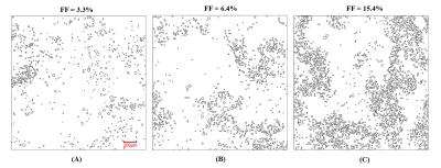 |
12 | Morphological Characterization of Hepatic Steatosis Using Stereology and Spatial Statistics
Jinyang Wang1, Changqing Wang1, Scott B. Reeder2,3,4,5,6, and Diego Hernando2,3
1School of Biomedical Engineering, Anhui Medical University, Heifei, China, 2Radiology, University of Wisconsin-Madison, Madison, WI, United States, 3Medical Physics, University of Wisconsin-Madison, Madison, WI, United States, 4Biomedical Engineering, University of Wisconsin-Madison, Madison, WI, United States, 5Medicine, University of Wisconsin-Madison, Madison, WI, United States, 6Emergency Medicine, University of Wisconsin-Madison, Madison, WI, United States Monte Carlo modeling enables characterization of MR signals in various tissues, and has been applied to liver MR in the presence of fat. However, Monte Carlo modeling requires accurate information about the underlying tissue properties. In this work, we investigate the size and clustering of fat droplets in the liver using stereology and spatial statistics for three human liver biopsy samples with steatosis. Results show that the generalized gamma distribution function can accurately determine the size and location distributions of fat droplets. This may enable analysis of the underlying biophysical mechanisms between fat fraction and R2* from microscopic magnetic sensitivity. |
||
3803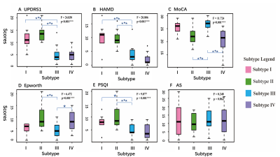 |
13 | Subtyping Parkinson’s disease by linking non-motor characteristics and brain functional connectome
Yao Zeng1, Chenfei Ye2, Junyan Sun3, Ting Ma1, and Tao Wu3
1Department of Electronic and Information Engineering, Harbin Institute of Technology at Shenzhen, Shenzhen, China, 2Peng Cheng Laboratory, Shenzhen, China, 3National Clinical Research Center for Geriatric Disorders, Xuanwu Hospital Capital Medical University, Beijing, China
Since the heterogeneity of non-motor disorders in Parkinson's patients, subtyping research will facilitate the development of clinical diagnosis and treatment. We applied the sparse canonical correlation analysis method on clinical scales and resting-state brain fMRI data, to extract the typical variables for subtyping. In this data-driven study, we found that four PD subtypes with varying cognitive and psychological phenotypes demonstrate unique patterns of brain functional connectivity, which may underly the mechanism of PD heterogeneous non-motor symptoms.
|
||
3804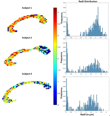 |
14 | Axon radii estimation from dMRI data acquired from a non-fancy MRI scanner
Debdut Mandal1, Lipeng Ning2, and Yogesh Rathi2
1Electronics and Electrical Communication Engineering, Indian Institute of Technology Kharagpur, Kharagpur, India, 2Harvard Medical School, Boston, MA, United States
Noninvasive measurement of axon radii has always been challenging using in vivo diffusion MRI (dMRI) data. Although dMRI allows estimation of the effective radius, a very high gradient strength scanner is required to acquire the data. In our proposed method, we overcome this limitation by using analytical methods to accurately predict the signal at high b-values from reasonably lower b-value data that can be obtained from a Prisma-like scanner. Our findings, both in synthetic data and in vivo dMRI data, show that estimating axon radii reliably is possible using dMRI data with a certain SNR level.
|
||
3805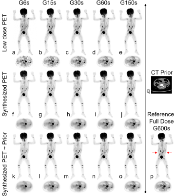 |
15 | Improving image quality of ultralow-dose pediatric total-body PET/CT using deep learning technique Video Permission Withheld
Qiyang Zhang1,2, Zizheng Xiao3, Xu Zhang3, Yingying Hu3, Yumo Zhao3, Jingyi Wang4, Jiatai Feng4, Yun Zhou4, Yongfeng Yang1, Xin Liu1, Hairong Zheng1, Wei Fan3, Dong Liang1, and Zhanli Hu1
1Lauterbur Research Center for Biomedical Imaging, Shenzhen Institute of Advanced Technology, Chinese Academy of Sciences, Shenzhen, China, 2National Innovation Center for High Performance Medical Devices, Shenzhen, China, 3Department of Nuclear Medicine, Sun Yat-sen University Cancer Center, Guangzhou, China, 4Central Research Institute, United Imaging Healthcare Group, Shanghai, China
Young children are more sensitive to radiation doses than adults, and their absorption of effective doses can be 4-5 times that of adults. When performing PET imaging, the use of low-dose imaging agents for high-quality imaging is of clinical importance. Here, we use artificial intelligence techniques combined with prior CT information to improve the quality of total-body PET/CT images in ultralow-dose pediatric FDG scans, and the results show that the equivalent quality of 600s acquisition data can be obtained using 15s acquisition.
|
||
3806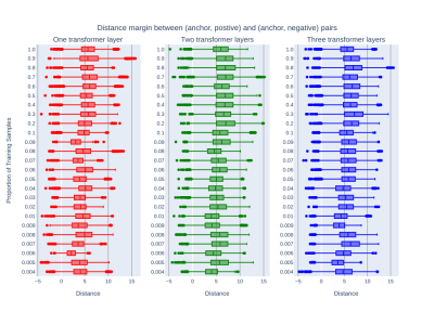 |
16 | Learning White Matter Streamline Representations Using Transformer-based Siamese Networks with Triplet Margin Loss
Shenjun Zhong1, Zhaolin Chen1, and Gary Egan1
1Monash Biomedical Imaging, Monash University, Australia, Melbourne, Australia
Robust latent representation of white matter streamlines are critical for parcellating streamlines. This work introduced a novel transformer-based siamese network with triplet margin loss, that learns to embed any lengths of streamlines into fixed-length latent representations. Results showed that a minimum of two layers of transformer encoders were sufficient to model streamlines with a very limited number of training data.
|
||
3807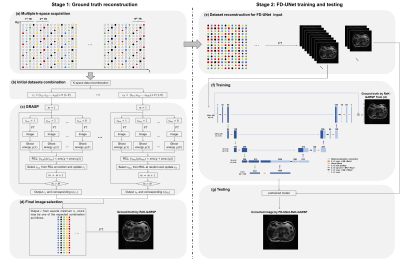 |
17 | Free-breathing MRI enabled by FD-UNet with ground truth from repeated k-t-subsampling and greedy randomized adaptive search procedure
Lu Wang1, Xiaorui Xu1, Liyuan Liang1, and Hing-Chiu Chang2
1Department of Diagnostic Radiology, University of Hong Kong, Hong Kong, China, 2Department of Biomedical Engineering, Chinese University of Hong Kong, Hong Kong, China
Deep learning solutions have been proposed to correct motion-induced artefact in free-breathing abdominal MRI that can overcome some challenges in conventional methods, such as requiring respiratory modeling, external motion monitoring devices, or extremely long computation time. However, ground truth data for training can only be acquired with breath-holding or using those conventional methods. In this study, we proposed to use FD-UNet for the motion correction of free-breathing abdominal MRI. The high-quality and artifact-free ground truth data were produced from repeated k-t-subsampling and greedy randomized adaptive search procedure (ReK-GRASP), without relying on respiratory modeling, external devices, or long computation time.
|
||
3808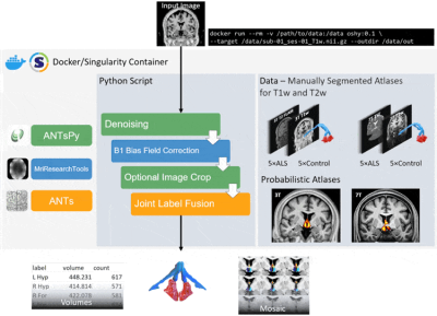 |
18 | Open-Source Hypothalamic-ForniX (OSHy-X) Atlases and Segmentation Tool for 3T and 7T
Jeryn Chang1, Frederik Steyn1,2,3,4, Shyuan Ngo2,3,4,5, Robert Henderson2,3,4, Christine Guo6, Steffen Bollmann7,8, Jurgen Fripp9, Markus Barth7,8, and Thomas B Shaw2,7,8
1School of Biomedical Sciences, The University of Queensland, St Lucia, Australia, 2Department of Neurology, Royal Brisbane and Women's Hospital, Brisbane, Australia, 3Wesley Medical Research, The Wesley Hospital, Brisbane, Australia, 4Centre for Clinical Research, The University of Queensland, Brisbane, Australia, 5Australian Institute of Bioengineering and Nanotechnology, The University of Queensland, St Lucia, Australia, 6QIMR Berghofer Medical Research Institute, Brisbane, Australia, 7School of Information Technology and Electrical Engineering, The University of Queensland, St Lucia, Australia, 8Centre for Advanced Imaging, The University of Queensland, Brisbane, Australia, 9Health and Biosecurity, The Commonwealth Scientific and Industrial Research Organisation (CSIRO), Brisbane, Australia Segmentation and volumetric analysis of the hypothalamus and fornix plays a critical role in improving the understanding of degenerative processes that might impact the function of these structures. We present Open-Source Hypothalamic-ForniX (OSHy-X) atlases and tool for multi-atlas fusion segmentation for 3T and 7T. The atlases are based on 20 manual segmentations, which we demonstrate have high interrater agreement. The versatility of the OSHy-X tool allows segmentation and volumetric analysis for different field strengths and contrasts. We also demonstrate that OSHy-X segmentation has higher Dice overlaps (3T and 7T inputs: p<0005, p<0.005) than a deep-learning segmentation method for the hypothalamus. |
||
3809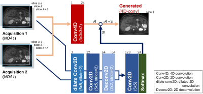 |
19 | The usefulness of 4D convolution in deep-learning-based noise reduction for low-SNR body DWI
Yasuhiko Tachibana1, Hiroki Tsuchiya1, Riwa Kishimoto1, Tokuhiko Omatsu1, Shinichiro Mori1, Takayuki Obata1, and Tatsuya Higashi1
1National institutes for Quantum science and Technology, Chiba, Japan Deep-learning-based slice-by-slice noise reduction may not be suitable for low-SNR body DWI that contains insufficient information in the original single slice. Moreover, averaging multiple acquisitions after denoising to avoid this problem is insufficient because it causes blurring owing to a mismatch between acquisitions. Herein, we designed a neural network that utilises 4D convolution to incorporate adjacent slices and multiple acquisitions simultaneously for a slice to achieve adequate denoising. The results support the utility of the proposed method in comparison with the usual slice-by-slice method and averaging. |
||
3810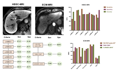 |
20 | Identification and diagnosis of hepatocellular carcinoma in high-risk patients with an abbreviated-protocol MR screening: a pilot study
Shang Wan1, Yi Wei1, Hehan Tang1, Lisha Nie2, Xiaocheng Wei2, and Bin Song3
1Radiology, West China Hospital, Sichuan University, Cheng Du, China, 2GE Healthcare Beijing China, Beijing, China, 3West China Hospital, Sichuan University, Cheng Du, China
Early detection and diagnosis of hepatocellular carcinoma (HCC) is essential for patients’ prognosis, however, the imaging hallmarks for tumor detection and diagnosis has remained same for years despite the use of many new imaging methods. Thus, in this study, we aimed to prospectively evaluated the detection performance of various MR sequences and abbreviated MRI (aMRI) protocols in different clinical settings and further compared the different imaging criteria for the diagnosis of HCC using either extracellular contrast agent (ECA) or hepatobiliary specific contrast (HBSC) MR imaging, and the developed diagnostic criteria for detection and diagnosis of HCC may aid clinical diagnosis.
|
||
 Back to Meeting Home
 Back to the Program-at-a-Glance
The International Society for Magnetic Resonance in Medicine is accredited by the Accreditation Council for Continuing Medical Education to provide continuing medical education for physicians.
 Back to Meeting Home
 Back to the Program-at-a-Glance
 View the Presentation