Online Gather.town Pitches
Processing & Analysis III
Joint Annual Meeting ISMRM-ESMRMB & ISMRT 31st Annual Meeting • 07-12 May 2022 • London, UK

Online Gather.town Pitches
Processing & Analysis III
| Booth # | ||||
|---|---|---|---|---|
3898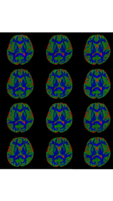 |
1 | Inter-site reliability of diffusion microstructure measurements: A 3-tissue constrained spherical deconvolution study Video Permission Withheld
Richard L Huang1, Benjamin T Newman1, and T Jason Druzgal1
1University of Virginia School of Medicine, Charlottesville, VA, United States
As large multi-site neuroimaging and diffusion MRI (dMRI) microstructure studies become more common, it is necessary to understand factors affecting reliability of outcome measurements collected across different sites. In this study, we analyze dMRI collected from 3 subjects traveling to 10 different sites with identical MRI scanners, sequence protocols, and software. We perform a detailed microstructural analysis in 212 grey matter and white matter brain regions and find that measurements are generally reliable across sites. However, there remains variation in specific locations that may suggest caution when interpreting small effects in small or hard to measure brain regions.
|
||
3899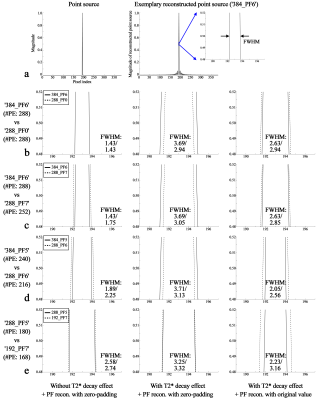 |
2 | The Spatial Resolution of Partial-Fourier-Accelerated EPI to Delineate a Submillimetre Voxel at 7T: Use of an Intensive Partial Fourier Factor
Seong Dae Yun1, Avdo Celik1, Michael Schöneck1, and N. Jon Shah1,2,3,4
1Institute of Neuroscience and Medicine 4, INM-4, Forschungszentrum Juelich, Juelich, Germany, 2Institute of Neuroscience and Medicine 11, INM-11, JARA, Forschungszentrum Juelich, Juelich, Germany, 3JARA - BRAIN - Translational Medicine, Aachen, Germany, 4Department of Neurology, RWTH Aachen University, Aachen, Germany
The employment of the partial Fourier technique for submillimetre-resolution EPI has been demonstrated in many recent MR studies. However, the performance of the resulting spatial resolution has not yet been thoroughly investigated. This work aims to evaluate the spatial resolution of high-resolution EPI protocols configured with various partial Fourier factors at 7T. The obtained results show that the spatial resolution of partial-Fourier EPI is mainly determined by the number of actually sampled points (matrix size × PF-factor). Here, the use of an intensive PF-factor (5/8) for high spatial resolution was also demonstrated with a relatively large matrix size.
|
||
3900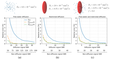 |
3 | Unbiasing orientationally-averaged diffusion MRI signal
Tomasz Pieciak1, Maryam Afzali2,3, Antonio Tristán-Vega1, and Santiago Aja-Fernández1
1Laboratorio de Procesado de Imagen (LPI), Universidad de Valladolid, Valladolid, Spain, 2Leeds Institute of Cardiovascular and Metabolic Medicine, University of Leeds, Leeds, United Kingdom, 3Cardiff University Brain Research Imaging Centre (CUBRIC), School of Psychology, Cardiff University, Cardiff, United Kingdom
The orientationally-averaging or the so-called power-averaging is a common approach to reduce the impact of macroscopic anisotropy in diffusion MRI (dMRI). This paper analytically derives two new closed-form unbiased orientationally-averaging diffusion MRI signal estimators and shows that the noise-related bias could be significantly suppressed with these new findings. The new formulas might be applied to retrospectively orientationally-averaged dMRI signals with minimal computational effort.
|
||
3901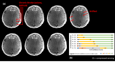 |
4 | The Performance of Image Quality Metrics Depends on the Diagnostic Task: A Case Study in Stroke MRI
Michelle Pryde1,2, Sarah Reeve2,3, Taylor Bouchie2,4, Elena Adela Cora5,6, David Volders 5,6, Matthias Schmidt5,6, Mohamed Abdolell5, Chris Bowen2,3,5, James Rioux2,3,5, and Steven Beyea1,2,3,5
1School of Biomedical Engineering, Dalhousie University, Halifax, NS, Canada, 2Biomedical Translational Imaging Centre, QEII Health Sciences Centre, Halifax, NS, Canada, 3Physics and Atmospheric Science, Dalhousie University, Halifax, NS, Canada, 4Medicine, Dalhousie University, Halifax, NS, Canada, 5Diagnostic Radiology, Dalhousie University, Halifax, NS, Canada, 6Diagnostic Imaging, Nova Scotia Health, Halifax, NS, Canada
Image Quality Metrics (IQMs) have allowed for objective analysis of MR images in order to optimize protocols or reconstruction algorithms, for example. However, the performance of IQMs depends on the diagnostic task. Therefore, the aim of this study was to explore how well leading IQMs correlate with, or predict, neuroradiologists’ diagnostic confidence in acute and chronic stroke diagnostic tasks. We observed that, although the IQMs in question calculated for T2 FLAIR images could be used to predict neuroradiologists’ diagnostic confidence scores for the chronic stroke diagnostic task, they did not correlate with diagnostic confidence scores for acute stroke.
|
||
3902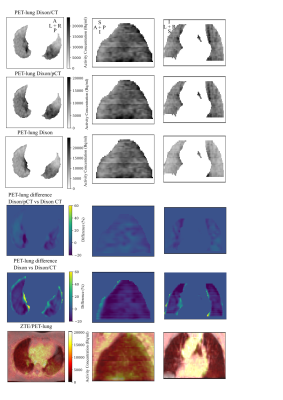 |
5 | PET attenuation correction in lung with ZTE based pseudo-CT generation
Chang Sun1, Roido Manavaki1, Jason Tarkin2, Christopher Wall2, James HF Rudd2, Fiona J Gilbert1, and Martin J Graves1
1Department of Radiology, University of Cambridge, Cambridge, United Kingdom, 2Division of Cardiovascular Medicine, University of Cambridge, Cambridge, United Kingdom
Attenuation correction methods in body PET/MRI do not consider lung density variations within and between participants, which can significantly affect PET image quantification in thoracic areas. We developed a hybrid ZTE-based/Dixon method for MR-based attenuation correction, which resulted in a pseudo-CT image (Dixon/pCT) and used for attenuation correction in the lung. PET images corrected with the Dixon/pCT attenuation map were compared to images produced with manufacturer’s Dixon attenuation map and a hybrid Dixon/CT image. Overall, the Dixon/pCT method reduced the absolute SUV error in the lung by 3%, when compared to the Dixon attenuation correction.
|
||
3903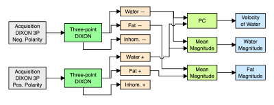 |
6 | Simultaneous acquisition of water-fat concentrations and velocity images using a Phase-contrast Three-point Dixon method
Esteban Denecken1,2,3, Cristobal Arrieta1,3, Hernán Mella1,3, and Sergio Uribe1,3,4
1Biomedical Imaging Center, Pontificia Universidad Católica de Chile, Santiago de Chile, Chile, 2Department of Electrical Engineering, School of Engineering, Pontificia Universidad Católica de Chile, Santiago de Chile, Chile, 3ANID – Millennium Science Initiative Program – Millennium Nucleus for Cardiovascular Magnetic Resonance, Santiago de Chile, Chile, 4Department of Radiology, School of Medicine, Pontificia Universidad Católica de Chile, Santiago de Chile, Chile
Phase-contrast in areas with a significant fat signal is subject to chemical shift artifacts where the fat signal interferes with the neighboring water signal. The purpose of this work is to present a novel three-point Dixon method that preserves the phase information in water and fat images and combines this method with phase-contrast to obtain water-fat concentration and velocity images from a single acquisition. We validate our method using a numerical phantom and MRI acquired in volunteers. Phase-contrast three-point Dixon and standard methods showed equivalent results comparing different ROIs of the PDFF using NRMSE.
|
||
3904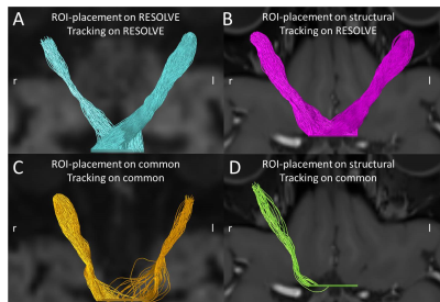 |
7 | Spatial alignment of structural images on common and RESOLVE diffusion images in the optic nerve
Markus Janko1,2, Patrick Rose1,2, Vanessa Ines Schoeffling1, Oliver Korczynski1, Katharina Ponto3, Esther Hoffmann3, Marc Brockmann1, Wolfgang Kleinekofort2, and Andrea Kronfeld1
1Neuroradiology, University Medical Center of the Johannes Gutenberg University Mainz, Mainz, Germany, 2Applied Physics & Medical Engineering, Hochschule RheinMain, Rüsselsheim, Germany, 3Ophthalmology, University Medical Center of the Johannes Gutenberg University Mainz, Mainz, Germany
A successful use of anatomical templates in the evaluation of functional neuroimaging-studies is only possible, if anatomical and functional intra-individual data can be registered with sufficient accuracy. We wanted to investigate the possibility to register structural on common and RESOLVE diffusion-images of the optic nerve. Three volunteers were examined and the structural images were registered to the two diffusion-datasets. Two observers defined seeds for tractography of the optic nerve on the structural and on the diffusion images. Tractography worked with seeds defined on the structural images registered to RESOLVE. Therefore RESOLVE is suitable for the use in template-based studies.
|
||
3905 |
8 | NOise Reduction with Distribution Corrected (NORDIC) PCA improves signal-to-noise and functional connectivity in rodent resting-state fMRI
Sarah Y. Wu1, Russell W. Chan1,2, Yixi Xue1, Emily L. Tse1, Giles Hamilton-Fletcher1, Steen Moeller3, and Kevin C. Chan1,4
1Department of Ophthalmology, New York University Grossman School of Medicine, New York, NY, United States, 2Neuroscience Institute, New York University Grossman School of Medicine, New York, NY, United States, 3Center for Magnetic Resonance Research (CMRR), University of Minnesota, Minneapolis, MN, United States, 4Department of Radiology, New York University Grossman School of Medicine, New York, NY, United States
The relatively poor temporal signal-to-noise ratio (tSNR) in resting-state fMRI (rsfMRI) serves to be a pressing area of improvement. NOise Reduction with Distributed Corrected (NORDIC) PCA can effectively increase tSNR. However, it has yet to be examined in rodent rsfMRI studies. Here, we applied NORDIC-correction to mouse rsfMRI, and evaluated the tSNR and functional connectivity against standard preprocessing data. NORDIC was able to significantly increase tSNR and reduce functional connectivity variability. NORDIC can also denoise rsfMRI signals at higher frequencies. Taken together, NORDIC can potentially become an important preprocessing step in future rodent rsfMRI studies.
|
||
3906 |
9 | dMRIQCpy: a python based toolbox for diffusion MRI quality control and beyond
Guillaume Theaud1,2 and Maxime Descoteaux1,2
1Sherbrooke Connectivity Imaging Laboratory (SCIL), Université de Sherbrooke, Canada, Sherbrooke, QC, Canada, 2Imeka Solutions Inc, Sherbrooke, QC, Canada
Diffusion MRI data suffers from many artifacts and parameters such as Echo and Repetition Time. From raw data to bundle segmentation, acquisition/processing issues in processing can happen and bias analyses. That is why quality control (QC) at each step of a study is important. We present a python-based library: dMRIQCpy that supports QC of raw data, intermediate DWI and T1 preprocessing, metrics from DWI, tractogram and bundles screenshots. dMRIQCpy enables users to save/load QC reports to permit collaboration between users. dMRIQCpy was tested on 3 datasets and highlighted a faster QC (50 % faster) than reviewing screenshots in a folder.
|
||
3907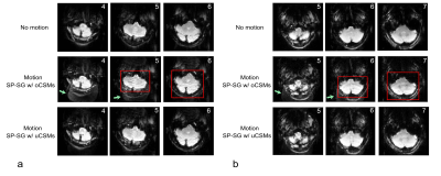 |
10 | Brain SMS-EPI reconstructions with prospective motion correction: improved quality with updated coil sensitivity maps
Bo Li1, Ningzhi Li2, Radu Balan3, Ze Wang1, and Thomas Ernst1
1Diagnostic Radiology and Nuclear Medicine, University of Maryland, Baltimore, Baltimore, MD, United States, 2U.S. Food Drug Administration, Silver Spring, MD, United States, 3Department of Mathematics, University of Maryland, College Park, College Park, MD, United States
We investigated prospective motion correction (PMC) for brain SMS-EPI using camera-based motion tracking and coil sensitivity interpolation. Phase compensation for the receiver was performed to eliminate motion-induced aliasing artifacts. Coil sensitivity profiles were interpolated and extrapolated with Makima piecewise cubic Hermite algorithm to remove residual SMS reconstruction artifacts caused by the misregistration of coil sensitivities between updated (mobile) SMS slices and stationary single-slice reference profiles.
|
||
3908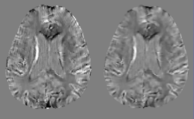 |
11 | Motion Artifact Reduction in Quantitative Susceptibility Mapping using Deep Neural Network
Chao Li1, Hang Zhang1, Jinwei Zhang1, Pascal Spincemaille1, Thanh Nguyen1, and Yi Wang1
1Cornell university, New York, NY, United States
An approach to reduce motion artifacts in Quantitative Susceptibility Mapping using deep learning is proposed. We use an affine motion model with randomly created motion profiles to simulate motion-corrupted QSM images. The simulated QSM image is paired with its motion-free reference to train a neural network using supervised learning. The trained network is tested on unseen simulated motion-corrupted QSM images, in healthy volunteers and in Parkinson’s disease patients. The results show that motion artifacts, such as ringing and ghosting, were successfully suppressed.
|
||
3909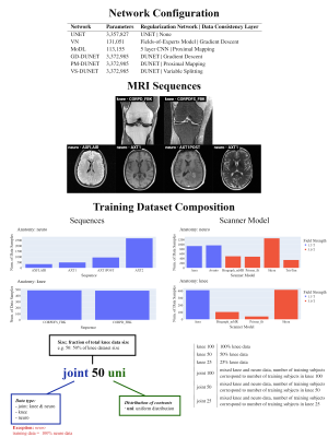 |
12 | Understanding domain shift in learned MRI reconstruction: A quantitative analysis on fastMRI knee and neuro sequences
Shizhe He1,2, Veronika Anne Zimmer1, Daniel Rueckert1,3, and Kerstin Hammernik1,3
1Lab for Artificial Intelligence in Healthcare and Medicine, Technical University of Munich, Munich, Germany, 2Otto-von-Taube-Gymnasium, Gauting, Germany, 3Department of Computing, Imperial College London, London, United Kingdom
In this work, we investigate the problem of domain shift in the context of state-of-the-art MRI reconstruction networks with respect to variations in training data. We provide visualization tools and support our findings with statistical analysis for the networks evaluated on fastMRI knee and neuro data. We observe that the signal-to-noise ratio of the examined sequences plays an essential role, and we statistically prove the hypothesis that the type/amount of training data is less important for low acceleration factors. Finally, we provide a visualization tool facilitating the examination of the networks’ performance on each individual subject of the fastMRI data.
|
||
3910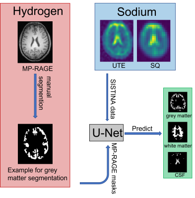 |
13 | A U-Net based Approach to the Prediction of Regions-of-Interest for Metabolic Sodium Imaging
Wieland A. Worthoff1, Yannic Sommer1, Zaheer Abbas1, and N. Jon Shah1,2,3,4
1Institut of Neuroscience and Medicine - 4, Forschungszentrum Jülich GmbH, Jülich, Germany, 2Institut of Neuroscience and Medicine - 11, Forschungszentrum Jülich GmbH, Jülich, Germany, 3Department of Neurology, RWTH Aachen University, Aachen, Germany, 4JARA-BRAIN - Translational Medicine, Jülich-Aachen Research Alliance, Aachen, Germany
Sodium MRI yields metabolic information about the brain and might indicate existing or emerging pathologies. Often this information is to be determined in a certain region-of-interest (ROI). These ROIs can be, for example, all grey or white matter regions, or more specific sub-regions thereof and it is important to predict these ROIs without bias. Here, an approach to obtain well segmented ROIs is presented based on a deep neural network architecture.
|
||
3911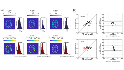 |
14 | Oxygen Transport Modelling for Mapping Brain Oxygen Extraction Fraction with Single Gas Calibrated fMRI
Antonio Maria Chiarelli1, Michael Germuska2, Hannah Chandler2, Rachael Stickland3, Eleonora Patitucci2, Emma Biondetti1, Daniele Mascali1, Neeraj Saxena2, Sharmila Khot2, Jessica Steventon2, Catherine Foster4, Ana E Rodríguez-Soto5, Erin Englund6, Kevin Murphy2, Valentina Tomassini1,2,7, Felix W Wehrli8, and Richard Wise1,2
1Department of Neuroscience, Imaging and Clinical Sciences, University G. D'Annunzio of Chieti Pescara, Chieti Scalo, Italy, 2Cardiff University Brain Research Imaging Centre (CUBRIC), Cardiff, United Kingdom, 3Physical Therapy and Human Movement Sciences, Feinberg School of Medicine, Northwestern University, Chicago, IL, United States, 4Cardiff University, Cardiff, United Kingdom, 5University of California, San Diego, La Jolla, CA, United States, 6University of Colorado, Colorado, CO, United States, 7MS Centre, Dept of Clinical Neurology, SS. Annunziata University Hospital, Chieti, Italy, 8Department of Radiology, University of Pennsylvania, Philadelphia, PA, United States
Dual-calibrated functional MRI (dc-fMRI) can map brain oxygen extraction fraction (OEF) by measuring BOLD-ASL signal changes during arterial O2 and CO2 modulations. Two modulations are required to decouple OEF and the deoxyhemoglobin-sensitive blood volume. Here, we propose a single gas calibrated approach that integrates a model of oxygen transport that links blood volume and CBF to OEF. Simulations demonstrated the method’s viability. In-vivo application with hypercapnia provided estimates of grey matter OEF in agreement with dc-fMRI and with whole-brain OEF derived from signal phase measures in the superior sagittal sinus. The simplified calibrated fMRI method holds promise for clinical application.
|
||
3912 |
15 | Real-time survey base estimation of gestational age to guide a fetal MRI scan
Maneesha Singh1, Sara Neves Silva1, Alena Uus1, Irina Grigorescu1, Mary Rutherford1, Jo Hajnal1, and Jana Hutter1
1Centre for the developing Brain, King's College London, London, United Kingdom
Fetal MRI is an excellent tool for estimating the fetal gestational age using the head circumference measurements. This study measures the biometry information using fast 30 sec survey scans for the measurement of fetal head circumference using 3D UNet, Bi-parietal diameter, Frontal-occipital diameter for the estimation of the fetal gestational age in the second half of gestation. The results show high accuracy in gestational age measurements which correlates well with the clinical data.
|
||
3913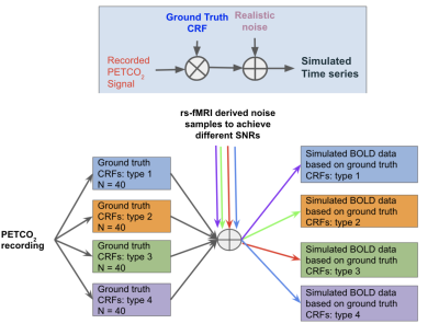 |
16 | A comparison of model-based deconvolution methods for estimating the carbon-dioxide response function in resting-state fMRI Video Not Available
Seyedmohammad Shams1, Prokopis C Prokopiou2, Georgios Mitsis3, and J. Jean Chen4
1Rotman Research Institute, Baycrest, Toronto, ON, Canada, 2Gordon Center for Medical Imaging, Massachusetts General Hospital, Harvard Medical School, Boston, MA, United States, 3McGill University, Montreal, ON, Canada, 4Rotman Research Institute, Baycrest Health Sciences, Toronto, ON, Canada
The resting-state fMRI response to carbon-dioxide (CO2) fluctuations is useful for denoising and for physiological mapping. In this work, we compare model-based deconvolution methods for estimating this response function (CRF) in a wide range of signal-to-noise conditions and with various assumed ground-truth CRF shapes. We also propose an improved method using canonical correlation analysis to identify the optimal CRF adaptively. The best choice of method depends on the desired CRF parameter, such as timing (dynamics) or area (static cerebrovascular reactivity). The inverse-Logit and adaptive CCA methods provided the highest accuracy and robustness.
|
||
 Back to Meeting Home
 Back to the Program-at-a-Glance
The International Society for Magnetic Resonance in Medicine is accredited by the Accreditation Council for Continuing Medical Education to provide continuing medical education for physicians.
 Back to Meeting Home
 Back to the Program-at-a-Glance
 View the Presentation