Online Gather.town Pitches
Processing & Analysis V
Joint Annual Meeting ISMRM-ESMRMB & ISMRT 31st Annual Meeting • 07-12 May 2022 • London, UK

Online Gather.town Pitches
Processing & Analysis V
| Booth # | ||||
|---|---|---|---|---|
4483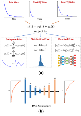 |
1 | Improved Myelin Water Fraction Estimation Integrating Learned Probabilistic Subspaces and Low-Dimensional Manifolds
Yudu Li1,2, Rong Guo1,2, Yibo Zhao1,2, Yao Li3, and Zhi-Pei Liang1,2
1Department of Electrical and Computer Engineering, University of Illinois at Urbana-Champaign, Urbana, IL, United States, 2Beckman Institute for Advanced Science and Technology, University of Illinois at Urbana-Champaign, Urbana, IL, United States, 3School of Biomedical Engineering, Shanghai Jiao Tong University, Shanghai, China
Multi-echo gradient-echo (mGRE)-based myelin water fraction (MWF) mapping is increasingly used for studying myelin integrity. The basic multi-exponential fitting method often suffers from severe ill-conditionedness of the exponential model. To address this problem, a number of more advanced estimation methods have been proposed, incorporating a priori constraints and machine learning. This work presents a new learning-based method to further improve MWF estimation. The proposed method represents different water components as low-rank subspaces through which both pre-learned subspace and manifold structures are synergistically integrated. Both simulation and experimental results demonstrate significantly improved performance over existing MWF estimation methods.
|
||
4484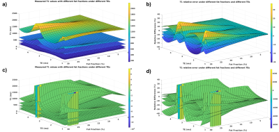 |
2 | A simulation study of the negative impact of fat on the accuracy of T1 measurements
Zhitao Li1, Shreyas S Vasanawala1, and Li Feng2
1Department of Radiology, Stanford University, Palo Alto, CA, United States, 2Biomedical Engineering and Imaging Institute and Department of Radiology, Icahn School of Medicine at Mount Sinai, New York, NY, United States
A simulation study examining the effects of fat in the accurate assessment of T1. The study looks at both the variable flip angle and Look-Locker methods, and different fat fractions and different T1 values are used in the simulation. The results indicated that while both VFA and Look-Locker methods’ accuracy are affected by fat, VFA method’s biases increases more rapidly as fat fraction goes up, and the bias for larger spin species with larger T1s are more severely impacted by the presence of fat.
|
||
4485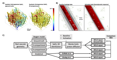 |
3 | Comparison of analytical BOLD-fMRI models against Two-Photon Microscopy and Monte Carlo simulations
Jordan Charest1, Pierre-Olivier Schwarz1, Mathieu Walsh1, Élie Genois2, Louis Gagnon3, and Michèle Desjardins1
1Physics, Engineering Physics and Optics, Laval University, Quebec City, QC, Canada, 2Physics, University of Sherbrooke, Sherbrooke, QC, Canada, 3Radiology and Nuclear Medicine, Laval University, Quebec City, QC, Canada
BOLD fMRI arises from a complex physiological and physical cascade of events taking place at the level of the cortical microvasculature which constitutes a medium with complex geometry. Several analytical models of the BOLD contrast have been developed but these have not been compared directly against detailed bottom-up modeling methods. Using a recent 3D modeling method based on Two-Photon Microscopy and Monte Carlo simulations, we tested the accuracy of two analytical models to predict the amplitude of the BOLD response at 1.5T, 3T, 4.7T and 7T for both gradient echo and spin echo acquisition protocols.
|
||
4486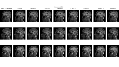 |
4 | Adaptive-CS-Net to Accelerate 3D T1-weighted Imaging: Brain Volume Measures for clinical use
Sandeep Ganji1,2, Brian Johnson3,4, John Penatzer3, and Johannes M. Peeters5
1MR R&D, Philips Healthcare, Rochestr, MN, United States, 2Mayo Clinic, Rochester, MN, United States, 3Philips Healthcare, Gainesville, FL, United States, 4University of Texas Southwestern Medical Center, Dallas, TX, United States, 5Philips Healthcare, Eindhoven, Netherlands
Despite long scan times, 3D T1-weighted (T1w) MRI is routinely used for MRI studies to provide high resolution structural and volumetric information of brain. Volumetric analysis can serve as a biomarker and aid in clinical diagnosis of certain diseases such as Alzheimer’s, mild cognitive impairment, and atrophy, however, the standardized 3D T1-weighted scans suffer from long acquisition times well over 5 minutes. We compared the results of volumetric brain analysis for 3D T1w images acquired over a range of compressed SENSE acceleration factors with and without Adaptive-CS-Net reconstruction against a standard clinical 3D T1w MRI protocol.
|
||
4487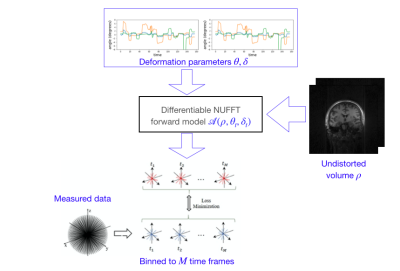 |
5 | A K-space based learning approach to head motion correction for 4D (3D+time) radial sequences
Parker David Evans1, Curtis A Corum2, John H Keller3, Vincent Magnotta4, and Mathews Jacob5
1AMCS, University of Iowa, Iowa City, IA, United States, 2Champaign Imaging LLC, Minneapolis, MN, MN, United States, 3Radiology, University of Iowa, Iowa City, IA, United States, 4Radiology, University of Iowa, Iowa city, IA, United States, 5ECE, University of Iowa, Iowa City, IA, United States A novel k-space learning based framework is introduced to compensate for bulk motion artifacts in UTE/ZTE radial acquisition schemes. The motion during the scan is modeled as rigid, and is parametrized by time varying translation and rotation parameters. The time varying parameters are used to define a forward model, which transforms the undistorted image to distorted k-t space data. The error between the measured and computed k-t space data is used to optimize the image and the deformation parameters using ADAM optimization. A multi-scale approach is used to minimize the computational complexity.
|
||
4488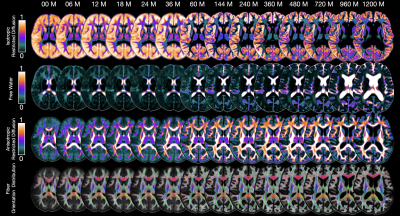 |
6 | Intravoxel Architecture Atlases (IAA) Across the Human Lifespan
Ye Wu1, Sahar Ahmad1, and Pew-Thian Yap1
1Department of Radiology and Biomedical Research Imaging Center (BRIC), University of North Carolina at Chapel Hill, Chapel Hill, NC, United States
Human brain atlases integrate multifaceted features of the brain in common coordinate spaces, allowing systematic investigation of brain development and maturation. Here, we introduce a set of intravoxel architecture atlases (IAA) covering changes of tissue microstructure from birth to 100 years of age.
|
||
4489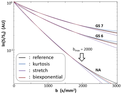 |
7 | Optimizing Prostate DWI Acquisitions for non-Gaussian models using an Activity MRI [aMRI] library
Xin Li1, Ryan P Kopp2,3, Eric M Baker1, Brendan Moloney1, Charles S Springer1, Mark G Gartotto2,3, and Fergus V Coakley4
1Advanced Imaging Research Center, Oregon Health & Science University, Portland, OR, United States, 2Portland VA Medical Center, Portland, OR, United States, 3Urology, Oregon Health & Science University, Portland, OR, United States, 4Diagnostic Radiology, Oregon Health & Science University, Portland, OR, United States Characterizing the non-Gaussian signature of the prostate DWI b-space decay often employs the kurtosis-, stretched-, and bi-exponential models. However, the optimal acquisition strategy in terms of maximum b value sampled and the number of b-values needed remains a research topic. Using the best-matched curves from a DWI library created with Monte Carlo random walk simulations of contracted Voronoi-cell ensembles, this work shows that, for prostate DWI, a maximum b-value near 2000 s/mm2 is optimal and the number of b-values is not as crucial as long as it is larger than the number of model parameters. |
||
4490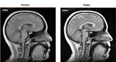 |
8 | 3D Dynamic Magnetic Resonance Imaging: A Tool for Describing Velopharyngeal Function
Imani R. Gilbert1, Fangxu Xing2, Riwei Jin3, Ryan K. Shosted3, Jonghye Woo2, Bradley P. Sutton3, and Jamie L. Perry1
1East Carolina University, Greenville, NC, United States, 2Massachusetts General Hospital, Boston, MA, United States, 3University of Illinois Urbana-Champaign, Champaign,, IL, United States
Direct visualization of velopharyngeal structures and musculature during speech is best attained using 3D dynamic magnetic resonance imaging. Through innovative MR imaging and atlasing methods, we successfully describe velopharyngeal contours as represented on MR statistical atlases and individual subject images. Manual linear measurements of velum configurations during /p/ across two different speech stimuli reveal slight velar differences, reflecting major influences of neighboring speech sounds on velar movements.
|
||
4491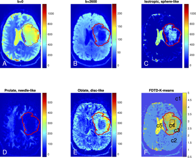 |
9 | Measurement of model-free diffusion tensor distribution using Connectome MRI and application in an anaplastic astrocytoma
Yiqiao Song1,2, Ina Ly3, Qiuyun Fan1,4, Aapo Nummenmaa1, Maria Martinez-Lage5, William T. Curry6, Jorg Dietrich3, Deborah A Forst3, Bruce R Rosen1, Susie Y Huang1, and Elizabeth R Gerstner3
1Athinoula A. Martinos Center for Biomedical Imaging, Department of Radiology, MGH, Charlestown, MA, United States, 2John A Paulson School of Engineering and Applied Sciences, Harvard Univ, Cambridge, MA, United States, 3Stephen E. and Catherine Pappas Center for Neuro-Oncology, MGH, Boston, MA, United States, 4Department of Biomedical Engineering, College of Precision Instruments and Optoelectronics Engineering, Tianjin University, Tianjin, China, 5Department of Pathology, MGH, Boston, MA, United States, 6Department of Neurosurgery, MGH, Boston, MA, United States The availability of the Connectome MRI scanner with gradient strengths up to 300 mT/m enables resolution of a wider range of diffusion coefficients. This is particularly important for capturing the complexity and heterogeneity of biological tissues. Here, we outline a framework for analyzing diffusion MRI data to obtain a model-free diffusion tensor distribution (FDTD) with a wide variety of diffusion tensor structures and test it on a healthy subject. We apply this method and use K-means clustering to identify features in FDTD to visualize and characterize tissue heterogeneity in a subject with a World Health Organization grade 3 anaplastic astrocytoma. |
||
4492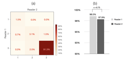 |
10 | Deep Learning-enabled Prostate Segmentation: Large Cohort Evaluation with Inter-Reader Variability Analysis
Yongkai Liu1, Miao Qi1,2, Chuthaporn Surawech1,3, Haoxin Zheng1, Dan Nguyen4, Guang Yang5, Steven Raman1, and Kyunghyun Sung1
1Department of Radiological Sciences, University of California, Los Angeles, Los Angeles, CA, United States, 2Department of Radiology, The First Affiliated Hospital of China Medical University, Shenyang, China, 3Department of Radiology, King Chulalongkorn Memorial Hospital, Bangkok, Thailand, 4Department of Radiation Oncology, UT Southwestern Medical Center, Los Angeles, CA, United States, 5National Heart and Lung Institute, Imperial College London, London, United Kingdom
Whole-prostate gland (WPG) segmentation plays a significant role in prostate volume measurement, treatment, and biopsy planning. This study evaluated a previously developed automatic WPG segmentation, deep attentive neural network (DANN), on a large, continuous patient cohort to test its feasibility in a clinical setting.
|
||
4493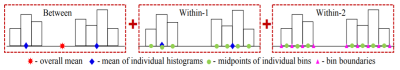 |
11 | Incorporating data heterogeneity for improved regression models: application to stroke
Anuja Sharma1 and Edward DiBella1
1Dept. of Radiology and Imaging Sciences, University of Utah, Salt Lake City, UT, United States
Symbolic data regression provides a systematic way to bring together heterogenous data from imaging and non-imaging sources in the form of histograms, intervals and scalar-valued observations. Classic multiple linear regression is adapted to mixed symbolic features and applied to data from diffusion spectrum images and clinical measurements for stroke recovery prediction. By utilizing the implicit variability within observations and natural grouping within features, the amount of information available to the modelling process is increased. This provides increased stability for model parameters over traditional regression and is especially beneficial with low sample sizes.
|
||
4494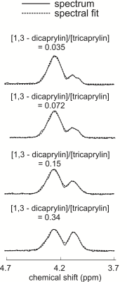 |
12 | Determining Relative Diglyceride to Triglyceride Levels with STEAM MRS at 3 T
Elliot Saive1, Logan Fairgrieve-Park1, and Atiyah Yahya1,2
1Department of Oncology, University of Alberta, Edmonton, AB, Canada, 2Department of Medical Physics, Cross Cancer Institute, Edmonton, AB, Canada
Diglyceride levels have been found elevated with some disease. Previously, STEAM (mixing time=TM=20ms) with an echo time (TE) of 100ms was shown to resolve triglyceride glycerol resonances from that of water of 3T while yielding adequate glycerol signal. The purpose of this work is to determine if STEAM with a TE of 100ms facilitates relative quantification of diglyceride/triglyceride levels at 3 T. Spectra were measured from phantoms containing 1,3-dicaprylin/tricaprylin with varying weight/weight contents of 2.5%/97.5%, 5%/95%, 10%/90% and 20%/80%. Concentration ratios of 1,3-dicaprylin/tricaprylin estimated from STEAM (TM=20ms, TE=100ms) resulted in a linear correlation with expected concentration ratios (R2 > 0.99).
|
||
4495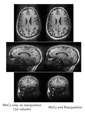 |
13 | Prospective Motion Correction and Data Re-acquisition at 7 Tesla using E/M Trackers
W. Scott Hoge1,2, Onur Afacan2,3, Simon K. Warfield2,3, Amir Roth4, and Erez Nevo4
1Radiology, Brigham and Women's Hospital, Boston, MA, United States, 2Dept of Radiology, Harvard Medical School, Boston, MA, United States, 3Computational Radiology Laboratory, Boston Children's Hospital, Boston, MA, United States, 4Robin Medical, Inc., Baltimore, MD, United States
We evaluated the utility of electro-magnetic (E/M) based tracking to correct for motion at 7 Tesla. A 3D Turbo-FLASH sequence was modified to include prospective field-of-view (FOV) steering and track motion events. Custom software provided FOV updates to the TFL sequence from E/M sensor location measurements. Motion events recorded by the software indicated time-periods when acquired data was likely corrupted due to motion. Logic within the sequence re-acquired motion-corrupted k-space data segments to yield high-quality high-spatial-resolution images. Results demonstrate the viability of E/M tracking at 7T.
|
||
4496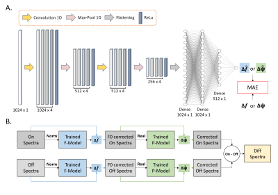 |
14 | Magnetic Resonance Spectroscopy Frequency and Phase Correction Using Convolutional Neural Networks
David Ma1, Hortense Le1, Yuming Ye1, Andrew Laine1, Jeffrey Lieberman2, Douglas Rothman3, Scott Small2,4,5, and Jia Guo2,6
1Department of Biomedical Engineering, Columbia University, New York, NY, United States, 2Department of Psychiatry, Columbia University, New York, NY, United States, 3Radiology and Biomedical Imaging of Biomedical Engineering, Yale University, New Haven, CT, United States, 4Department of Neurology, Columbia University, New York, NY, United States, 5Taub Institute Research on Alzheimer's Disease and the Aging Brain, Columbia University, New York, NY, United States, 6Mortimer B. Zuckerman Mind Brain Behavior Institute, Columbia University, New York, NY, United States
Frequency and Phase Correction (FPC) is an essential technique to resolve frequency and phase shifts that arise in Magnetic Resonance Spectroscopy (MRS). As of today, a deep learning method using multilayer perceptrons has been developed to correct these shifts. However, a more robust network such as convolutional neural networks (CNN) can be considered as this approach more accurately obtains spatial information and extract key features of the given data. In this study, we aim to investigate the feasibility and utility of CNNs for FPC of single voxel MEGA-PRESS MRS simulated and in vivo data.
|
||
 Back to Meeting Home
 Back to the Program-at-a-Glance
The International Society for Magnetic Resonance in Medicine is accredited by the Accreditation Council for Continuing Medical Education to provide continuing medical education for physicians.
 Back to Meeting Home
 Back to the Program-at-a-Glance
 View the Presentation