Online Gather.town Pitches
Processing & Analysis III
Joint Annual Meeting ISMRM-ESMRMB & ISMRT 31st Annual Meeting • 07-12 May 2022 • London, UK

Online Gather.town Pitches
Processing & Analysis III
| Booth # | ||||
|---|---|---|---|---|
4969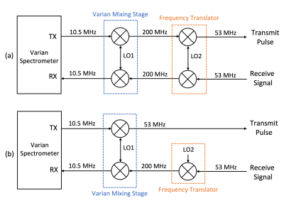 |
1 | Phase Correction Approach for Receive-Only Frequency Translation Studies
Jue Hou1, Courtney Bauer1, Mary McDougall1,2, and Steve Wright1,2
1Electrical and Computer Engineering, Texas A&M University, College Station, TX, United States, 2Biomedical Engineering, Texas A&M University, College Station, TX, United States
23Na and 31P spectroscopy are powerful tools in assessing muscles in Duchenne Muscular Dystrophy studies. Frequency translation has been previously introduced as a means to facilitate multichannel studies on systems equipped with only 1H receiver arrays. Many approaches to frequency translation require mixing on both transmit and receive, which bypasses the need for phase correction. Presented here is an approach to receive-only translation of acquired data to the 1H frequency, that allows for post-processing phase correction using signals coupled from the host system’s and translator’s local oscillators.
|
||
4970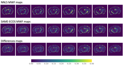 |
2 | Application of SAME-ECOS to 7T gradient-echo based myelin water imaging: a comparison of model-free and model-based approaches
Hanwen Liu1,2, Vladimir Grouza1,2, Marius Tuznik1,2, and David Rudko1,2,3
1McConnell Brain Imaging Centre, Montreal Neurological Institute and Hospital, Montreal, QC, Canada, 2Department of Neurology and Neurosurgery, McGill University, Montreal, QC, Canada, 3Department of Biomedical Engineering, McGill University, Montreal, QC, Canada Multi-echo gradient echo (GRE) based, myelin water imaging (MWI) is subject to data quality constraints related to signal to noise ratio (SNR), B0 field inhomogeneities and gradient imperfections. These constraints pose challenges for conventional model-free, non-negative least squares (NNLS) fitting. As an alternative, a three-pool parametric model can be applied, with nonlinear least squares(NLLS) fitting to process MWI data. The three-pool model confers stability, but has the disadvantage of making prior assumptions. This study investigated the feasibility of using a novel, model-free approach called spectrum analysis for multiple exponentials via experimental condition oriented simulation (SAME-ECOS) for the GRE MWI analysis. |
||
4971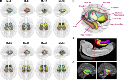 |
3 | Subcortical Parcellation of the Human Brain via Automated Voxel Annotation with Fiber Clusters
Ye Wu1, Sahar Ahmad1, and Pew-Thian Yap1
1Department of Radiology and Biomedical Research Imaging Center (BRIC), University of North Carolina at Chapel Hill, Chapel Hill, NC, United States
Connectivity-based parcellation of subcortical structures typically relies on the pre-parcellation of the cortex. Here, we propose an unsupervised data-driven approach for subcortical parcellation based on diffusion tractography without relying on the annotation of cortical regions or fiber tracts.
|
||
4972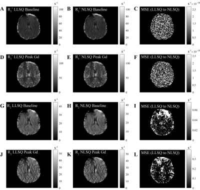 |
4 | Rapid Parameter Estimation for Combined Spin and Gradient Echo (SAGE) Imaging
Nicholas John Sisco1, Elizabeth G Keeling1, Aimee Borazanci1, Richard D Dortch1, and Ashley M Stokes1
1Division of Neuroimaging Research, Barrow Neurological Institute, Phoenix, AZ, United States
In this study, we propose a generalized linear solution to estimate dynamic relaxation parameters R2* and R2 from combined spin- and gradient-echo (SAGE) MRI. We compared linear least squares (LLSQ) to nonlinear least squares (NLSQ) using Monte-Carlo simulated data and in vivo whole-brain data with varying signal-to-noise ratios (SNR). We show that using the LLSQ is both computationally more efficient and retains NLSQ precision, producing nearly the same estimates of R2* and R2 in a fraction of the time. This approach is widely extendable to other multi-echo, multi-contrast MRI methods, with applications including both perfusion MRI and functional MRI.
|
||
4973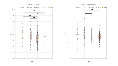 |
5 | Hippocampus Shape Characterization with 3D Zernike Transformation in Clinical Alzheimer’s Disease Progression
David C Zhu1, Chih-Ying Gwo2, An-Wen Deng2, Norman Scheel1, Mari A Dowling1, and Rong Zhang3
1Michigan State University, East Lansing, MI, United States, 2Chien Hsin University of Science and Technology, Taoyuan, Taiwan, 3University of Texas Southwestern Medical Center, Dallas, TX, United States Alzheimer’s disease (AD) is a neurodegenerative disease and the most common cause of dementia among older adults. We implemented 3D Zernike transformation to characterize the shape changes of hippocampus in 428 older subjects with high-quality T1-weighted volumetric brain scans. The hippocampus shape features characterized with 3D Zernike transformation, in complement to volume measures, may serve as a novel imaging marker to monitor clinical AD progression. |
||
4974 |
6 | Estimating Arterial Transit Time (ATT) From ASL MRI Acquired at A Single Post-Labeling-Delay Time
Aldo Camargo1 and Ze Wang2
1UMB, Baltimore, MD, United States, 2Radiology, University of Maryland of Baltimore, Baltimore, MD, United States
We proposed a novel method to compute arterial transit time from ASL MRI acquired at a single post-labeling-delay time. Using the method, we found significant difference between NC and AD subjects.
|
||
4975 |
7 | COMEDI: A Toolkit for Lifespan Computational Diffusion MRI
Ye Wu1, Sahar Ahmad1, Khoi Minh Huynh1, Tiantian Xu1, and Pew-Thian Yap1
1Department of Radiology and Biomedical Research Imaging Center (BRIC), University of North Carolina at Chapel Hill, Chapel Hill, NC, United States
Processing and analyzing diffusion MRI (dMRI) data is important for uncovering the neural underpinnings of white matter (WM) development and degeneration across the human lifespan. Here, we introduce a robust and integrative toolkit, called Computational Medical Imaging (COMEDI), for processing, analyzing, and visualizing lifespan dMRI data.
|
||
4976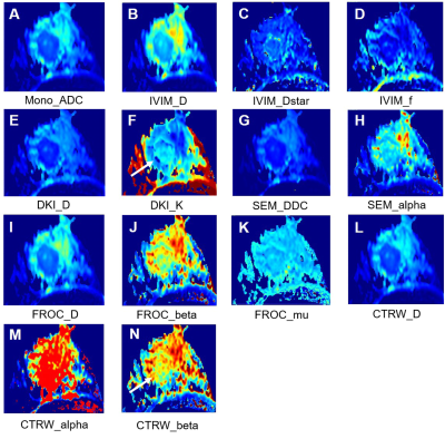 |
8 | Evaluate Values of First-order Features based on Advanced DWI Models in Predicting the HER-2 Expression in Breast Invasive Ductal Cancer
Siyao Du1, Mengfan Wang1, Shasha Liu1, Xiaoqian Bian1, Xinyue Chen1, Liangcun Guo1, Guoliang Huang1, Ruimeng Zhao1, Can Peng1, Wenhong Jiang1, Qinglei Shi2, Xu Yan2, Guang Yang3, and Lina Zhang1
1Department of Radiology, The First Affiliated Hospital of China Medical University, Shenyang, China, 2MR Scientific Marketing, Siemens Healthineers Ltd., Beijing, China, 3Shanghai Key Laboratory of Magnetic Resonance, East China Normal University, Shanghai, China
In this study, we compared first-order features of six DWI models in predicting the HER-2 expression in breast invasive ductal carcinoma, including conventional mono-exponential model derived parameter ADC, IVIM derived parameters (D, Dstar, f), DKI derived parameters (D, K), SEM derived parameters (DDC, α), FROC derived parameters (D, β, mu), CTRW derived parameters (D, α, β), and a comprehensive diagnostic logistic regression model was established to improve the diagnostic performance. We found that CTRM and IVIM derived first-order features demonstrated the powerful performance, and their combination reached the highest performance. The finding has a great value in breast cancer treatment.
|
||
4977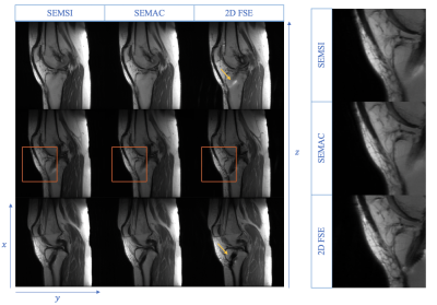 |
9 | Spectrally encoded multi-spectral imaging (SEMSI) for off-resonance correction near metal with multi-spin echo and parallel imaging
Xuetong Zhou1,2, Philip K. Lee2,3, Daehyun Yoon2, and Brian A. Hargreaves1,2,3
1Bioengineering, Stanford University, Stanford, CA, United States, 2Radiology, Stanford University, Stanford, CA, United States, 3Electrical Engineering, Stanford University, Stanford, CA, United States
Metal implants induce severe B0 field inhomogeneities, causing severe distortion near the implant. In this work, we combined multi-echo spin echo and parallel imaging to an approach that uses echo shifting to encode spectral components and resolve the metal-induced artifacts. Phantom and in vivo experiments were conducted and indicated that the proposed method provides comparable artifact reduction and image quality to existing multispectral imaging approaches in comparable scan time.
|
||
4978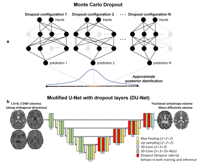 |
10 | Quantifying the uncertainty of neural networks using Monte Carlo dropout for safer and more accurate deep learning based quantitative MRI
Mehmet Yigit Avci1,2, Ziyu Li3, Qiuyun Fan2,4,5, Susie Huang2,4, Berkin Bilgic2,4, and Qiyuan Tian2,4
1Electrical and Electronics Engineering, Bogazici University, Istanbul, Turkey, 2Athinoula A. Martinos Center for Biomedical Imaging, Charlestown, MA, United States, 3Nuffield Department of Clinical Neurosciences, University of Oxford, Oxford, United Kingdom, 4Department of Radiology, Harvard Medical School, Boston, MA, United States, 5Department of Biomedical Engineering,College of Precision Instruments and Optoelectronics Engineering, Tianjin University, Tianjin, China Neural networks reduce the data requirement for deep learning-based quantitative MRI, nonetheless their uncertainty/confidence has rarely been characterized. We implemented Monte Carlo dropout, a Bayesian approximation of Gaussian process, using U-Net that include dropout layers (active during training and inference) to address this. The uncertainty was calculated as the variance of predictions from 100 different dropout configurations. The estimates were calculated as the average of predictions. The proposed method also achieved higher accuracy in estimating FA and MD from only 3 diffusion-weighted images compared to standard U-Net, which was readily usable for other MRI applications (reconstruction, super-resolution, denoising, segmentation, classification). |
||
4979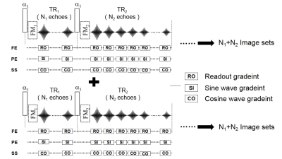 |
11 | Highly Accelerated Multiple Parametric MR Imaging with Wave-CAIPI and MULTIPLEX
Haifeng Wang1, Yongquan Ye2, Congcong Liu1, Jingyuan Lv2, Zhilang Qiu1, Xin Liu1, Jian Xu2, Dong Liang1, and Hairong Zheng1
1Shenzhen Institute of Advanced Technology, Chinese Academy of Sciences, Shenzhen, China, 2UIH America, Inc., Houston, TX, United States
A novel single-scan hybrid 3D imaging method using Wave-CAIPI and MULTIPLEX technologies, named as WAMP, is proposed for 3D high-resolution rapid imaging. One single scan of the proposed rapid imaging method not only generates simultaneous B1t and T1 maps, but also qualitative images of T1W, PDW, T2*W, augmented T1W (aT1W), SWI and MRA, as well as parametric maps of T2* (R2*), PD and QSM. The in vivo experiments have showed that the proposed method could rapidly achieve high image quality and high quantification accuracy at the high acceleration factors in clinical applications.
|
||
4980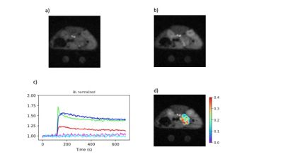 |
12 | Abdominal DCE-MRI in mice with stack of stars sampling and KWIC image reconstruction
Stephen Pickup1, Miguel Romanello Giroud Joaquim1, Hoon Choi1, Mamta Gupta1, Cynthia Clendenin1, Hee Kwon Song1, and Rong Zhou1
1Department of Radiology, University of Pennsylvania, Philadelphia, PA, United States
Dynamic contrast enhanced MRI data in the abdomens of small animal models is often corrupted due to the effects respiratory and peristaltic motion. Here a DCE protocol that employs stack of stars sampling throughout was implemented and was shown to be robust with respect to motion artifacts. The sampling scheme also facilitates image reconstruction methods that employ view sharing, notably KWIC, yielding images with high temporal and spatial resolution. The protocol was demonstrated in an orthotopic murine model of pancreatic cancer. The resulting data were analyzed using the reference tissue method and provided high quality Ktrans and ve parameter maps.
|
||
4981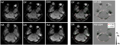 |
13 | Signal dropout reduction in PSF mapping based reverse gradient fMRI with reversed partial-Fourier acquisition
Myung-Ho In1, Daehun Kang1, Hang Joon Jo2, Uten Yarach3, Nolan K Meyer1,4, Joshua D Trzasko1, John Huston III1, Matt A Bernstein1, and Yunhong Shu1
1Department of Radiology, Mayo Clinic, Rochester, MN, United States, 2Department of Physiology, College of Medicine, Hanyang University, Seoul, Korea, Republic of, 3Department of Radiologic Technology, Faculty of Associated Medical Sciences, Chiang Mai University, Chiang Mai, Thailand, 4Mayo Clinic Graduate School of Biomedical Sciences, Mayo Clinic, Rochester, MN, United States
An interleaved, reverse gradient fMRI (RG-fMRI) with a point-spread-function mapping-based approach was recently proposed to minimize geometric distortion and signal dropout in echo-planar-imaging (EPI) by acquiring a pair of EPIs with the opposite (forward and reverse) phase-encoding gradient polarity and combining them after distortion correction. This study showed that the effective echo-shift map can be reliably obtained from PSF mapping to predict local signal dropouts. The predicted echo-shifting can provide guidance for protocol optimization for RG-fMRI using partial Fourier (PF) acquisition.
|
||
4982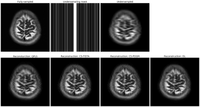 |
14 | MIRTorch: A PyTorch-powered Differentiable Toolbox for Fast Image Reconstruction and Scan Protocol Optimization
Guanhua Wang1, Neel Shah2, Keyue Zhu2, Douglas C. Noll1, and Jeffrey A. Fessler2
1Biomedical Engineering, University of Michigan, Ann Arbor, MI, United States, 2EECS, University of Michigan, Ann Arbor, MI, United States
MIRTorch (Michigan Image Reconstruction Toolbox for PyTorch) is an image reconstruction toolbox implemented with native Python/PyTorch. It provides fast iterative and data-driven image reconstruction on both CPUs and GPUs. Researchers can rapidly develop new model-based and learning-based methods (i.e., unrolled neural networks) with its convenient abstraction layers. With the full support of auto-differentiation, one may optimize imaging protocols, such as sampling patterns and image reconstruction parameters with gradient-based methods.
|
||
 Back to Meeting Home
 Back to the Program-at-a-Glance
The International Society for Magnetic Resonance in Medicine is accredited by the Accreditation Council for Continuing Medical Education to provide continuing medical education for physicians.
 Back to Meeting Home
 Back to the Program-at-a-Glance
 View the Presentation