Online Gather.town Pitches
Novel Image Reconstruction Techniques I
Joint Annual Meeting ISMRM-ESMRMB & ISMRT 31st Annual Meeting • 07-12 May 2022 • London, UK

Online Gather.town Pitches
Novel Image Reconstruction Techniques I
| Booth # | ||||
|---|---|---|---|---|
| 3280 | 1 | Automatic parameter selection for quantitative susceptibility mapping (QSM) with regard to Shearlet/TGV-regularization Video Permission Withheld
Janis Stiegeler1,2 and Sina Straub1
1Medical Physics in Radiology, German Cancer Research Center (DKFZ), Heidelberg, Germany, 2Department of Physics and Astronomy, University of Heidelberg, Heidelberg, Germany In this work noise is assumed to be a random vector and the method of unbiased predictive risk estimator (UPRE) is used to select suitable data/regularization parameters to solve the local phase to susceptibility deconvolution problem of quantitative susceptibility mapping (QSM). The proposed algorithm is tested on the simulated multi-echo data provided at the 2019 QSM Reconstruction Challenge. This work is a further development of the algorithm presented at the ISMRM Meeting 2021 and its purpose is to show that the method of UPRE can be applied advantageously to a shearlet /TGV based susceptibility reconstruction. |
||
3281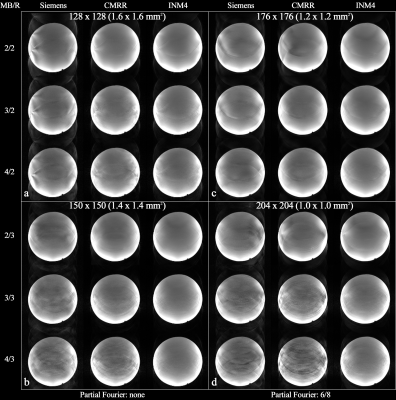 |
2 | An Evaluation of High-Resolution EPI sequences and Enhanced Image Reconstruction for fMRI at 7T: Siemens-EPI, CMRR-EPI and INM4-EPI
Seong Dae Yun1 and N. Jon Shah1,2,3,4
1Institute of Neuroscience and Medicine 4, INM-4, Forschungszentrum Juelich, Juelich, Germany, 2Institute of Neuroscience and Medicine 11, INM-11, JARA, Forschungszentrum Juelich, Juelich, Germany, 3JARA - BRAIN - Translational Medicine, Aachen, Germany, 4Department of Neurology, RWTH Aachen University, Aachen, Germany
EPI is of great importance in the MR community due to its wide applicability in various dynamic MR applications. Recent advances in EPI techniques extend its use for the depiction of neuronal activation with a cortical depth-dependence. Therefore, enhanced performance with reliable image quality is highly desirable in EPI. This work presents an in-house designed EPI sequence and its corresponding image reconstruction software at 7T. Results show that our EPI sequence outperforms two widely used sequences: Siemens-EPI and CMRR-EPI. In addition, our image reconstruction routine significantly improves the reconstructed image quality of Siemens and CMRR data.
|
||
3282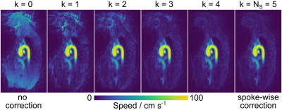 |
3 | Radial Maxwell correction for real-time phase-contrast MRI using spoke clustering
Jost M. Kollmeier1 and Jens Frahm1
1Biomed NMR, Max-Planck-Institut für biophysikalische Chemie, Göttingen, Germany
In undersampled radial phase-contrast imaging, Maxwell terms can vary for individual radial projections (spokes). Integrated into a model-based image reconstruction, a computationally expensive spoke-by-spoke correction has been proposed, for which the reconstruction times scale with the number of spokes per frame. To make this approach practical for large numbers of spokes, this work proposes to use k-means clustering of Maxwell terms in the spoke dimension and thereby reduce the computational costs of the model-based image reconstruction for phase-contrast MRI with radial Maxwell correction.
|
||
3283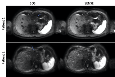 |
4 | SENSE-like reconstruction of multi-average body DWI to remove motion-induced signal loss: application in liver DWI
Anh Tu Van1, Sean McTavish1, Johannes K. J. Raspe1, Felix Harder1, Johannes M. Peeters2, Kilian Weiss3, Marcus R Makowski1, Rickmer F. Braren1, and Dimitrios C Karampinos1
1Department of Diagnostic and Interventional Radiology, Technical University of Munich, Munich, Germany, 2Philips Healthcare, Best, Netherlands, 3Philips GmbH Market DACH, Hamburg, Germany
Motion-induced signal loss and phase maps were generated and used in a SENSE-like reconstruction routine to solve for a single DWI from a multi-average liver diffusion-weighted imaging experiment. The proposed reconstruction yields homogeneous liver signal and possibly improves diagnostic values.
|
||
3284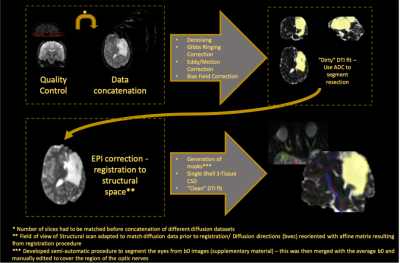 |
5 | Applying the single shell 3-Tissue method to investigate long-term degeneration of the visual system following hemidisconnection surgery
Luis Miguel Lacerda1, Alki Liasis2,3, Sian Handley3, Martin Tisdall4, Helen Cross5, Faraneh Vargha-khadem5, and Chris Clark1
1Developmental Imaging and Biophysics Section, UCL Great Ormond Street Institute of Child Health, LONDON, United Kingdom, 2Children’s Hospital of Pittsburgh, University of Pittsburgh Medical Centre, Pittsburgh, KS, United States, 3Clinical and Academic Department of Ophthalmology, Great Ormond Street Hospital for Children NHS Foundation Trust, London, United Kingdom, 4Neurosurgery, Great Ormond Street Hospital for Children NHS Foundation Trust, London, United Kingdom, 5Clinical Neurosciences, UCL Great Ormond Street Institute of Child Health, LONDON, United Kingdom
There is a need to map microstructural changes over long time periods and develop/apply methods that work with legacy data. In this study, we applied the novel single shell 3-Tissue method to data from a cohort of 4 patients who were scanned 20-years following childhood hemidisconnection surgery and presented degeneration of the visual system. We believe this study suggests that diffusion MRI can be used to monitor the integrity of the visual system following hemispherectomy and if extended to larger cohorts and a greater number of time-points, provide a clearer picture of the natural history of visual system degeneration.
|
||
3285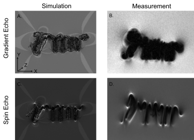 |
6 | Numerical approach to quantify MR imaging artifacts at metallic orthopedic implants at 1.5T, 3T, and 7T
Tobias Spronk1,2,3, Oliver Kraff1, Gregor Schaefers3,4, and Harald H. Quick1,2
1Erwin L. Hahn Institute for MR Imaging, University of Duisburg-Essen, Essen, Germany, 2High-Field and Hybrid MR Imaging, University Hospital Essen, University Duisburg-Essen, Essen, Germany, 3MRI-STaR Magnetic Resonance Institute for Safety, Technology and Research GmbH, Gelsenkirchen, Germany, 4MR:comp GmbH, Testing Services for MR Safety & Compatibility, Gelsenkirchen, Germany
This study evaluates a numerical approach to simulate artifacts due to presence of orthopedic metallic implants in the MR environment. Further to previously published studies, the numerical approach is validated by comparing simulations and measurements of orthopedic implants at three different field strengths (1.5T, 3T, and 7T). Artifact simulations and measurements of the implants show high correlation at all field strengths. The method potentially can be applied to improve the artifacts testing procedure for medical implants according to ASTM F2119. Additionally, the influence of different imaging parameters (echo time and bandwidth) on the artifact size is quantified via numerical simulations.
|
||
3286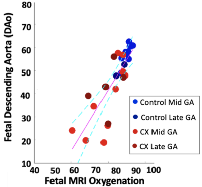 |
7 | Validation of non-invasive MR measurement of feto-placental oxygen saturation in a sheep model of human pregnancy
Dimitra Flouri1,2, Jack RT Darby3, Stacey L Holman3, Sunthara R Perumal4, Sebastien Ourselin1, Anna L David5,6, Andrew Melbourne1,2, and Janna L Morrison3
1School of Biomedical Engineering & Imaging Sciences, King's College London, London, United Kingdom, 2Department of Medical Physics & Biomedical Engineering, University College London, London, United Kingdom, 3Early Origins of Adult Health Research Group, University of South Australia, Adelaide, Australia, 4Preclinical Imaging and Research Laboratories, South Australian Health and Medical Research Institute, Adelaide, Australia, 5Institute for Women's Health, University College London, London, United Kingdom, 6NIHR Biomedical Research Center, University College London Hospitals, London, United Kingdom Abnormal placental development is postulated as one of the leading causes of fetal growth restriction (FGR). Advances in MRI technology enable non-invasive measurement of fetal oxygen saturation, but not all have yet been validated. Due to the invasiveness of tests required, validation in human subjects is not possible. Preclinical models such as pregnant sheep allow invasive methods to validate MRI measurements. Here we show that multi-compartment modelling of non-invasive placental MRI can be used to estimate the oxygen saturation of feto-placental oxygenation in normal sheep pregnancy and pregnancies affected by early gestation onstet FGR. |
||
3287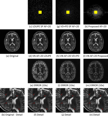 |
8 | Alternating Joint Learning Approach for Variational Networks and Sampling Pattern in Parallel MRI
Marcelo Victor Wust Zibetti1, Florian Knoll2, and Ravinder Regatte1
1Radiology, NYU Langone Health, New York, NY, United States, 2Department of Artificial Intelligence in Biomedical Engineering, FAU Erlangen-Nuremberg, Erlangen, Germany
We propose a new alternating learning approach to jointly learn the sampling pattern (SP) and the parameters of a variational network (VN) for acquisition and reconstruction on 3D Cartesian parallel MRI problems. This approach is composed of alternating short training with BASS algorithm to learn the SP, and ADAM algorithm to learn the parameters of the VN, both with forced monotonicity. The results illustrate that this approach provides reduced error when compared to other joint learning approaches, and surpasses VN trained with recently developed fixed SPs.
|
||
 Back to Meeting Home
 Back to the Program-at-a-Glance
The International Society for Magnetic Resonance in Medicine is accredited by the Accreditation Council for Continuing Medical Education to provide continuing medical education for physicians.
 Back to Meeting Home
 Back to the Program-at-a-Glance
 View the Presentation