Online Gather.town Pitches
Imaging Nerves, Head & Neck
Joint Annual Meeting ISMRM-ESMRMB & ISMRT 31st Annual Meeting • 07-12 May 2022 • London, UK

Online Gather.town Pitches
Imaging Nerves, Head & Neck
| Booth # | ||||
|---|---|---|---|---|
4527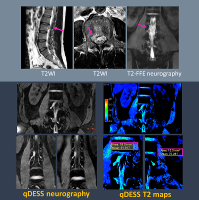 |
1 | Rapid High-resolution Quantitative MR Neurography in the Lumbosacral Plexus using Accelerated Quantitative Double Echo Steady-State (qDESS)
Takumi Ogawa1, Kayoko Abe2, Yutaka Hamatani1, Yasuhiro Goto1, Masami Yoneyama3, Quin Lu4, Isao Shiina1, Kazuo Kodaira1, Mana Kato1, Michinobu Nagao2, and Shuji Sakai2
1Department of Radiological Services, Tokyo Woman's Medical University, Tokyo, Japan, 2Department of Diagnostic imaging & Nuclear Medicine, Tokyo Woman's Medical University, Tokyo, Japan, 3Philips Japan, Tokyo, Japan, 4Philips Healthcare NA, San Francisco, CA, United States
Double Echo Steady-State (DESS) with water-selective excitation is a useful sequence to depict small nerves such as intracranial nerves, it would also be promising for visualizing lumbar nerves. Recently, accelerated qDESS sequence combined with compressed sensing sensitivity encoding (Compressed SENSE) has been proposed for further rapid knee morphological and quantitative imaging. In this study, we evaluate the feasibility of the accelerated qDESS sequence for visualization and quantification of the lumbosacral plexus within a short scan time.
|
||
4528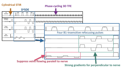 |
2 | Improved visualization of MR Neurography in the lumbosacral and sciatic nerves using double-diffusion sensitized driven-equilibrium (D2SDE)
Daichi Murayama1, Masami Yoneyama2, Takayuki Sakai1, Hajime Yokota3, Shigehiro Ochi1, and Atsuya Watanabe4,5
1Department of Radiology, Eastern Chiba Medical Center, Chiba, Japan, 2Philips Japan, Tokyo, Japan, 3Diagnostic Radiology and Radiation Oncology, Graduate School of Medicine, Chiba University, Chiba, Japan, 4Orthopaedic Surgery, Eastern Chiba Medical Center, Chiba, Japan, 5General Medical Services, Graduate School of Medicine, Chiba University, Chiba, Japan
We developed a new prepulse scheme, D2SDE, inspired by filter-probe double diffusion scheme for improved visualization of MR Neurography in the lumbosacral and sciatic nerves. This technique may be useful for arriving at an accurate diagnosis of lumbosacral plexus pathology.
|
||
4529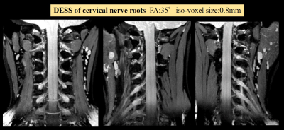 |
3 | Improved Visualization of MR Neurography in the Cervical Nerve Roots using Accelerated Quantitative Double Echo Steady-State (qDESS)
Yutaka Hamatani1, Kayoko Abe2, Yasuhiro Goto1, Masami Yoneyama3, Quin Lu4, Isao Shiina1, Kazuo Kodaira1, Takumi Ogawa1, Mana Kato1, Michinobu Nagao2, and Shuji Sakai2
1Department of Radiological Services, Tokyo Women's Medical University, Tokyo, Japan, 2Department of Diagnostic imaging & Nuclear Medicine, Tokyo Women's Medical University, Tokyo, Japan, 3Philips Japan, Tokyo, Japan, 4Philips Healthcare NA, San Francisco, CA, United States
Accelerated quantitative double echo steady state (qDESS), which is combined with Compressed SENSE is expected to improve visualization of the cervical nerve roots and brachial plexus. The purpose of this study was to optimize flip angle (FA) and spatial resolution (3D voxel size) of accelerated qDESS to visualize the small nerve roots and brachial plexus in the cervical spine. As a result, a combination of FA: 35 degree and voxel size: 0.8mm3 was the most appropriate parameters to demonstrate them while suppressing the background signals including the muscles, blood vessels, and cerebrospinal fluid.
|
||
4530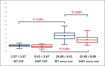 |
4 | Feasibility of inhomogeneous magnetization transfer for the peripheral nerve of the brachial plexus
Takayuki Sada1, Hajime Yokota2, Ryuna Kurosawa1, Takafumi Yoda1, Koji Matsumoto1, Takashi Namiki3, Masami Yoneyama3, Guillaume Gilbert4, Yoshitada Masuda1, and Takashi Uno2
1Department of Radiology, Chiba university hospital, Chiba, Japan, 2Diagnostic Radiology and Radiation Oncology, Graduate School of Medicine, Chiba University, Chiba, Japan, 3Philips Japan, Tokyo, Japan, 4Philips Canada, Mississauga, ON, Canada
Magnetization transfer (MT) has been conventionally used for the determination of myelin. However, the problem with the MT is that it reflects bound water that is not related to myelin. Inhomogeneous Magnetization Transfer (ihMT) can act specifically on myelin, it has been reported to be a useful method for quantifying myelin in brain and spinal cord regions. In this study, we compared conventional MT and ihMT for the brachial plexus, and ihMT might be able to reflect more accurate information about myelin than MT.
|
||
4531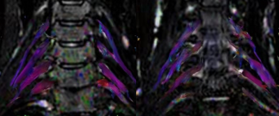 |
5 | The evaluation of brachial plexus injury by using diffusion tensor imaging (DTI)
Jianxiu Lian*1, Liyang Zhao*2, Zhiwei Shen1, Xiaofang Xu1, Zhigang Peng2, and Xiaona Li2
1Philips Healthcare, Beijing, China, 2The Third Hospital of Hebei Medical University, Shijiazhuang, China
Quantifying the functional parameters of brachial plexus injury can provide more valuable information for clinic. This article aims to apply diffusion tensor imaging (DTI)to brachial plexus injury and reveal the pathological causes of the disease from Magnetic Resonance Imaging (MRI). Our study revealed that patients with brachial plexus injury had higher FA and lower ADC values when compared with the healthy control, and FA showed the better diagnostic efficiency (AUC:0.763; sensitivity: 94.1%; specificity: 44.1%).
|
||
4532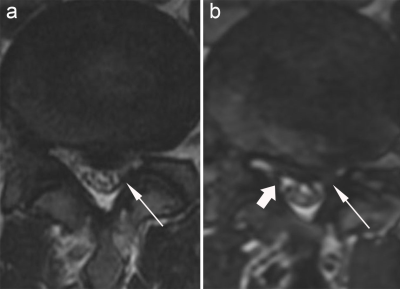 |
6 | Quantitative assessment of lumbar nerve root compression using contrast-enhanced 3D-FIESTA imaging sequence
Kun Ou1, Hui Gao1, Weiyn Vivian Liu2, and Kun Zhang1
1First Affiliated Hospital of Hunan University of Chinese Medicine, Changsha, China, 2GE Healthcare, Beijing, China
Actual contact surface between the extraforaminal nerve root and the adjacent structures is commonly missing on the conventional MR scanning, possibly leading to misdiagnosis. In our study, the detection rate of nerve root compression was significantly higher on the 3D-FIESTA-C images than T2WI. The shortest distance between the extraforaminal nerve root and the disc of L4/5 and L5/S1 in the healthy group was significantly longer than that in the lumbar disc herniation (LDH) group. The spatial relationship between nerve roots and surrounding tissues could be better described using 3D-FIESTA-C sequence and was helpful for the detection of nerve root compression.
|
||
4533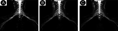 |
7 | Accelerated brachial Plexus MRI using deep learning constrained compressed SENSE reconstruction
Yi Xiao1, Sixian Hu1, Chunchao Xia1, Zhenlin Li1, Xiaoyong Zhang2, and Chuntang Ling3
1Radiology, West China Hospital Of Sichuan University, Chengdu, China, 2Clinical Science, Philips Healthcare, Chengdu, China, 3Application, Philips Healthcare, Chongqing, China
This study presents an accelerated brachial Plexus MR imaging technique using deep learning constrained compressed SENSE reconstruction (CS-AI-SENSE). The results showed that CS-AI-SENSE could enable comparable diagnostic image quality to the conventional SENSE-accelerated 3D T2-TSE approaches while significantly reduced the data acquisition time, which has potential to enhance the workflow of brachial Plexus MR examinations in clinics.
|
||
4534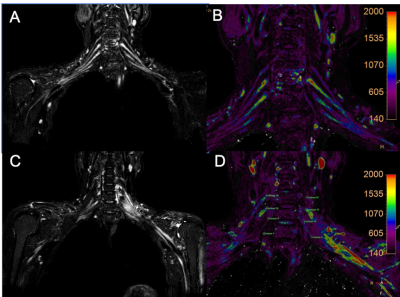 |
8 | To assess the patients with brachial plexus injury and the changes of T2 value after injury by using T2 mapping
Jianxiu Lian*1, Luyao Duan*2, Zhiwei Shen1, Xiaofang Xu1, Zhigang Peng2, and Xiaoman Yu2
1Philips Healthcare, Beijing, China, 2The Third Hospital of Hebei Medical University, Shijiazhuang, China
T2 mapping can provide quantitative parameter T2 values without radiation and damage, which can be used to quantitatively evaluate tissue inflammation and edema. The aim of this study was to evaluate patients with brachial plexus injury by using T2 mapping. And results showed that the T2 value would be prolonged when brachial plexus injured, and the T2 value would be shortened as patients recovers.
|
||
4535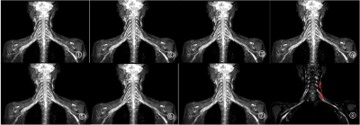 |
9 | The effect of compressed sensing with multiple acceleration factors on brachial plexus 3D NerveVIEW
Haonan Zhang1, Renwang Pu1, and Qingwei Song1
1Department of Radiology, the First Affiliated Hospital of Dalian Medical University, Dalian, Dalian, China
The 3D-NerveVIEW sequence with suppression of lipid and blood signals and a long echo time can obtain high-quality images for visualization of brachial plexus, while the long scan time may limit its clinical application. Compressed SENSE (CS) is a newly developed technique in MRI that enables accelerated acquisition with maintained image quality. By comparing results of 3D-NerveVIEW for brachial plexus imaging with acceleration by the conventional SENSE and the advanced CS with dierent acceleration factors. We found that the 3D-NerveVIEW for brachial plexus imaging with a CS acceleration factor of 6 can obtained favorable images within signicantly reduced scan time.
|
||
4536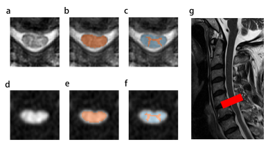 |
10 | Utility of advanced diffusion model in early detection of cervical spondylotic myelopathy: a comparison between DTI and NODDI Video Permission Withheld
Meng-Ze Zhang1, Han-Qiang Ou-Yang1,2,3, Dan Jin1, Chun-Jie Wang1, Jian-Fang Liu1, Qiang Zhao1, Xian-Chang Zhang4, Xiao-Guang Liu1,2,3, Zhong-Jun Liu1,2,3, Ning Lang1, Xing-Wen Sun1, Liang Jiang1,2,3, and Hui-Shu Yuan1
1Peking University Third Hospital, Beijing, China, 2Engineering Research Center of Bone and Joint Precision Medicine, Beijing, China, 3Beijing Key Laboratory of Spinal Disease Research, Beijing, China, 4MR collaboration, Siemens Healthineers Ltd, Beijing, China
This study compared the sensitivity of diffusion tensor imaging (DTI) and neurite orientation dispersion and density imaging (NODDI) to detect cervical spondylotic myelopathy (CSM) at an early stage. The results showed that NODDI-based indicators can distinguish patients with CSM without T2-weighted increased signal intensity from healthy controls during internal validation, while DTI-based indicators cannot. These findings suggest that NODDI is a promising method to detect CSM at an early stage.
|
||
4537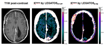 |
11 | High-spatial assessment of low-level permeability within normal-appearing brain tissue using dual-temporal resolution DCE-MRI
Ka-Loh Li1, Daniel Lewis2, Xiaoping Zhu1, Timothy Cootes1, and Alan Jackson1
1Division of Informatics, Imaging and Data Sciences, University of Manchester, Manchester, United Kingdom, 2Department of Neurosurgery, Salford Royal NHS Foundation Trust, Manchester, United Kingdom Quantifying blood-brain barrier permeability in normal-appearing brain through dynamic contrast-enhanced MRI (DCE-MRI) is challenging. We evaluated a new method, which combines the hybrid first-pass Patlak-plot (FP-PP) model with a high spatiotemporal resolution DCE approach termed LEGATOS. Dual-temporal resolution DCE-MRI data in twelve patients with neurofibromatosis type II (NF2) imaged at 1.5T were analyzed, and estimates of Ktrans within normal-appearing grey matter (NAGM) and white matter (NAWM) were compared. This new method permitted high-spatial resolution assessment of low-level permeability in normal-appearing brain and in our NF2 patient cohort a significant positive correlation between tumor volume and NAGM/NAWM Ktrans values was observed. |
||
4538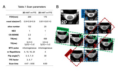 |
12 | Visible "Butterfly" of the Cervical Spinal Cord: A Pilot Study Using 3D and 2D m-FFE with Inhomogeneous Magnetization Transfer Pulse
Takafumi Yoda1, Hajime Yokota2, Takayuki Sada1, Ryuna Kurosawa1, Koji Matsumoto1, Takashi Namiki3, Masami Yoneyama3, Guillaume Gilbert4, Yoshitada Masuda1, and Takashi Uno2
1Department of Radiology, Chiba University Hospital, Chiba, Japan, 2Department of Diagnostic Radiology and Radiation Oncology, Graduate School of Medicine, Chiba University, Chiba, Japan, 3Philips Japan, Tokyo, Japan, 4Philips Canada, Mississauga, ON, Canada
The purpose of this study is to find the parameters that provide the optimal trade-off in Signal-to-noise-ratio (SNR) and contrast-to-noise ratio (CNR) for 3D ihMT m-FFE (3D-ihMT) and to explore the feasibility of that for the segmentability of gray and white matters in the cervical spinal cord compared with 2D ihMT m-FFE (2D-ihMT). We recommend a Flip angle of 7 and TFE factor of 5 as an optimal trade-off between segmentability and artifact level, at least 16 nr repetitions are required for robust segmentation. The segmentability of 3D-ihMT could be increased to a level close to that of 2D-ihMT.
|
||
4539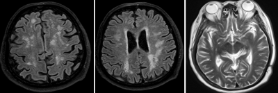 |
13 | White matter hyperintensities and brain volume changes in Cushing disease and related risk factors
Yue Wu1, Weiwei Wang1, Shiman Wu1, Qing Li2, Zhenwei Yao1, and Zengyi Ma3
1Department of Radiology, Huashan Hospital, Shanghai Medical College, Fudan University, Shanghai, China, 2MR Collaborations, Siemens Healthineers Ltd., Shanghai, China, 3Department of Neurosurgery, National Center for Neurological Disorders (NCND), Shanghai Pituitary Tumor Center, Huashan Hospital, Shanghai Medical College, Fudan University, Shanghai, China
Cushing's disease(CD) has significantly more white matter hyperintensities and enlarged perivascular spaces, compare to same-aged healthy subjects. Age, diabetes, hypokalemia was significantly related with white matter hyperintensities in CD. The gray matter volume of CD patients was significantly smaller than the healthy subjects. Compared with patients with type 2 diabetes alone, the mean age of CD with diabetes were younger with shorter course of disease, but with more severe white matter damage. the severity of white matter hyperintensities were ranked as belowed: CD with type 2 diabetes, CD without type 2 diabetes, type 2 diabetes.
|
||
4540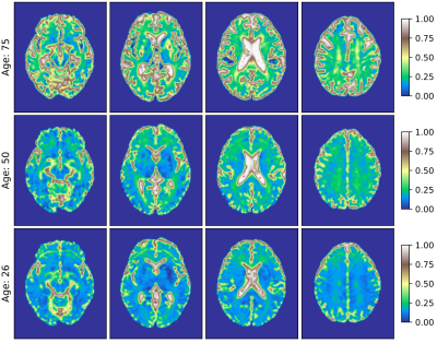 |
14 | Free-water volume fraction increases nonlinearly across the lifespan in healthy human brain white matter areas
Tomasz Pieciak1, Guillem París1, Dani Beck2,3, Ivan I. Maximov3,4, Antonio Tristán-Vega1, Rodrigo de Luis-García1, Lars T. Westlye2,3,5, and Santiago Aja-Fernández1
1Laboratorio de Procesado de Imagen (LPI), Universidad de Valladolid, Valladolid, Spain, 2Department of Psychology, University of Oslo, Oslo, Norway, 3NORMENT, Division of Mental Health and Addiction, Oslo University Hospital & Institute of Clinical Medicine, University of Oslo, Oslo, Norway, 4Western Norway University of Applied Sciences, Bergen, Norway, 5KG Jebsen Centre for Neurodevelopmental Disorders, University of Oslo, Oslo, Norway
The free-water (FW) volume fraction $$$f$$$ is a sensitive diffusion MRI biomarker correlated with microstructural alterations in the human brain. This paper studies the variations in the FW volume fraction $$$f$$$ in healthy human brain white matter (WM) across the lifespan from a set of 248 female and 140 male subjects. Our study reveals positive nonlinear correlations of the volume fraction $$$f$$$ with age over the WM regions and suggests a heteroscedastic nature and age-dependent intra-region-of-interest variability of $$$f$$$ across the lifespan.
|
||
4541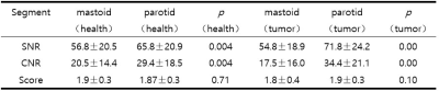 |
15 | Demonstrating the Facial Nerve in the Parotid Gland using 3 Dimension Fast Field Echo imaging: a Pilot Study
Yihua Wang1, Lijun Wang1, Xiaoxiao Zhang2, and Ailian Liu1
1Department of Radiology, the First Affiliated Hospital of Dalian Medical University, Dalian, China, 2Philips Healthcare, Beijing, China, Beijing, China
Facial nerve MRI is a clinical challenge that it is difficult to differentiate parotid gland tumors from facial nerve on conventional imaging. It is great demand to develop new diagnostic technology to accurately display peripheral nerve and tumor for avoiding intraoperative injury. In this study, 3 dimension fast field echo imaging(T2WI-3D-FFE) is potentially a valuable sequence in displaying the intraparotid facial nerve and localizing the tumor.
|
||
 Back to Meeting Home
 Back to the Program-at-a-Glance
The International Society for Magnetic Resonance in Medicine is accredited by the Accreditation Council for Continuing Medical Education to provide continuing medical education for physicians.
 Back to Meeting Home
 Back to the Program-at-a-Glance
 View the Presentation