Online Gather.town Pitches
Imaging Head, Spinal Cord & Nerves
Joint Annual Meeting ISMRM-ESMRMB & ISMRT 31st Annual Meeting • 07-12 May 2022 • London, UK

Online Gather.town Pitches
Imaging Head, Spinal Cord & Nerves
| Booth # | ||||
|---|---|---|---|---|
 |
3873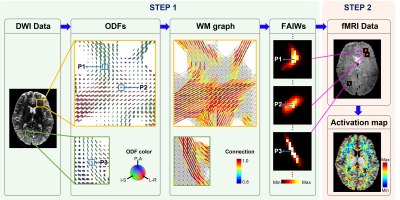 |
1 | Model-free mapping of neural activation in brain white matter based on local fiber architecture
Yu Zhao1, Yurui Gao1, Zhongliang Zu1, Muwei Li1, Kurt Schilling1, Adam W. Anderson1, Zhaohua Ding1, and John C. Gore 1
1Vanderbilt University Medical Center, Nashville, TN, United States
We propose a model-free method to detect BOLD signal changes in brain white matter (WM) without an assumption of a hemodynamic response function or a linear shift-invariant response as assumed in conventional general linear models. Instead, neural activations are measured by the synchrony of BOLD signals under constraints of the anisotropic architecture of WM fibers. A 60 subject sub-group sourced from the Human Connectome Project database was used to validate the proposed method at a group level. Our results demonstrate that the proposed method can probe neural activations in WM with high sensitivities and high specificities.
|
|
3874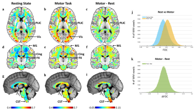 |
2 | Local BOLD correlations in white matter measured under constraints of fiber orientation
Yurui Gao1, Yu Zhao2, Kurt G Schilling2, Muwei Li2, Adam W Anderson1, Zhaohua Ding1, and John C Gore2
1VANDERBILT UNIVERSITY, Nashville, TN, United States, 2Vanderbilt University Medical Center, Nashville, TN, United States
Correlations between BOLD signals from adjacent voxels in white matter (WM) are anisotropic and appear similar to metrics that describe functional connectivity in gray matter. We propose a method to measure the fiber-oriented correlation (FOC) between BOLD signals in WM. This method was implemented on both resting state and motor task fMRI data across 64 HCP subjects. The FOC map revealed WM areas apparently engaged in default mode network and visual function in both rest and task conditions. The motor task map also delineates the corticospinal tract. The proposed metric FOC may be valuable for investigating functional WM pathways.
|
||
3875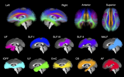 |
3 | White Matter Tract Microstructure Influences Semantic Memory & Emotion Perception Performance in the HCP Dataset
Leo R. Zekelman1,2, Fan Zhang3, Nikos Makris4,5, Jianzhong He3, Yuqian Chen3,6, Tengfei Xue3,6, Daniela Liera7, Daniel L. Drane8,9,10, Yogesh Rathi3,11, Alexandra J. Golby1,3, and Lauren J. O'Donnell3
1Department of Neurosurgery, Brigham and Women’s Hospital, Boston, MA, United States, 2Speech and Hearing Bioscience and Technology, Harvard Medical School, Boston, MA, United States, 3Department of Radiology, Brigham and Women’s Hospital, Boston, MA, United States, 4Department of Anatomy and Neurobiology, Boston University School of Medicine, Boston, MA, United States, 5Department of Psychiatry and Neurology, A. Martinos Center for Biomedical Imaging, Boston, MA, United States, 6School of Computer Science, University of Sydney, Sydney, Australia, 7Harvard College, Cambridge, MA, United States, 8Department of Neurology, Emory University School of Medicine, Atlanta, GA, United States, 9Department of Pediatrics, Emory University School of Medicine, Atlanta, GA, United States, 10Department of Neurology, University of Washington School of Medicine, Seattle, WA, United States, 11Psychiatry Neuroimaging Laboratory, Department of Psychiatry, Brigham and Women's Hospital, Boston, MA, United States
Using dMRI and behavioral data from 809 participants in the Human Connectome Project, we examined how the microstructure of the white matter (WM) association tracts influences performance on assessments of semantic memory and emotion perception. WM tracts were extracted and measured using an automated WM tract atlas. We found the microstructure of left hemisphere tracts influenced semantic memory, while that of right hemisphere tracts influenced emotion perception performance. Furthermore, the microstructure of the left arcuate fasciculus influenced performance on both assessments. Our findings suggest WM tract microstructure relates to the opposing hemispheric dominance of language and theory of mind processing.
|
||
3876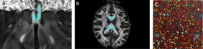 |
4 | Estimation of axon-bundle parallel-diffusivity on clinical settings with linear-encoding DW-MR data
Pablo Antonio Stack-Sanchez1, Alonso Ramirez-Manzanares1, Arturo Gonzalez-Vega2, Jose Luis Marroquin1, and Luis Concha3
1Computer Science, Centro de Investigación en Matemáticas A.C., Guanajuato, Mexico, 2Department of Chemical, Electronic and Biomedical Engineering, Division of Sciences and Engineering, University of Guanajuato, Guanajuato, Mexico, Leon, Mexico, 3Institute of Neurobiology, Universidad Nacional Autonoma de Mexico, Juriquilla, Mexico
The estimation of reliable parameters of multi-compartment models of water diffusivity is a challenging open problem, especially when low-SNR clinical-datasets are analyzed. The ill-posedness of the models requires fixing a subset of parameters in order to estimate the rest of them. We propose a simple and fast method where only averaged parameters are required (not voxelwise information is required). The method is based on the processing of powder-averaged data to estimate a robust value for the axon-bundle parallel-diffusivity. We test the robustness of the method on synthetic experiments and show the results on in-vivo human and ex-vivo rodent datasets.
|
||
3877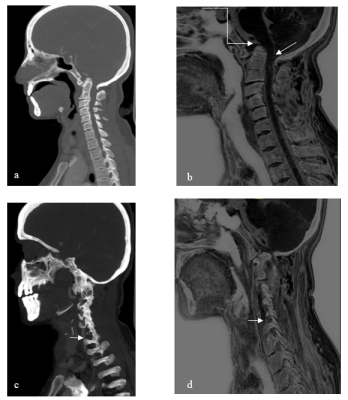 |
5 | Exploring the Application of Compressed-SENSE Accelerated Fast MR Based Bone Imaging Method in CVJ Anomalies: Preliminary Experience
Sabha Ahmed1, Jitender Saini1, Nupur Pruthi2, Alok Mohan Uppar2, Rashmi Rao3, Kumar Madan3, Narayan Krishna Rolla3, and Indrajit Saha3
1Neuroimaging and interventional radiology, NIMHANS, Bangalore, India, 2Neurosurgery, NIMHANS, Bangalore, India, 3Philips India Ltd, Bangalore, India
Imaging analysis of craniovertebral junction anomalies comprises of bony, ligament, vascular and spinal cord evaluation. MDCT falls short in the evaluation of the latter while conventional MRI does not offer optimal bone resolution. Novel Compressed SENSE accelerated Fast field echo resembling a CT using restricted echo-spacing (CS-FRACTURE) technique is a bid to overcome this shortcoming in routine clinical setup and we aim to explore the same.
|
||
3878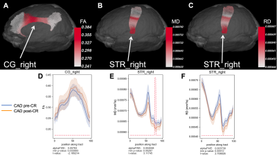 |
6 | Probing evidence of brain macrostructural disruptions in coronary artery disease: A diffusion MRI tractometry study
Stefan E. Poirier1,2, Neville Suskin3, Keith S. St. Lawrence1,2, Christopher McIntyre1,2,4, Jonathan D. Thiessen1,2, J. Kevin Shoemaker5, and Udunna C. Anazodo1,2
1Lawson Imaging, Lawson Health Research Institute, London, ON, Canada, 2Medical Biophysics, Western University, London, ON, Canada, 3Cardiology, Western University, London, ON, Canada, 4Lilibeth Caberto Kidney Clinical Research Unit, London Health Sciences Centre, London, ON, Canada, 5School of Kinesiology, Western University, London, ON, Canada
White matter (WM) breakdown is linked to cognitive impairment in coronary artery disease (CAD). We used diffusion MRI tractometry to assess the effects of cardiac disease, brain aging, and cardiac rehabilitation (CR) on regional WM macrostructure in brains of CAD patients. WM disruptions were found in CAD patients at baseline (pre-CR) compared to controls and in old controls compared to young controls. WM improvements, especially in regions linked to cognition, were observed in CAD patients following CR. Both cardiac disease and brain aging may contribute to WM dysfunction in CAD, and these WM disruptions may be favourably modified by CR.
|
||
3879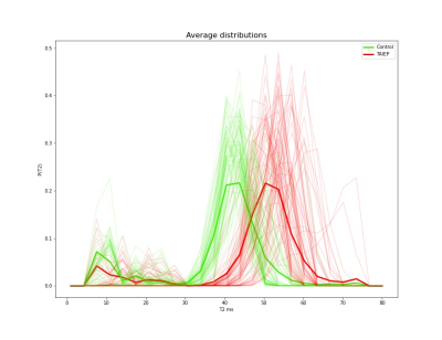 |
7 | A Learning Method to Estimate Multi-compartmental T2 Distributions with low Data Requirements
Daniel Vallejo-Aldana1, Arturo Gonzalez-Vega2, Victor H Hernandez2, Valeria Piazza3, Milvia Alata3, Jonathan Rafael-Patiño4,5, Thomas Yu4,6, Luis Concha7, and Alonso Ramirez-Manzanares8
1Mathematics Department, Universidad de Guanajuato, Guanajuato, Mexico, 2Department of Chemical, Electronic and Biomedical Engineering, Division of Sciences and Engineering, University of Guanajuato, Leon, Mexico, 3Center of Research in Optics, Leon, Mexico, 4Signal Processing Lab 5 (LTS5), Ecole Polytechnique Fédérale de Lausanne, Lausanne, Switzerland, 5Radiology Department, Centre Hospitalier Universitaire Vaudois and University of Lausanne, Lausanne, Switzerland, 6Medical Image Analysis Laboratory, Center for Biomedical Imaging (CIBM), University of Lausanne, Lausanne, Switzerland, 7Institute of Neurobiology, Universidad Nacional Autonoma de Mexico, Juriquilla, Mexico, 8Computer Science, Centro de Investigación en Matemáticas A.C., Guanajuato, Mexico
The estimation of intravoxel distributions of T2 values based on multi-echo MR data is a challenging task. Interestingly, the information above is quite useful to detect damage on brain tissue, e.g. to estimate myelin-water-fraction changes associated with demyelination processes. Currently available methods typically require a long train of echoes, which are not always feasible to acquire. In this work we tackle this problem using state-of-the-art supervised learning convolutional networks to build a robust prediction model on very limited data ( 5 echoes and 4 TR). The methodology identifies myelin abnormalities in a rodent model of a neurological disorder with demyelination.
|
||
3880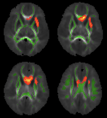 |
8 | Folate Intake during Pregnancy is Associated with Greater White Matter Development in the Newborn Brain
Natalie E. Phelan1, Rajikha Raja2, Aline Andres3,4, and Xiawei Ou2,3,4,5
1College of Medicine, University of Arkansas for Medical Sciences, Little Rock, AR, United States, 2Department of Radiology, University of Arkansas for Medical Sciences, Little Rock, AR, United States, 3Department of Pediatrics, University of Arkansas for Medical Sciences, Little Rock, AR, United States, 4Arkansas Children's Nutrition Center, Little Rock, AR, United States, 5Arkansas Children's Research Institute, Little Rock, AR, United States
This study examined potential relationships between maternal folate intake during pregnancy and neonatal brain white matter development. Healthy pregnant women were recruited at early pregnancy and their newborns underwent a brain MRI examination at ~2 weeks of age, including diffusion tensor imaging to assess white matter development. We found that folate intake during the second trimester of pregnancy positively correlated with fractional anisotropy (FA) values in brain white matter regions including the genu of corpus callosum and the external capsule. Our findings suggest positive impact of maternal folate intake during pregnancy on offspring brain white matter development.
|
||
3881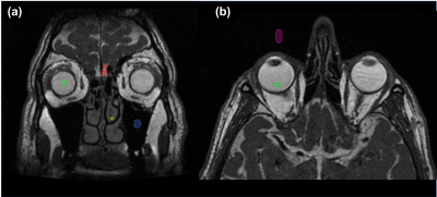 |
9 | Can a sub 2-minute MRI exam compete with CT for imaging sinus anatomy?
Sarah Reeve1,2, Mark Parker2,3, James Rioux1,2,4, Elena Adela Cora4,5, Chris Bowen1,2,4, Steven Beyea1,2,4,6, and David Volders4,5
1Physics and Atmospheric Science, Dalhousie University, Halifax, NS, Canada, 2Biomedical Translational Imaging Centre, QEII Health Sciences Centre, Halifax, NS, Canada, 3Medicine, Dalhousie University, Halifax, NS, Canada, 4Diagnostic Radiology, Dalhousie University, Halifax, NS, Canada, 5Diagnostic Imaging, Nova Scotia Health, Halifax, NS, Canada, 6School of Biomedical Engineering, Dalhousie University, Halifax, NS, Canada
MRI of the paranasal sinuses with bSSFP sequences is not typically performed due to prohibitive exam times and the prevalence of artifacts caused by off-resonance effects. We have shown that bSSFP images acquired in under 2 minutes using high-performance gradients at low field resist banding artifacts and demonstrate SNR and resolution sufficient to delineate relevant structures. This is supported by high SNR values in tissues and fluid-filled spaces, and low SNR in the air-filled maxillary sinus. 2-minute scan times compete with CT for clinical work flow and demonstrate the potential of MRI for high throughput sinus pathology evaluation.
|
||
3882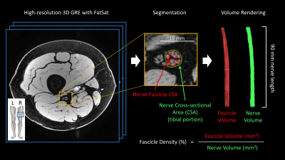 |
10 | Can Proximal Nerve Morphometrics Predict Disease Severity in Polyneuropathies?
Yongsheng Chen1, Alexandar Bezanovski1, Sadaf Saba1, Vince Marceau1, Yang Xuan2, Mariam Tariq1, Xue Yang1, and Jun Li1
1Neurology, Wayne State University School of Medicine, Detroit, MI, United States, 2Radiology, Wayne State University School of Medicine, Detroit, MI, United States Human sciatic nerve morphometrics were measured for nerve volume, fascicle volume, and fascicle density using a 3D gradient echo sequence with an in-plane resolution of 0.15 x 0.15 mm2. Metrics from healthy controls showed a moderate correlation of nerve and fascicle volumes with body mass index but not with age. There was a strong correlation between nerve volume and the clinical score of disease severity in patients with hereditary neuropathy with liability to pressure palsies. |
||
3883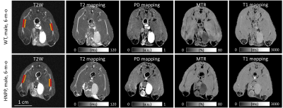 |
11 | Quantitative MRI Reveals Nerve Pathology in a HNPP Mouse Model
Yongsheng Chen1, Yimin Shen2, Daniel Moiseev1, Zafar Wazir1, Bo Hu1, and Jun Li1
1Neurology, Wayne State University School of Medicine, Detroit, MI, United States, 2Radiology, Wayne State University School of Medicine, Detroit, MI, United States
Quantitative MRI (qMRI) methods were developed to measure T1, T2, proton density, and MTR of mouse sciatic nerve in-vivo. Pmp22+/- mouse, an animal model of hereditary neuropathy with liability to pressure palsies (HNPP), was imaged at 3- and 6-month of age. The imaging demonstrated an increase of MTR, and a decrease of proton density and T1 in the Pmp22+/- nerves, compared with those in wild-type nerves. These qMRI changes correlate with the growth of HNPP pathology – tomacula formed by excessive myelin folding over time. Ongoing experiment is to determine the pathological and molecular substrates responsible for the qMRI findings.
|
||
3884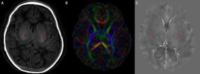 |
12 | Quantitative susceptibility mapping of early myelination in the posterior limb of internal capsule in neonates
Brian Ma1, Lara Bartels1,2, Christian Kames1,2, Yuting Zhang3, Alexander Weber1,4, and Alexander Rauscher1,2,4,5
1UBC MRI Research Centre, University of British Columbia, Vancouver, BC, Canada, 2Department of Physics and Astronomy, University of British Columbia, Vancouver, BC, Canada, 3Department of Radiology, Children's Hospital of Chongqing Medical University, Chongqing, China, 4Division of Neurology, Department of Pediatrics, University of British Columbia, Vancouver, BC, Canada, 5Department of Radiology, University of British Columbia, Vancouver, BC, Canada
We performed quantitative susceptibility mapping (QSM) of the posterior limb of internal capsule (PLIC) in human neonates. As the PLIC is one of the earliest structures to myelinate, it is clearly visible as a hypointense structure on QSM. This work investigates the potential as a marker for brain health in neonates. There were no significant differences in magnetic susceptibility of the PLIC between healthy controls, injured preterm, and injured term neonates.
|
||
3885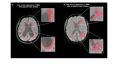 |
13 | Reduced white matter venous density is associated with neurodegeneration and cognitive impairment: a susceptibility weighted imaging study
Chenyang Li1, Henry Rusinek1, Jingyun Chen1,2, Louisa Bokacheva2, Alok Vedvyas2, Arjun Masurkar2, Thomas Wisniewski2, E.Mark Haacke3, and Yulin Ge1
1Department of Radiology, NYU Grossman School of Medicine, New York, NY, United States, 2Department of Neurology, NYU Grossman School of Medicine, New York, NY, United States, 3Department of Radiology, Wayne State University School of Medicine, Detroit, MI, United States
High resolution SWI images provide unique contrast to small venous vasculature. The conspicuity of small veins on SWI venography, such as deep medullary vein in white matter (WM), is susceptible to venous blood oxygenation level changes. This study demonstrates a significant association between WM venous density and neurodegenerative feature characterized by brain atrophy in the elderly, but no significant association of WM venous density to white matter hyperintensities (WMHs) load. Further clinical correlative analysis revealed a significant correlation of WM venous density to semantic fluency and different stages of cognitive status.
|
||
 Back to Meeting Home
 Back to the Program-at-a-Glance
The International Society for Magnetic Resonance in Medicine is accredited by the Accreditation Council for Continuing Medical Education to provide continuing medical education for physicians.
 Back to Meeting Home
 Back to the Program-at-a-Glance
 View the Presentation