Online Gather.town Pitches
fMRI & Connectivity in Healthy Conditions
Joint Annual Meeting ISMRM-ESMRMB & ISMRT 31st Annual Meeting • 07-12 May 2022 • London, UK

Online Gather.town Pitches
fMRI & Connectivity in Healthy Conditions
| Booth # | ||||
|---|---|---|---|---|
2980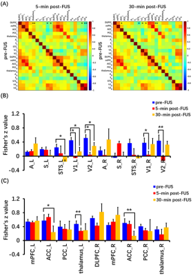 |
1 | Focused ultrasound-modulated left dorsolateral prefrontal cortex of the non-human primate assessed by functional MRI Video Permission Withheld
Yu Xu1, Tingting He2, Haiming Wang2, Xiao Yu1, Boyi Qu2, Ssu-Ju Li3, and Hsin-Yi Lai4
1Interdisciplinary Institute of Neuroscience and Technology, School of Medicine, Zhejiang University, Hangzhou, China, 2College of Biomedical Engineering & Instrument Science, Zhejiang University. Interdisciplinary Institute of Neuroscience and Technology, Zhejiang University, Hangzhou, China, 3Department of Biomedical Engineering, National Yang Ming University, No. 155, Section 2, Linong Street, Taipei, 112, Taiwan, ROC. Department of Biomedical Engineering, National Yang Ming Chiao Tung University, No. 1001 Ta-Hsueh Rd., Hsinchu, 300, Taiwan, ROC, Taiwan, China, 4College of Biomedical Engineering & Instrument Science, Zhejiang University. Department of Neurology of the Second Affiliated Hospital, Interdisciplinary Institute of Neuroscience and Technology, School of Medicine, Zhejiang University. Department of Neurology of Sir Run Run Shaw Hospital, School of Medicine, Zhejiang University, Hangzhou, China Focused ultrasound (FUS) has shown its unique advantages in the field of non-invasive neuromodulation. The dorsolateral prefrontal cortex (DLPFC) is a key node in brain networks involved in cognitive, emotional and sensory processing. Here we evaluated the immediate and latency effects of DLPFC-FUS by functional magnetic resonance imaging (fMRI). The results showed that DLPFC-FUS induced a 3% increase in positive BOLD signal, and decreased FCs in bilateral primary and secondary visual cortices, supported by visual stimuli-evoked BOLD change. Moreover, FCs significantly decreased in left thalamus at 5-min post-FUS and in bilateral anterior cingulate cortex (ACC) at 30-min post-FUS. These results may help to elucidate the effect of FUS neuromodulation. |
||
2981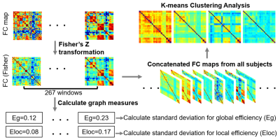 |
2 | Analysis of Dynamic Function Connectivity in a Normal Rat Model After Focused Ultrasound-Neuromodulation in VPM/VPL
Yu-Chieh Hung1, Yi-Cheng Wang1, Hao-Li Liu2, and Hsu-Hsia Peng1
1Department of Biomedical Engineering and Environmental Sciences, National Tsing Hua University, Hsinchu, Taiwan, 2Department of Electrical Engineering, National Taiwan University, Taipei, Taiwan Focused ultrasound (FUS) has been considered as a noninvasive neuromodulation method. In addition to evaluate the functional connectivity (FC) by an average of BOLD signals across the whole scan, a dynamic FC (dFC) can help to comprehend the instant alternation of FC. We aimed to investigate the alterations of dFC after FUS-neuromodulation at VPM/VPL in a normal rat model. We used k-means and dynamic network analyses to evaluate changes of dFC at Pre-FUS, Sham, 35-min, 3-hr, and 3-day for rats with FUS sonication at VPM/VPL. We found an instant effect of FUS-neuromodulation (35-min) and a recovering trend in 3-day. |
||
2982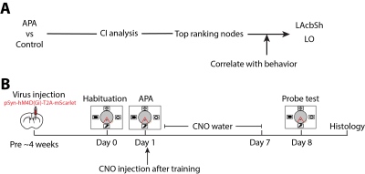 |
3 | Establishing causal relationship between resting-state network and behavior
Zengmin Li1, Dilsher Athwal1, and Kai-Hsiang Chuang1,2
1Queensland Brain Institute, The University of Queensland, St Lucia, Australia, 2Centre for Advance Imaging, The University of Queensland, St Lucia, Australia
|
||
2983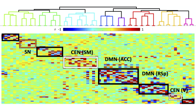 |
4 | Causal Evidence for a Mouse Homologue of the Human Salience Network via Default Mode Network Inhibition Video Not Available
Evan Houldin1, Zengmin Li1, and Kai-Hsiang Chuang1
1University of Queensland, Brisbane, Australia
Recent evidence indicates a putative mouse homologue of the human triple-network model, for which a salience network (SN) actively engages either internally-oriented default mode network (DMN) or externally-oriented central executive network (CEN). The interaction amongst these homologues has yet to be validated. This study probed SN responses to DMN inhibition in mice, during functional magnetic resonance imaging. We found that SN radically adjusts its connectivity profile with triple-network components, depending on which DMN hub is inhibited. This indicates that triple-network must be considered holistically, with SN serving an important role in overall triple-network connectivity.
|
||
2984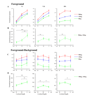 |
5 | 7 T CBV fMRI reveal cortical microcircuits of bottom-up saliency in the human brain
Chengwen Liu1, Chen Liu2, Li Zhaoping3, and Peng Zhang2
1Second Xiangya Hospital,Central South University, Changsha, China, 2Institute of Biophysics, Chinese Academy of Sciences, Beijing, China, 3University of T ̈ubingen, Max Planck Institute for Biological Cybernetics, T ̈ubingen, Germany Using a slab-selective VASO sequence at 7 T, we investigate cortical depth-dependent CBV fMRI activity to orientation-defined saliency stimuli in early visual and parietal cortices of the human brain. Results show that the fMRI response of salient foreground bars is strongest in the superficial depth of V1, and peaks in the middle cortical depth of V2/V3 and IPS. These findings support the hypothesis that bottom-up saliency is initially created by iso-feature suppressions through lateral connections in V1 superficial layers, and then feedforward to parietal cortex to generate the attention priority map. |
||
2985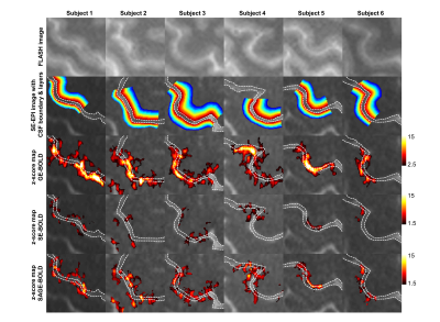 |
6 | Improving laminar functional specificity and sensitivity using combined spin- and gradient-echo EPI at 7 T Video Permission Withheld
SoHyun Han1,2, Seulgi Eun1,2, HyungJoon Cho3, Kâmil Uludaǧ1,2, and Seong-Gi Kim1,2
1Center for Neuroscience Imaging Research, Suwon, Korea, Republic of, 2Department of Biomedical Engineering, Sungkyunkwan University, Suwon, Korea, Republic of, 3Department of Biomedical Engineering, Ulsan National Institute of Science and Technology, Ulsan, Korea, Republic of
Both spatial specificity and sensitivity are important for high spatial resolution fMRI. In this study, we applied a filter designed based on the ΔR2*/ΔR2 ratio to enhance both the sensitivity and specificity of the BOLD signal using SAGE-EPI sequence. fMRI experiments during fist-clenching with touching with 0.8 mm isotropic resolution were performed and we directly compared the layer profile of SAGE-BOLD with that of GE- and SE-BOLD in the primary sensory and motor cortices. Vessel-size tuned SAGE-BOLD fMRI provided enhanced laminar specificity and sensitivity in the gray matter region.
|
||
2986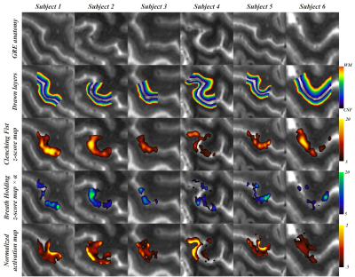 |
7 | Laminar BOLD normalization using breath-hold challenge
Seulgi Eun1,2,3, SoHyun Han1,2, Kâmil Uludaǧ1,2,4, and Seong-Gi Kim1,2
1Center for Neuroscience Imaging Research, Institute for Basic Science (IBS), Suwon, Korea, Republic of, 2Department of Biomedical Engineering, Sungkyunkwan University, Suwon, Korea, Republic of, 3Department of Biomedical Engineering, Kyung Hee University, Yongin, Korea, Republic of, 4Techna Institute & Koerner Scientist in MR Imaging, University Health Network, Toronto, ON, Canada
Limitations of the several developed laminar fMRI acquisition methods and pial and intra-cortical ascending vein contamination of the gradient-echo (GE) restrict the neural specificity of the depth profiles. Preserving the advantage of high sensitivity of the GE-EPI sequence, we propose a simple and intuitive model to normalize laminar BOLD with breath holding. Our normalization method increased specificity of the BOLD activation map and emphasized laminar BOLD signal changes in gray matter regions by suppressing signal changes from CSF. This approach takes into account the different vascular sensitivity across layers but may not fully account for the draining vein contamination.
|
||
2987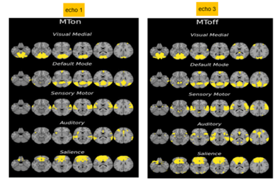 |
8 | Use of Arterial Blood Contrast (ABC) to detect resting state networks at short TE
Fatemeh Taheriyan1, Zahrah Fazal1, Kevin Klein Gunnewiek1, Jenni Schulz1, Jose Marques1, and David G Norris1
1Radboud University, Donders Institute, Nijmegen, Netherlands
Arterial blood contrast increase the cerebral blood volume contribution to brain activation studies by suppressing grey matter signal using on-resonance magnetisation transfer. An increase in CBV will lead to an increase in signal, which is then complementary to BOLD. We performed multi-echo resting state fMRI in a group of 16 subjects and compared the results at TE=28ms with MT-off (standard BOLD) and TE=6.9 MT-on (ABC). Z-scores were higher for standard BOLD at all TEs but with higher variance in the z-score value. Dual regression results never the less showed high similarity activation maps for standard resting state networks.
|
||
2988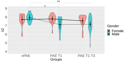 |
9 | Multiplex network analysis reveals disrupted brain processing speed in children with prenatal alcohol exposure Video Not Available
Xiaoyun Liang1,2, Chun-Hung Yeh3,4, and Peter J. Anderson1,5
1Victorian Infant Brain Study (VIBeS), Murdoch Children's Research Institute, Melbourne, Australia, 2Florey Department of Neuroscience and Mental Health, University of Melbourne, Heidelberg, Australia, 3Institute for Radiological Research, Chang Gung University and Chang Gung Memorial Hospital, Taoyuan, Taiwan, 4Department of Psychiatry, Chang Gung Memorial Hospital, Taoyuan, Taiwan, 5Turner Institute for Brain and Mental Health, Monash University, Clayton, Australia
In this study, we applied a method that integrates dynamic functional connectivity using the multiplex network approach to enhance the sensitivity to subtle alterations of functional connectivity on participants subject to PAE. Our results demonstrated its capability in characterizing subtle brain network changes in children with low-moderate PAE. The lower values of the PAE T1-T3 group indicates that the information processing speed could have been compromised due to PAE. In line with literature, the distinct findings between male and female groups revealed that PAE may induce gender-dependent brain disruptions and males are more susceptible to PAE.
|
||
2989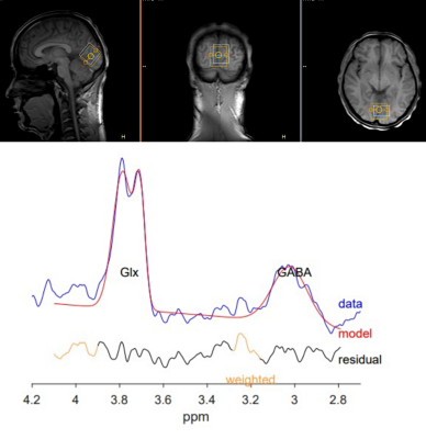 |
10 | Relationship between GABA+ level in occipital lobe and topological characters of cerebral EEG functional connectivity network in young adults
Yanting Liu1, Sihui Zhao1, Yanan Gao1, Hui Steve2,3, Mikkelsen Mark2,3, A.E.Edden Richard2,3, Chen Zhang4, and Bing Yu1
1China Medical University, Shenyang, China, China, 2Russell H. Morgan Department of Radiology and Radiological Science, The Johns Hopkins University School of Medicine, Baltimore, MD, USA, Baltimore, MD, United States, 3F. M. Kirby Center for Functional Brain Imaging, Kennedy Krieger Institute, Baltimore, MD, USA, Baltimore, MD, United States, 4MR Scientific Marketing, Siemens Healthineers, Beijing, China
We carried investigation to explore the relationship of GABA level of occipital lobe on topological and organizational properties of the entire brain using Hadamard Encoding and Reconstruction of Mega-Edited Spectroscopy (HERMES) method. Synchronous EEG-MRS data were obtained from forty healthy volunteers. GABA+ level and the global topological topological characters of the EEG functional connectivity network were calculated. The results demonstrate that the GABA+ level of the occipital lobe showed a significant negative correlation with the small world index of EEG functional connectivity network in the EEG α band.
|
||
2990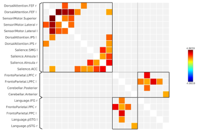 |
11 | Understanding auditory cognition and its underlying acoustic characteristics
Himanshu Singh1, S Senthil Kumaran1, and Ankeeta A1
1Department of NMR, All India Institute of Medical Sciences, Delhi, India
Auditory working memory is associated with sound pressure level for perception of information. However, its acoustic characteristics that represent information is not well understood. We used 1-back and 2-back auditory working memory tasks and acoustic characteristics (including pure tone assessment, audiometric threshold) to understand cognitive connectome with respect to frequency. This study explores the association of frequency characteristics underlying semantics necessary to understand auditory cognition.
|
||
2991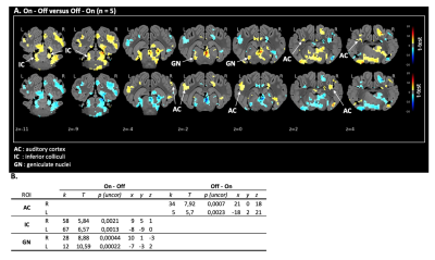 |
12 | Auditory functional MRI in the conventional pig at 1.5T
Paul COUDERT1,2, Pierre Antoine ELIAT3,4, Yann SERRAND3, Nicolas COQUERY3, Gwenole TANGUY1,2, Stéphane LAURENT1,5, Hervé SAINT-JALMES4,6, David VAL LAILLET3, and Benoit GODEY1,2
1ENT, CHU of Rennes, Rennes, France, 2INSERM, MediCIS, University Rennes 1, Rennes, France, 3INRAE, NuMeCan 1341, University Rennes 1, Rennes, France, 4PRISM, CNRS, INSERM, BIOSIT, Univ Rennes, Rennes, France, 5School of audioprosthesis of Fougères, Université Rennes 1, Fougères, France, 6INSERM, LTSI UMR 1099, University Rennes 1, Rennes, France
This work presents a functional MRI model to study the activation of central auditory pathways in a pig model at 1.5T. The conventional pig is a good model due to its anatomical similarity to humans, and therefore could be relevant to study and understand brain responses to auditory stimulations.
|
||
2992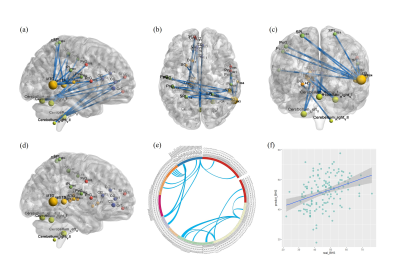 |
13 | Functional connectome-based brain features are related to hopelessness in healthy adolescents and young adults
Xin Xu1, Han Lai1, Cheng Yang1, John Sweeney1, and Qiyong Gong1
1Huaxi MR Research Center (HMRRC), Department of Radiology, West China Hospital of Sichuan Uinversity, Chengdu, China
The neural correlations that characterize hopelessness may help identify brain mechanisms and individuals at risk of depression and suicide. Here, we examined the functional connectivity (FC) patterns of resting state associated with hopelessness in healthy later adolescents and young adults by using CPM. We found that the level of hopelessness was negatively correlated with the FC between the right MTG and the bilateral PoG and PrG, as well as the FC between the right cerebellum VI and the left thalamus. The finding suggested that cortical-cerebellum networks underlying negative future expectation processing characterized hopelessness.
|
||
2993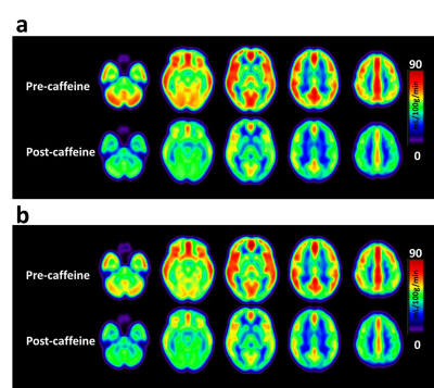 |
14 | Effect of caffeine on cerebral hemodynamic response: The modulation of dietary caffeine consumption
Shin-Lei Peng1, Lok Wang Lauren Chu1, and Feng-Yi Su2
1Department of Biomedical Imaging and Radiological Science, China Medical University, Taichung, Taiwan, China Medical University, Taichung, Taiwan, 2Department of Medical Imaging, China Medical University Hospital, Taichung, Taiwan
The main aim of this study was to characterize the acute effect of caffeine on cerebral hemodynamic responses in participants with different caffeine consumption habits. The non-habitual group exhibited a larger degree of vasoconstriction and thus diminished the ability to dilate upon stimulation. As the vessel dilation ability has been considered as a covariate to explain variabilities in fMRI signals, our results may suggest that the suppressed BOLD response to a visual stimulation in low-caffeine-level users could be partially attributed to the decreased vascular reactivity altered by the baseline perfusion.
|
||
2994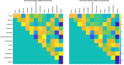 |
15 | The structure-function coupling of aging brain
Hui Zhang1,2, Peng Cao1, Henry K.F. Mak2,3, and Edward S. Hui4
1Diagnostic Radiology, The University of Hong Kong, Pokfulam, Hong Kong, 2Alzheimer's Disease Research Network, The University of Hong Kong, Pokfulam, Hong Kong, 3The University of Hong Kong, Pokfulam, Hong Kong, 4Rehabilitation Sciences, The Hong Kong Polytechnic University, Hung Hom, Hong Kong
We aim to investigate the effect of aging, sex, years of education, total cognition, and the disease burden of small vessel disease, the most cause of vascular dementia, on not only the coupling of the entire brain, but also the coupling of intra and inter-functional networks. We have demonstrated varying effect of sex, years of education and total cognition on global and intra/inter-network couplings.
|
||
 Back to Meeting Home
 Back to the Program-at-a-Glance
The International Society for Magnetic Resonance in Medicine is accredited by the Accreditation Council for Continuing Medical Education to provide continuing medical education for physicians.
 Back to Meeting Home
 Back to the Program-at-a-Glance
 View the Presentation