Online Gather.town Pitches
fMRI & Connectivity in Disease
Joint Annual Meeting ISMRM-ESMRMB & ISMRT 31st Annual Meeting • 07-12 May 2022 • London, UK

Online Gather.town Pitches
fMRI & Connectivity in Disease
| Booth # | ||||
|---|---|---|---|---|
3603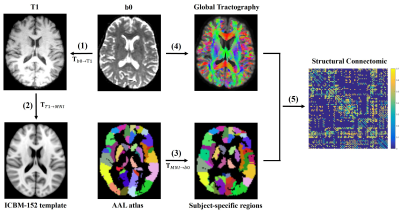 |
1 | Radiation-induced structural network alterations in patients with nasopharyngeal carcinoma: a longitudinal study
Xinyuan Zhang1, Xiaofei Lv2, Pu Xu1, Yuhao Lin1, Long Qian3, and Yanqiu Feng1
1School of Biomedical Engineering, Southern Medical University, Guangzhou, China, 2Department of Medical Imaging, Sun Yat-sen University Cancer Center, State Key Laboratory of Oncology in South China, Collaborative Innovation Center for Cancer Medicine, Guangdong Key Laboratory of Nasopharyngeal Carcinoma Diagnosis and Therapy, Guangzhou, China, 3MR Research, GE Healthcare, Beijing, China
We conducted a longitudinal study to explore the radiation-induced alterations of structural brain network for nasopharyngeal carcinoma (NPC) patients using diffusion tensor imaging (DTI) technique and graph theory. The dose-dependent effect was also investigated using Spearman's Rank Correlation Coefficient. The structural brain connectivity was found to be altered at both 0~3 (acute) and 6 months (early-delayed) after radiotherapy (RT) and showed a tendency of recovery at 12 months (late-delayed) after RT. Also, the change of nodal properties were related to the temporal dose. These findings provide new insights into the pathophysiological mechanism of radiation-induced brain injuries.
|
||
3604 |
2 | The Brain Functional Connectivity can Predict Response to Treatment with Transcutaneous Auricular Vagus Nerve Stimulation in Primary Insomnia Video Not Available
Wenting Luo1, Yue Zhang2, Xiaoyan Hou2, Menghan Feng1, Chengwei Fu1, Weicui Chen2, Xian Liu2, Zhaoxian Yan2, Kan Deng3, Biyun Xu4, and Bo Liu2
1The Second Clinical College, Guangzhou University of Chinese Medicine, Guangzhou, China, 2Department of Radiology, The Second Affiliated Hospital of Guangzhou University of Chinese Medicine, Guangzhou, China, 3Philips Healthcare, Guangzhou, China, 4Department of Sleep Disorder, Fangcun Branch, The Second Affiliated Hospital of Guangzhou University of Chinese Medicine, Guangzhou, China
The individual response to treatment with the transcutaneous auricular vagus nerve stimulation ( taVNS ) in primary insomnia varies greatly, while there is lack of objective markers for patient’s treatment outcome. In this work, we demonstrated that the baseline functional connectivity combined with machine learning algorithms can predict response to treatment with taVNS in primary insomnia. The functional connectivity values within and between brain networks such as the default mode network, affective network, visual network, and cerebellar network maybe potential objective markers of patient’s treatment outcome.
|
||
3605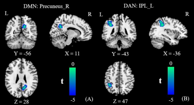 |
3 | Dysfunctional interaction between the dorsal attention network and the default mode network in Patients with Type 2 Diabetes Mellitus
Yumeng Lei1, Dongsheng Zhang1, Fei Qi1, Jie Gao1, Min Tang1, Kai Ai2, Xuejiao Yan1, Xiaoyan Lei1, Zhirong Shao3, Yu Su1, and Xiaoling Zhang1
1Department of MRI, Shaanxi Provincial People’s Hospital, Xi’an, China, 2Philips Healthcare, Xi'an, China, 3Xi'an Medical University, Xi’an, China
To investigate the specific neural substrate of type 2 diabetes mellitus (T2DM) -related cognitive impairment, independent component analysis (ICA) and functional network connection analysis (FNC) methods were applied to resting-state fMRI images from 44 patients with T2DM and 47 healthy controls (HCs). This study found the inherent neural mechanism between the dorsal attention network (DAN) and default mode network (DMN) was eliminated, and these abnormal changes were correlated with the scores of multiple neuropsychological assessments (P < 0.05), indicated that the abnormal changes of the DAN-DMN may be the neural basis of T2DM-related cognitive deficits.
|
||
3606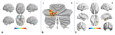 |
4 | Altered Brain Regional Homogeneity at Resting-State in Spinocerebellar Ataxia Type 3 Patients
Bing Liu1,2, Linwei Zhang3, Wenwen Gao1, Lei Du1, Aocai Yang1, Yue Chen1, Yige Wang1, Xiuxiu Liu1, Kuan Lv1, and Guolin Ma1
1Department of Radiology, China Japan Friendship Hospital, Beijing, China, 2Graduate School of Peking Union Medical College, Beijing, China, 3Department of Neurology, China Japan Friendship Hospital, Beijing, China
To explore the neurofunctional change in spinocerebellar ataxia type 3 patients, regional homogeneity values were analyzed from this resting-state functional magnetic imaging study. The altered brain regional homogeneity mainly appeared in the left cerebellum and right cerebrum. And positive correlations were shown between regional homogeneity values for the right cerebrum and the clinical ataxia scores.
|
||
3607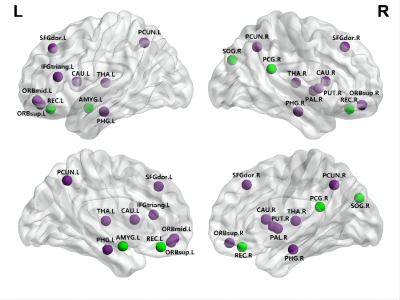 |
5 | Disrupted small-world networks in children with attention-deficit/hyperactivity disorder: a DTI-based network analysis
Liuhui Wu1, Yingqian Chen1, Shu Su1, Zhiyun Yang1, Yan Dai1, and Long Qian2
1Diagnostic Radiology, The First Affiliated Hospital of Sun Yat-sen University, Guangzhou, Guangdong, China, 2MR Research, GE Healthcare, Beijing, China
Attention-deficit/hyperactivity disorder (ADHD) is the most common neurodevelopmental disorder in school-age children, the pathogenesis is still unclear. In current study, 49 pediatric with ADHD and 51 age- and gender-matched typically developing (TD) children were included. Diffusion tensor imaging (DTI) and graph theory approaches was used. At the global level, patients with ADHD showed increased characteristic Lp, γ, σ and decreased Eglob; regionally, altered nodal profiles were mainly in the default mode, central executive network and basal ganglia. In particular, nodal betweenness of left caudate was negatively associated with the clinical symptom severity.
|
||
3608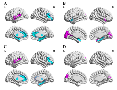 |
6 | Altered neurovascular coupling in drug-naïve attention-deficit/hyperactivity disorder: Evidence from a comprehensive fMRI analysis
shu su1, Yingqian Chen 1, Long Qian 2, Yan Dai 1, Zi Yan1, Liping Lin1, Mengsha Zou1, Qin Zhou1, Meina Liu1, Hongyu Zhang1, and Zhiyun Yang1
1First Affiliated Hospital, Sun Yat-sen University, guangzhou, China, 2MR Research, GE Healthcare, guangzhou, China
As the most common childhood-onset neurodevelopmental disorder, the pathogenesis of attention-deficit/ hyperactivity disorder (ADHD) is still unclear. Non-invasive neuroimaging methods open new avenues to characterize developmental changes in the human brain. Neurovascular coupling (NVC) reflects the close relationship between neuronal activity and cerebral blood flow (CBF), which provides a new mechanistic insight into developmental brain alteration. We aim to investigate the potential NVC alteration in ADHD children.
|
||
3609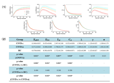 |
7 | Disrupted topological organization in cerebral small vessel disease: A resting-state functional brain networks and graph theory study
Haotian Xin1, Hongwei Wen2,3, Mengmeng Feng1, Lingfei Guo4, and Changhu Liang4
1Cheeloo College of Medicine, Shandong University, Jinan, China, 2Key Laboratory of Cognition and Personality (Ministry of Education), Chongqing, China, 3School of Psychology, Southwest University, Chongqing, China, 4Department of Radiology, Shandong Provincial Hospital Affiliated to Shandong First Medical University, Jinan, China
We aim to investigate functional topological brain networks changes in cerebral small vessel disease (CSVD) patients with and without cerebral microbleeds (CMBs). We divided the subjects into three subgroups: CSVD with CMBs (CSVD-c), CSVD without CMBs (CSVD-n) and control. Then, we analyzed topological disruptions between groups using graph theory and related these alterations to clinical parameters. CSVD-c showed significantly altered global, nodal topological properties and disrupted distribution of hub regions, involving the default mode network (DMN), attention, sensorimotor and visual functional areas. These findings extended our understanding of neurobiological mechanisms of CSVD.
|
||
3610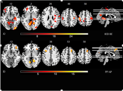 |
8 | Static and temporal dynamic changes of intrinsic brain activity in adolescents and adult OCD patients
Xu yin huan1, Zheng rui ping1, Wei ya rui1, Han shao qiang1, and Zhang yan1
1The First Affiliated Hospital of Zhengzhou University, Zhengzhou, China
This study used a novel statistical analysis-2x2 interaction effect to differentiate OCD patients from healthy controls in both adolescents and adults. We analyzed a classical index-low frequency fluctuation amplitude(ALFF) and drew a convincing conclusion that some regions had a correlation with people in different age and symptoms in OCD patients. Originally, we proposed a novel strategy of dALFF which allows estimating dynamics of intrinsic brain activity with patients. Compared to static indexes, dynamic indexes might be used as a powerful supplement differentiating patients from different age, helping us obtain a more comprehensive understanding of neural activity.
|
||
3611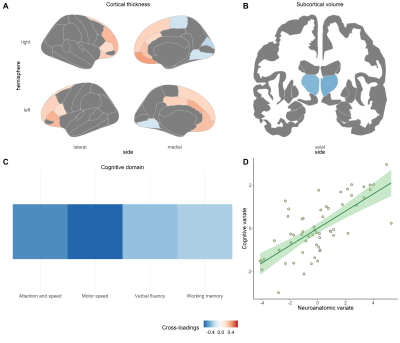 |
9 | Disparate Multivariate Relationships between Brain-Behavior Dimensions across Patients with Schizophrenia at Different Illness Stages
Qiannan Zhao1 and Su Lui1
1Huaxi MR Research Center (HMRRC), Functional and Molecular Imaging Key Laboratory of Sichuan Provinc, West China Hospital of Sichuan University, Chengdu, China
Clarifying brain-behavior associations in schizophrenia helps understand neurobiological mechanisms and explore biomarkers for patient stratification and cognition-targeted interventions. Sparse canonical correlation analysis (sCCA) is a method that maximizes the correlations between linear combinations of each high-dimensional data set, providing more information relative to traditional bivariate correlation analysis. In order to characterize multivariate brain-behavior associations in schizophrenia, we performed sCCA in patients at different illness stages. Disparate canonical correlations were found across FES patients and stable treated patients, involving cortical thickness and surface area contributed in each sample, consistent with their well-established differences in neurodevelopmental trajectories, genetics, and contributions to cognition.
|
||
3612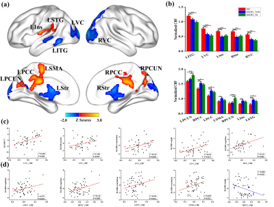 |
10 | Altered resting-state cerebral blood flow and functional connectivity mediate suicidal ideation in major depressive disorder
Dandan Fan1, Cancan He1, Xinyi Liu1, Feifei Zang1, Yao Zhu1, and Chunming Xie1,2,3
1Department of Neurology, Affiliated ZhongDa Hospital, School of Medicine, Southeast University, Nanjing, China, 2Neuropsychiatric Institute, Affiliated ZhongDa Hospital, Southeast University, Nanjing, China, 3The Key Laboratory of Developmental Genes and Human Disease, Southeast University, Nanjing, China
The relationship among cerebral blood flow (CBF), functional connectivity (FC), and suicidal ideation (SI) in MDD patients has been elusive. 175 participants including 47 MDD without SI (MDDNSI), 59 MDD with SI (MDDSI), and 69 controls (HC) underwent ASL, resting-state fMRI scans, and completed neuropsychological tests. Results showed that CBF was increased in MDDSI patients in the bilateral precuneus. MDDSI patients exhibited enhanced FC in the prefrontal-limbic system and decreased FC in the right precentral gyrus relative to MDDNSI patients. Mediation analyses identified that the FC could mediate the association between CBF and behavioral performance in subgroups.
|
||
3613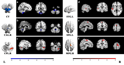 |
11 | Dynamic Alterations in SpontaneousNeural Activity in First-Episode, Drug-Naive Patients with Major Depressive Disorder.
Yingying Wang1, Jing Liu1, Hailong Li1, Hui Qiu1, Yingxue Gao1, Zilin Zhou1, Kaili Liang1, Weijie Bao1, Lianqing Zhang1, Qiyong Gong1, and Xiaoqi Huang1
1Huaxi MR Research Center (HMRRC), Functional and molecular imaging Key Laboratory of Sichuan Province, Department of Radiology, West China Hospital, Sichuan University, Chengdu, China
We collected resting-state fMRI data from 115 patients with major depressive disorder (MDD) and 127 normal controls (NC). The dynamic amplitude of low-frequency fluctuations (dALFF) and dynamic regional homogeneity (dReHo) in 115 MDD patients and 127 NC were compared. Correlation analyses between dALFF and dReHo in brain regions showing significant intergroup differences and clinical scores were calculated in MDD patients. We found aberrant dALFF and dReHo in MDD patients and their significant correlations with clinical scores, suggesting the pattern of intrinsic brain activity variability might be potential tools to assess MDD patients’ severity of illness.
|
||
3614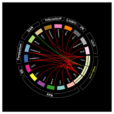 |
12 | Abnormal functional connectivity of default mode network subsystems in first-episode drug-naïve major depressive disorder
Hui Qiu1, Hailong Li1, Yingxue Gao1, Lingxiao Cao1, Jing Liu1, Weijie Bao1, Yingying Wang1, Zilin Zhou1, Suming Zhang1, Mengyue Tang1, Lianqing Zhang1, Lu Lu1, Xiaoqi Huang1, and Qiyong Gong1
1Huaxi MR Research Center (HMRRC), Department of Radiology, West China Hospital of Sichuan University, Chengdu, China
Most previous studies took DMN as a whole and measured its FC abnormalities on a network level while recently, researchers revealed DMN has three subsystems (core, dMPFC and MTL subsystem). Few studies had explored FC on subsystem levels in first-episode drug-naïve (FEDN) MDD patients. We used seed-based whole-brain functional connectivity analysis in a relatively large sample of FEDN MDD patients, we found hyper-connectivity between DMN three subsystems and frontoparietal network as well as cerebellum network, and hypo-connectivity between dMPFC subsystem and dorsal attention network. These findings might provide a more comprehensive understanding for DMN FC changes in MDD.
|
||
3615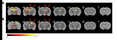 |
13 | Brain mapping of the temporal interference stimulation wth functional MRI
Zonghao Xin1, Yoshifumi Abe2, Akihiro Kuwahata3, Kenji F. Tanaka2, and Masaki Sekino1
1Department of Bioengineering, School of Engineering, The University of Tokyo, Tokyo, Japan, 2Division of Brain Sciences, Institute for Advanced Medical Research, Keio University School of Medicine, Tokyo, Japan, 3Department of Electrical Engineering, Graduate School of Engineering, Tohoku University, Sendai, Japan
Temporal interference stimulation (TIS) is a novel, non-invasive deep brain stimulation (DBS) methodology thatutilizes multiple external electric fields with amplitude modulation (AM) to stimulate deep brain regions without affectingthe superficial cortical areas. However, the clinical application of TIS is inhibited by its unproved response of the brain to the modulated stimulation. In this study, we investigated the instantaneous brain response to TIS using fMRI technique in rodents. The results demonstrated the compatibility of TIS with fMRI, as well as the feasibility of stimulating subcortical encephalic regions non-invasively and focally.
|
||
3616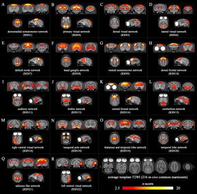 |
14 | Highly reliability resting-state networks in common marmoset brains. Video Permission Withheld
Yawara Haga1,2,3, Junichi Hata1,2,3,4,5, Takaaki Kaneko1, Kei Hagiya1, Yuji Komaki3,4, Fumiko Seki1,3,4, Daisuke Yoshimaru1,3,4,5, Kanako Muta1,2,5, Noriyuki Kishi1,4, Takako Shirakawa2, and Hideyuki Okano1,4
1Laboratory for Marmoset Neural Architecture, RIKEN CBS, Saitama, Japan, 2Graduate School of Human Health Sciences, Tokyo Metropolitan University, Tokyo, Japan, 3Live Animal Imaging Center, Central Institute for Experimental Animals, Kanagawa, Japan, 4Department of Physiology, Keio University School of Medicine, Tokyo, Japan, 5Division of Regenerative Medicine, The Jikei University School of Medicine, Tokyo, Japan
We identified the resting-state networks in common marmoset brains with a larger amount of rsfMRI data (60 hours). The group-ICA and single subject-ICA were performed in this study. As a results, 18 networks were detected with highly reliability. It included the default mode network, a network most likely involved with visual pathways, and the limbic network. In addition, our data suggests that the resting-state networks in marmoset may be similar to that in human and macaque monkey. Therefore, our study shows that the evaluation of the functional effects of the neurodegenerative diseases using common marmosets is highly useful.
|
||
3617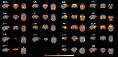 |
15 | How does midazolam affect resting-state networks in common marmosets? An investigation using a 9.4 T magnetic resonance imaging system. Video Permission Withheld
Kanako Muta1,2, Junichi Hata1,2,3,4, Yawara Haga1,3, Daisuke Yoshimaru2, Kei Hagiya3, Takaaki Kaneko3, Takako Miyabe-Nishiwaki5, Yuji Komaki6, Fumiko Seki6, Hirotaka James Okano2, and Hideyuki Okano3,4
1Tokyo Metropolitan University, Tokyo, Japan, 2The Jikei University School of Medicine, Tokyo, Japan, 3RIKEN Center for Brain Science, Saitama, Japan, 4Keio University, Tokyo, Japan, 5Kyoto University, Aichi, Japan, 6Central Institute for Experimental Animals, Kanagawa, Japan
The purpose of this study is to evaluate the effects of midazolam on resting-state networks in common marmosets. Functional data were collected using a 9.4 T MRI system in the sedative condition with midazolam, the awake condition as a positive control, and the anesthetic condition with isoflurane as a negative control. Independent component analysis and functional connectivity analysis were performed. These results were apparently altered by isoflurane from awake condition, but not largely by midazolam. These results in midazolam were different from a previous report in humans and it may cause different effects of midazolam in human and nonhuman primates.
|
||
3618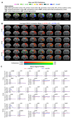 |
16 | Are topographically segregated excitatory neurons in visual thalamus functionally diverse? An optogenetic fMRI study
Linshan Xie1,2, Xunda Wang1,2, Teng Ma1,2,3, Pit Shan Chong4, Lee Wei Lim4, Peng Cao3, Pek-Lan Khong3, Ed X. Wu1,2, and Alex T. L. Leong1,2
1Laboratory of Biomedical Imaging and Signal Processing, The University of Hong Kong, Hong Kong SAR, China, 2Department of Electrical and Electronic Engineering, The University of Hong Kong, Hong Kong SAR, China, 3Department of Diagnostic Radiology, Li Ka Shing Faculty of Medicine, The University of Hong Kong, Hong Kong SAR, China, 4School of Biomedical Sciences, Li Ka Shing Faculty of Medicine, The University of Hong Kong, Hong Kong SAR, China
The dorsal lateral geniculate nucleus (dLGN) plays an essential role in visual processing. There are two types of topographically segregated excitatory neurons in dLGN with different outputs to visual cortex, suggesting functional differences when processing visual inputs at the subcortical thalamic level. However, their long-range functional pathways have yet to be reported. Here, we employed optogenetics in combination with fMRI to precisely target the two subdivisions of dLGN and examine whether these two types of neurons are truly functionally diverse at the systems level to facilitate various known complex visual processing functions.
|
||
 Back to Meeting Home
 Back to the Program-at-a-Glance
The International Society for Magnetic Resonance in Medicine is accredited by the Accreditation Council for Continuing Medical Education to provide continuing medical education for physicians.
 Back to Meeting Home
 Back to the Program-at-a-Glance
 View the Presentation