Online Gather.town Pitches
Structural & Functional Connectivity
Joint Annual Meeting ISMRM-ESMRMB & ISMRT 31st Annual Meeting • 07-12 May 2022 • London, UK

Online Gather.town Pitches
Structural & Functional Connectivity
| Booth # | ||||
|---|---|---|---|---|
| 4758 | 1 | Diffusion MRI harmonization and thresholding improve multicentre network analysis: a demonstration in cerebral small vessel disease
Bruno Miguel de Brito Robalo1,2, Alberto de Luca1,2, Christopher Chen3, Anna Dewenter4, Marco Duering5, Saima Hilal3, Huiberdina L. Koek6, Anna Kopczak4, Bonnie Yin Ka Lam7, Alexander Leemans2, Vincent Mok7, Laurien P. Onkenhout1, Hilde van den Brink1,
and Geert Jan Biessels1
1Neurology, Department of Neurology and Neurosurgery, UMC Utrecht Brain Center, University Medical Center Utrecht, Utrecht, Netherlands, 2Image Sciences Institute, University Medical Center Utrecht, Utrecht University, Utrecht, Netherlands, 3Memory, Aging and Cognition Center, Department of Pharmacology, National University of Singapore, Singapore, Singapore, 4Institute for Stroke and Dementia Research (ISD), University Hospital, LMU Munich, Munich, Germany, 5Medical Image Analysis Center (MIAC) and qbig, Department of Biomedical Engineering, University of Basel, Basel, Switzerland, 6Department of Geriatric Medicine, University Medical Center Utrecht, Utrecht, Netherlands, 7Department of Geriatric Medicine, University Medical Center Utrecht, Utrecht, The Netherlands 7Division of Neurology, Department of Medicine and Therapeutics, Gerald Choa Neuroscience Centre, Faculty of Medicine, Prince of Wales Hospital, The Chinese University of Hong Kong, Hong Kong, Hong Kong We investigated if network thresholding and diffusion MRI (dMRI) harmonization improve a) cross-site consistency of network architecture and b) precision and sensitivity to detect network connections disrupted in cerebral small vessel disease (SVD). Brain networks were reconstructed from dMRI in five cohorts. Consistency of network architecture was examined in age-matched controls whereas sensitivity and precision to detect disrupted connections was assessed in sporadic SVD patients. Network consistency, as well as sensitivity and precision to detect disrupted connections were improved by thresholding and harmonization. We recommend using these techniques in networks studies of SVD to leverage existing multicentre datasets. |
||
 |
4759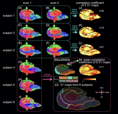 |
2 | Intra-hippocampal fiber tracking and connectome from submillimeter DTI of the human hippocampus in vivo and ex vivo
Yixin Ma1,2, Trong-Kha Truong1,2,3, Iain P. Bruce1,4, Alexandra Badea1,2,3,4, Simon Davis4, Chun-Hung Yeh5, Jeffrey R. Petrella1,2,3, and Allen W. Song1,2,3
1Brain Imaging and Analysis Center, Duke University, Durham, NC, United States, 2Medical Physics Graduate Program, Duke University, Durham, NC, United States, 3Department of Radiology, Duke University, Durham, NC, United States, 4Department of Neurology, Duke University, Durham, NC, United States, 5Institute for Radiological Research, Chang Gung University and Chang Gung Memorial Hospital, Taoyuan, Taiwan
The hippocampus plays an essential role in memory; the impairment in intra-hippocampal connectivity could result in memory loss in neurodegenerative dementias. We generate intra-hippocampal fiber tracts and connectomes from submillimeter isotropic DTI images for repeated scans from the same subjects and across different subjects. We characterize the fiber orientations and connectivity across hippocampal subfields by registering all in vivo scans onto the population template and we compare these results with those from an ex vivo human brain sample. This method can assess intra-hippocampal connectivity in vivo, which could be potentially used to diagnose neurodegenerative diseases.
|
|
4760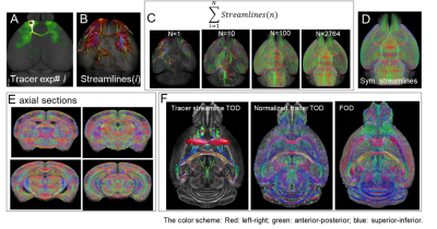 |
3 | Accurate Estimation of Fiber Orientations in the Mouse Brain from Diffusion MRI Signals: Learning from Histological Ground Truth
Zifei Liang1, Tanzil Mahmud Arefin1, Choong Heon Lee1, and Jiangyang Zhang1
1NYU Langone Health, New York, NY, United States
Although dMRI tractograophy has been successfully used to examine brain connectivity, its limitation, mainly in specificity, has also been reported. In this study, we generated a comprehensive mouse brain streamline database based on 2700+ viral tracer data from Allen Institute. The database was used as a ground truth to train a deep learning network to estimate fiber orientations from diffusion MRI data of the mouse brain. Compared to conventional methods, the deep learning network provided more accurate estimation of fiber orientation leading to improved tractography.
|
||
4761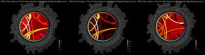 |
4 | Quantitative anisotropy-based fiber tractography reveals tracts moderating age-related decline in functional fitness
Paul B Camacho1,2,3,4, Nishant Bhamidipati1,5, Emily Erlenbach6, Veronica Garcia6, Edward McAuley1,6, Nicholas Burd6, Jessica Damoiseaux7,8, Brad P Sutton1,2,5, and Neha P Gothe1,6
1Beckman Institute for Advanced Science & Technology, University of Illinois at Urbana-Champaign, Urbana, IL, United States, 2Neuroscience Program, University of Illinois at Urbana-Champaign, Urbana, IL, United States, 3Interdisciplinary Health Sciences Institute, University of Illinois at Urbana Champaign, Urbana, IL, United States, 4Carle-Illinois Advanced Imaging Center, University of Illinois at Urbana-Champaign, Urbana, IL, United States, 5Bioengineering, University of Illinois at Urbana-Champaign, Urbana, IL, United States, 6Kinesiology & Community Health, University of Illinois at Urbana-Champaign, Urbana, IL, United States, 7Institute of Gerontology, Wayne State University, Detroit, MI, United States, 8Psychology, Wayne State University, Detroit, MI, United States
We present a moderation analysis of the effects of edge strengths from generalized q-sampling imaging-based tractography on the relationship between age and decline in functional fitness in older adults (n = 105, ages 55-79 years old, right-handed). The results of these moderation analyses suggest that the strengths of white matter structural connections involving the cerebellum, cingulum, and other areas involved in motor, sensory, and environmental perception may play a significant role in preserving functional fitness in older adults.
|
||
4762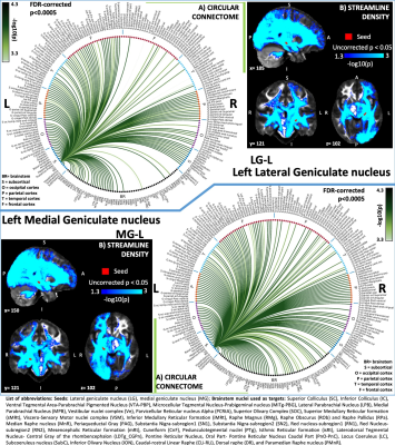 |
5 | Functional and structural connectomes of the lateral and medial geniculate nuclei in living humans using 7 Tesla MRI
Kavita Singh1, Maria Guadalupe Garcia Gomar1,2, Simone Cauzzo1,3,4, and Marta Bianciardi1,5
1Brainstem Imaging Laboratory, Department of Radiology, Athinoula A. Martinos Center for Biomedical Imaging, Boston, MA, United States, 2National Autonomous University of Mexico, Mexico, Mexico, 3Sant'Anna School of Advanced Studies, Institute of Life Sciences, Pisa, Italy, 4Research Center E. Piaggio, University of Pisa, Pisa, Italy, 5Division of Sleep Medicine, Harvard University, Division of Sleep Medicine, Boston, MA, United States
The connectivity of lateral and medial geniculate nuclei with cortical and sub-cortical areas involved in visual and auditory functions is known in humans, yet their recently postulated connectivity with neuromodulatory, visceral and sensory brainstem nuclei is understudied. This is due to limited resolution of clinical scanners and lack of brainstem atlas in living humans. To this end, we applied our recently developed in-vivo atlas of brainstem and diencephalic nuclei to 7 Tesla HARDI and resting-state fMRI, and mapped structural and functional connectomes of lateral and medial geniculate nuclei with brainstem nuclei along with cortical and sub-cortical regions.
|
||
4763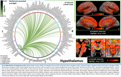 |
6 | Functional connectome of hypothalamus, nucleus accumbens and hippocampus in living humans from 7 Tesla resting-state fMRI
Simone Cauzzo1,2,3, Maria Guadalupe Garcia Gomar3,4, Kavita Singh3, and Marta Bianciardi3,5
1Institute of Life Sciences, Scuola Superiore Sant'Anna, Pisa, Italy, 2Research Center E. Piaggio, University of Pisa, Pisa, Italy, 3Department of Radiology, Athinoula A. Martinos Center for Biomedical Imaging (MGH), Charlestown, MA, United States, 4Escuela Nacional de Estudios Superiores, Juriquilla, Universidad Nacional Autónoma de México, Queretaro, Mexico, 5Division of Sleep Medicine, Harvard University, Boston, MA, United States The hypothalamus and nucleus accumbens promote respectively wakeful arousal and sleep, producing a stable cycle between states. The hippocampus is inherently connected to this cycle, which modulates memory encoding during wakefulness and sleep. While their connectivity to cortical regions is detailed in literature, their connectivity with brainstem nuclei is understudied in living humans. By using high spatial resolution 7 Tesla resting state fMRI and an in-vivo brainstem nuclei atlas, we provided a functional connectome of hypothalamus, nucleus accumbens and hippocampus with the rest of the brain, including 58 brainstem nuclei along with 148 cortical and 25 subcortical structures. |
||
4764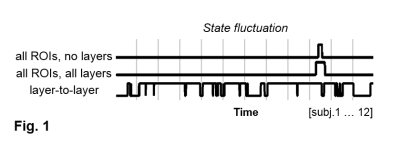 |
7 | High dynamicity of cortical depth-dependent connectivity states
Patricia Pais-Roldan1, Shukti Ramkiran1,2, Seong Dae Yun1, and Jon N. Shah1,3,4,5
1INM-4, Forschungszentrum Juelich, Juelich, Germany, 2Department of Psychiatry, Psychotherapy and Psychosomatics, RWTH Aachen University, Aachen, Germany, 3INM-11, Forschungszentrum Juelich, Juelich, Germany, 4Translational Medicine, JARA-BRAIN, Aachen, Germany, 5Department of Neurology, RWTH Aachen University, Aachen, Germany
We assessed resting-state fMRI acquired with high spatial resolution with a dynamic functional connectivity analysis involving multiple cortical ROIs and depths. Two states relevant to the normalized global laminar connectivity were identified in healthy volunteers (superficial vs. deep layer connectivity predominance). Mean laminar connectivity states were much more dynamic than the states identified in a routine ROI-based dynamic connectivity analysis, suggesting the potential of high-spatial-resolution fMRI to reveal time-varying features in the human brain.
|
||
4765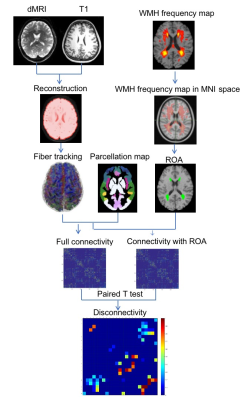 |
8 | Disconnectome Associated with Progressive White Matter Hyperintensities in Brain Ageing
Meng Li1,2, Mohamad Habes3, and John Detre 1
1Department of Neurology, University of Pennsylvania, Philadelphia, PA, United States, 2College of Medicine and Biological Information Engineering, Northeastern University, Shenyang, China, 3Biggs Alzheimer’s Institute, University of Texas San Antonio, San Antonio, TX, United States
White matter hyperintensities (WMH) are commonly seen in older adults and are associated with an increased risk of cognitive decline and dementia, though the mechanism by which WMH cause cognitive decline are incompletely characterized. In this study, we estimated changes in the structural connectome due WMH with aging using WMH lesion frequency maps derived from a population based study as regions of avoidance for DTI tractography on healthy subject DTI data. We found that subcortical connections to frontal cortex are affected in the 50’s and 60’s, with more widespread disconnection increasing more rapidly in the 70’s and 80’s. However, even in the oldest subjects, most edges were only partially disconnected.
|
||
4766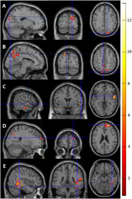 |
9 | Effect of Meditation on Brain Function During an Attention Task Using ASL and BOLD fMRI
Yakun Zhang1, Shichun Chen1, Zongpai Zhang1, Wenna Duan1, Li Zhao2, George Weinschenk1, Wen-Ming Luh3, Adam Anderson4, and Weiying Dai1
1Department of Computer Science, State University of New York at Binghamton, Binghamton, NY, United States, 2College of Biomedical Engineering & Instrument Science, Zhejiang University, Hangzhou, China, 3National Institute on Aging, National Institutes of Health, Baltimore, MD, United States, 4Department of Human Development, Cornell University, Ithaca, NY, United States
The effect of meditation on brain functional activation when engaged in an attention task was evaluated longitudinally using dASL and BOLD fMRI in nine healthy subjects. Functional activation before and after meditation practice was compared and the change of functional activation was correlated with practice time. Using dASL, functional activation in the occipital region was significantly reduced; more practice time was associated with more reduced activation in the mediofrontal, temporal, and precuneus regions. Using BOLD fMRI, no significant activation was found. The findings suggest that dASL has superior performance in detecting task performance and that meditation can improve brain efficiency.
|
||
4767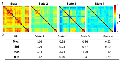 |
10 | Longitudinal Recovery of Dynamic Functional Connectivity After Focused Ultrasound-Neuromodulation in V2L of Normal Rat Model
Yu-Chieh Hung1, Yi-Cheng Wang1, Hao-Li Liu2, and Hsu-Hsia Peng1
1Department of Biomedical Engineering and Environmental Sciences, National Tsing Hua University, Hsinchu, Taiwan, 2Department of Electrical Engineering, National Taiwan University, Taipei, Taiwan
Non-invasive focused ultrasound (FUS) offered attractive advantages to modulate neuronal activity. The functional connectivity (FC) between different brain regions is a dynamic process during a period of examinations. We aimed to explore the longitudinal effect of FUS-neuromodulation on secondary visual cortex lateral area (V2L) by dynamic FC. We assessed k-means analysis and dynamic network analysis of dynamic FC of Pre-FUS, Sham, 35-min, 3-hr, and 3-day after FUS sonication at V2L. We found the instant effect of FUS-neuromodulation (35-min) and the recovery in a longitudinal follow-up of 3-day, suggesting the potential usefulness of FUS-neuromodulation.
|
||
4768 |
11 | Evaluation of traumatic brain injury in pigs using a novel cross-group temporal correlation analysis
Wenwu Sun1, William Reeves1, Madison Fagan2, Christina Welch2, Kelly Scheulin2, Sydney Sneed2, Franklin West2, and Qun Zhao1
1Department of Physics and Astronomy, University of Georgia, Athens, GA, United States, 2Regenerative Bioscience Center and Department of Animal and Dairy Science, University of Georgia, Athens, GA, United States
Traumatic brain injury (TBI) can lead to dynamic changes in functional network activity that can be assessed by functional magnetic resonance imaging (fMRI). In this study, we utilized a novel temporal correlation analysis approach to evaluate a novel microbiome transplantation treatment effect on network connectivity in a translational porcine. Sparse dictionary learning (sDL) and independent component analysis (ICA) were applied to both a full dataset (TBI and sham) and the Sham-only dataset, resulting in four groups of results. Consistency was observed across the four results, indicating that evaluation of treatment effects can be achieved through the proposed temporal correlation analysis.
|
||
4769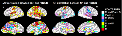 |
12 | Brain Mapping of Mindfulness Meditation, Slow Paced Breathing and Spontaneous Breathing
Suk-tak Chan1 and Kenneth Kwong K Kwong1
1Martinos Center, Radiology, Massachusetts General Hospital, Charlestown, MA, United States
A preliminary comparison of physiological and fMRI data from an experienced meditator during mindfulness, paced breathing at 6 breaths/min, and spontaneous breathing provided several characteristic findings related to the brain-body mechanisms of mindfulness: 1) a reduction of breathing rate together with indicators of improved respiratory gas exchange (RGE); 2) a significant correlation between the temporal oscillation of RGE metrics and cerebral hemodynamic fluctuations (CHF) in regions within the salience and default mode networks; 3) a difference in the coherence of CHF with RGE and with heart cycle duration (NN).
|
||
 Back to Meeting Home
 Back to the Program-at-a-Glance
The International Society for Magnetic Resonance in Medicine is accredited by the Accreditation Council for Continuing Medical Education to provide continuing medical education for physicians.
 Back to Meeting Home
 Back to the Program-at-a-Glance
 View the Presentation