Online Gather.town Pitches
Advanced Imaging in Neurology: Brain Tumors
Joint Annual Meeting ISMRM-ESMRMB & ISMRT 31st Annual Meeting • 07-12 May 2022 • London, UK

Online Gather.town Pitches
Advanced Imaging in Neurology: Brain Tumors
| Booth # | ||||
|---|---|---|---|---|
3619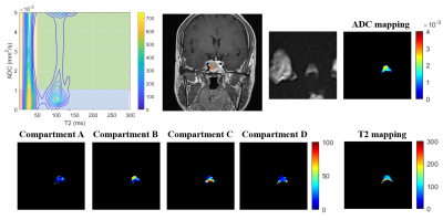 |
1 | Diffusion-Relaxation Correlation Spectrum Imaging for the Assessment of Pituitary Macroadenomas Consistency: A Preliminary Study
Chun-Qiu Su1, Wentao Hu2, Xun-Ning Hong1, Shan-Shan Lu1, and Yongming Dai2
1Department of Radiology, The First Affiliated Hospital of Nanjing Medical University, Nanjing, China, 2United Imaging Healthcare, Shanghai, China Preoperative evaluation of the consistency of pituitary macroadenomas plays a significant role in the determination of the surgical strategy. However, previous studies concerning the assessment of tumor consistency of macroadenomas were controversial1-4. Diffusion-relaxation correlation spectrum imaging (DR-CSI), which provides a correlation spectrum of both water molecular diffusion and T2 relaxation, could be used to resolve the information of tissue compositions, compartments, and heterogeneity at a sub-voxel level. The purpose of our study is to evaluate the utility of DR-CSI in assessing the consistency of pituitary macroadenomas. |
||
3620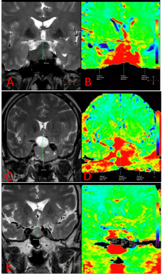 |
2 | Differentiation of hormone secretion related pituitary macroadenomas using Amide Proton Transfer weighted imaging
Wenjun Cui1, Jing Zhang1, Kai Ai2, Yurong Zheng1, Yuping Bai1, Tiejun Gan1, Rui Wang1, Tingli Yang1, Pengfei Wang1, Jie Zou1, Haoyuan Li1, and Yuan Ding1
1Department of Magnetic Resonance,Lanzhou University Second Hospital, Lan Zhou, China, 2Philips Healthcare, Xi'an, China
Advanced radiological modalities such as Amide Proton Transfer weighted imaging (APTw) may be useful in probing biological tissue properties of pituitary adenomas. The objective of the present study was to examine APTw derived APT value to noninvasively and quantitatively measuring pituitary adenomas. Moreover, the correlation analysis was performed between ATP value and hormone secretion to validate the efficiency of ATP value in differentiate hormone secretion related pituitary adenomas.
|
||
3621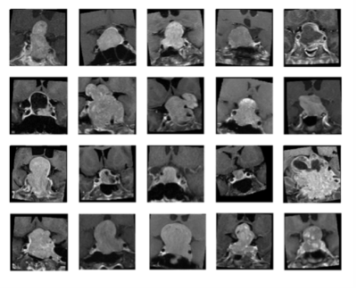 |
3 | Deep Learning for Prediction of Recurrence in Nonfunctioning Pituitary Macroadenomas: Combination of Clinical and MRI Features
Ching-Chung Ko1,2, Yan-Jen Chen3,4, Hsun-Ping Hsieh3, and Jeon-Hor Chen5,6
1Department of Medical Imaging, Chi Mei Medical Center, Tainan, Taiwan, 2Chia Nan University of Pharmacy and Science, Tainan, Taiwan, 3Department of Electrical Engineering, National Cheng Kung University, Tainan, Taiwan, 4Graduate Institute of Electronics Engineering, National Taiwan University, Taipei, Taiwan, 5Department of Radiological Sciences, University of California, Irvine, CA, United States, 6Department of Radiology, E-Da Hospital and I-Shou University, Kaohsiung, Taiwan
A subset of nonfunctioning pituitary macroadenomas (NFMAs) show early progression/recurrence (P/R) after surgery. In clinical practice, one of the main challenges in the treatment of NFMAs is to determine factors that associated with P/R. This study investigated the role of deep learning for the prediction of P/R in NFMAs. 78 patients diagnosed with NFMAs were included. The hybrid CNN-MLP model using both clinical and MRI features showed the best performance for prediction of P/R in NFMAs, with accuracy of 84%, precision of 88%, and AUC of 0.87.
|
||
3622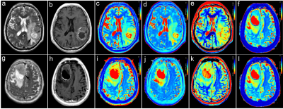 |
4 | Discrimination between High-Grade Gliomas and Solitary Brain Metastases in Peritumoral Brain Zone: Qualitative Analysis Using Relaxation Maps
Xin Ge1,2, Xueying Huang2, Kai Zhu2, Aijun Wang2, Xiaocheng Wei3, Min Li4, Ying Shen1,2, Wenxiao Liu1,2, Ruirui Lv1,2, Peng Yong1,2, Xuhong Yang1,2, and Xiaodong Wang2
1Ningxia Medical University, Yinchuan, China, 2General Hospital of Ningxia Medical University, Yinchuan, China, 3GE Healthcare, MR Research, Beijing, China, 4GE Healthcare, MR Enhancement Application, Beijing, China
This work sought to identify a new non-invasive means to differentiate high-grade gliomas (HGGs) from solitary brain metastases (SBMs). It was concluded that the T1native, T2native, PDnative, and ΔT1ratio values were measured in peritumoral brain zone from synthetic MRI can be used as quantitative imaging biomarkers for distinguishing between HGGs and SBMs. The ΔT1ratio values have higher discrimination abilities compared with other parameters, which worth further study.
|
||
3623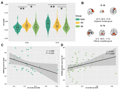 |
5 | The shared and disorder-specific temporal dynamics in degree centrality for patients with obsessive-compulsive disorder and schizophrenia
Yaxuan Wang1, Lekai Luo1, Qian Li1, Wanfang You1, Yuxia Wang1, Qian Zhang1, Su Lui1, Qiyong Gong1, and Fei Li1
1Huaxi MR Research Center (HMRRC), Department of Radiology, West China Hospital of Sichuan University, Chengdu 610041, Sichuan, P.R. China., Chengdu, China
Temporal dynamics in degree centrality (DC) were measured to detect alterations of dynamic topological organization in patients with obsessive-compulsive disorder (OCD) and schizophrenia (SZ). We found both patients with OCD and SZ showed decreased occurrence of a state manifested as the highest centrality in sensory systems. Moreover, we observed common and specific alteration of temporal variability of DC in some brain regions, which correlated with symptom severity of patients, respectively.
|
||
3624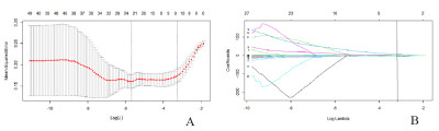 |
6 | A radiomics signature-based nomogram to predict TERT promoter mutation status and the prognosis of glioblastoma
Jun Lu1, Hailiang Li2, and Xiang Li1
1Department of Radiology, Affiliated Cancer Hospital of Zhengzhou University & Henan Cancer Hospital, Zhengzhou, China, 2Department of Radiology and Intervention, Affiliated Cancer Hospital of Zhengzhou University & Henan Cancer Hospital, Zhengzhou, China
This study aimed to establish and validate a radiomics signature-based nomogram with robust radiomics features from contrast enhanced MRI images. The radiomics features were selected using LASSO regression. A prediction model was constructed with multivariate logistic regression analysis. A nomogram combined radiomics signature and clinical factors were established, showing good performance for predicting the TERT mutation status. The clinical value of radiomics nomogram was further assessed by the prognosis analysis. In conclusion, the radiomics signature-based nomogram is a promising method for preoperatively predicting TERT promoter mutation status and has the potential to assess prognosis noninvasively in glioblastoma patients.
|
||
3625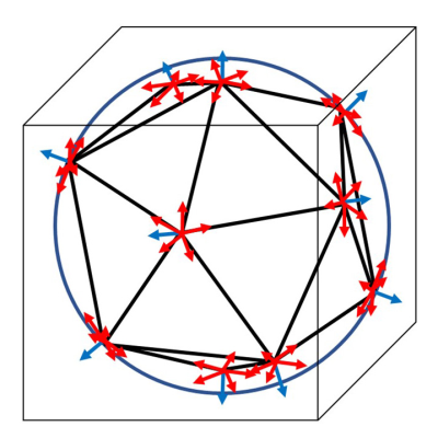 |
7 | Advanced diffusion-weighted imaging for differentiating between glioblastoma and primary central nervous system lymphoma
Kiyohisa Kamimura1, Masanori Nakajo1, Bohara Manisha1, Yoshihiko Fukukura1, Hiroyuki Uchida2, Takashi Iwanaga3, Thorsten Feiweier4, Hiroshi Imai5, and Takashi Yoshiura1
1Radiology, Kagoshima University, Kagoshima, Japan, 2Neurosurgery, Kagoshima University, Kagoshima, Japan, 3Radiological Technology, Kagoshima University Hospital, Kagoshima, Japan, 4Siemens Healthcare GmbH, Erlangen, Germany, 5Siemens Healthcare K.K., Tokyo, Japan
To determine whether time-dependent DWI parameters and microscopic diffusion anisotropy (μFA) are useful for differentiating between glioblastoma and primary central nervous system lymphoma (PCNSL), 51 patients with glioblastoma and 13 with PCNSL were examined. In addition to ADC at two different diffusion times, ADC difference (ΔADC) and ADC change ratio (rcADC) were significantly different between the two tumor types, while no difference was shown for μFA. rcADC showed the best diagnostic performance followed by ΔADC.
|
||
3626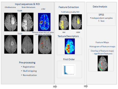 |
8 | Radiomics based characterization of peritumoral edema segmented from DCE MRI to differentiate glioblastoma from solitary brain metastasis
Suhail Parvaze P1, Rakesh K Gupta2, Rupsa Bhattacharjee3, Anup Singh4, Rakesh Kumar Singh5, Gaurav Khanna6, Amal Roy Chaudhary7, Rana Patir8, Sandeep Vaishya8, Jaladhar Neelavalli 9, and Tejas Shah9
1Philips Innovation Campus, Bangalore, India, 2Department of Radiology and Imaging, Fortis Memorial Research Institute, Gurugram, India, 3Indian Institute of Technology Delhi, Delhi, India, 4Department of Biomedical Engineering, Indian Institute of Technology Delhi, Delhi, India, 5Fortis Memorial Research Institute, Gurugram, India, 6SRL Diagnostics, Fortis Memorial Research Institute, Gurugram, India, 7Radiation Oncology, Fortis Memorial Research Institute, Gurugram, India, 8Department of Neurosurgery, Fortis Memorial Research Institute, Gurugram, India, 9BIU, Philips Innovation Campus, Bangalore, India
Edema in GB is characterized by the presence of tumor cells infiltration as compared to brain metastasis with only pure edema. Radiomics features extracted using FLAIR images in GB and BM are found to be exhibiting variation in some of the entropy and non-uniformity-based features, which could be used as signature to differentiate GB from brain metastasis.
|
||
3627 |
9 | Diffusion–Based Virtual MR Elastography of the asymmetric features of neonatal white matter
Yao Ge1, Xianjun Li2, Congcong Liu2, Miaomiao Wang2, Linlin Zhu2, Pengxuan Bai2, Na Zhang2, Yuying Feng2, and Jian Yang2
1Department of Radiology, The First Affiliated Hospital of Xi’an Jiaotong University, xi’an, China, 2Department of Radiology, The First Affiliated Hospital of Xi’an Jiaotong University, xi'an, China
Exploring the asymmetry of the neonatal brain white matter is of great significance for exploring the developmental rules and disease pathogenesis. Virtual MR elastography (VMRE) as a non-invasive technique has also enabled to relate maturation of cerebral structures to neonatal neurodevelopment in addition to conventional techniques.This study aims to explore the capability of diffusion–based VMRE in the characterization of the asymmetric of the neonatal white matter stiffness.The stiffness of neonatal white matter existed asymmetry, and there was a correlation between it and PMA. VMRE may enable to explore the developing brain at several levels.
|
||
3628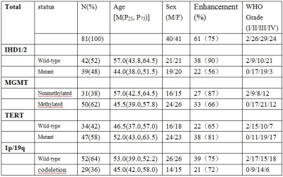 |
10 | Multiparametric MR radiomics in brain glioma: models comparation to predict biomarker status
Jinlong He1, Jianlin Wu1, Yang Gao2, Qiong Wu2, Jing Shen1, Shaoyu Wang3, Huapeng Zhang3, Jialiang Ren4, and Peng Wang5
1Tianjin Medical University Graduate School, Tianjin, China, 2Department Of Imaging Diagnosis, Affiliated Hospital Of Inner Monglia Medical University, Hohhot, China, 3MR Scientific Marketing, Siemens Healthineers, Shanghai, China, 4GE Healthcare, Shanghai, China, 5Inner Monglia Medical University, Hohhot, China
Objective:To compare the performance of clinical model, radiomics model, and combined model in predicting biomarker status (IDH, MGMT, TERT, 1p/19q) of glioma.Methods: 81 glioma patients confirmed by histology were enrolled in this study. The predictive performance of each model was validated by receiver operating characteristic curve (ROC) analysis and decision curve analysis (DCA).Results: The mixed model showed the highest performance in each genic phenotype (IDH AUC = 0.93, MGMT AUC=0.88, TERT AUC=0.76, 1p/19q AUC=0.71).Conclusion: The mixed model is an effective tool to distinguish genic phenotype of brain glioma which have highest diagnostic efficiency than other models.
|
||
3629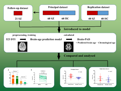 |
11 | Neuroimaging-based brain age prediction of first-episode schizophrenia and the alteration of brain age after early medication
Xu-Sha Wu1, Yi-Bin Xi1, and Hong Yin1
1Xi'an People's Hospital (Xi'an Fourth Hospital), Xi'an, China
Schizophrenia is related to accelerated brain ageing, while individualized biomarkers are scarce. We established a brain age prediction model based on diffusion tensor imaging sequence. The difference between brain age and chronological age of first-episode schizophrenia patients and healthy controls were compared, and the longitudinal studies were performed in follow-up patients after a short-term antipsychotic treatment. The results verified the theory of accelerated brain ageing and medication have an effect on ameliorating it. Structural brain changes in schizophrenia could reveal the accelerated aging and quantification of age discrepancy could contribute to clinical decision-making and find new targeted therapies.
|
||
3630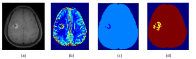 |
12 | Comparison of 2D- and 3D-generated Radiomics feature maps in glioblastoma tissue subtypes segmented from DCE perfusion MRI
Suhail Parvaze P1, Tejas Shah2, Jaladhar Neelavalli 3, Anup Singh4, and Rakesh K Gupta5
1Philips Innovation Campus, Bangalore, India, 2BIU, Philips Innovation Campus, Bangalore, India, 3BIU, Philips Innovation Campus, Gurugram, India, 4Center for Biomedical Engineering, Indian Institute of Technology Delhi, Bangalore, India, 5Department of Radiology and Imaging, Fortis Memorial Research Institute, Gurugram, India
Radiomics based feature value analysis and feature maps especially for first order and texture based features are being thoroughly investigated for diagnostic usability and relevance. Radiomics based feature map generation using 2D and 3D modes on different tumor sub-regions of glioblastoma were performed. 2D-based feature maps depicted accurately the texture variation with respect to other images from the slice of interest, whereas in 3D based maps anatomy and pathology of the neighboring slices induced influence resulting in over depiction of pathology.
|
||
3631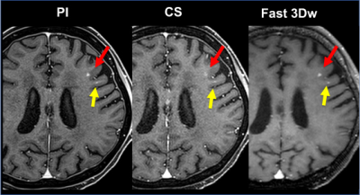 |
13 | Comparison of Brain Metastasis Detection Capability among 3D FFE with Parallel Imaging and CS and FASE MPV with Fast 3D Wheel Video Permission Withheld
Kazuhiro Murayama1, Yoshiharu Ohno2, Kaori Yamamoto3, Takahiro Matsuyama2, Seiichiro Ota2, Masao Yui3, Masato Ikedo3, Saki Takeda4, Akiyoshi Iwase4, Satomu Hanamatsu2, Hiroyuki Nagata2, Takahiro Ueda2, Hirotaka Ikeda2, and
Hiroshi Toyama2
1Joint Research Laboratory of Advanced Medical Imaging, Fujita Health University School of Medicine, Toyoake, Japan, 2Radiology, Fujita Health University School of Medicine, Toyoake, Japan, 3Canon Medical Systems Corporation, Otawara, Japan, 4Radiology, Fujita Health University Hospital, Toyoake, Japan
We hypothesize that the newly developed wheel encoding order (Fast 3D wheel: i.e. Fast 3Dw) method can reduce examination time as well as compressed sensing (CS) with parallel imaging (PI) technique (Compressed SPEEDER) and obtain contrast-enhanced MR examination without any degradation of image quality as compared with conventional PI technique in suspected brain metastases patients. The purpose of this study was to compare the capability for examination time reduction and image quality and diagnostic performance improvements among conventional PI, CS and Fast 3Dw methods on contrast-enhanced MR examination for brain metastases screening.
|
||
3632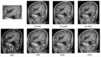 |
14 | The feasibility of contrast-enhanced 3D T1WI reconstructed with Compressed SENSE Artificial Intelligence in evaluation of brain metastases
Linna Li1, Zhongping Chen1, Renjie Lu2, Xin He1, Ying Qiu1, Dandan Guo1, Hongkun Shi1, Dan Tong1, Yi Zhu3, and Ke Jiang3
1Radiology, The First Hospital of Jilin Universty, Changchun, China, 2Ultrasound, The First Hospital of Jilin Universty, Changchun, China, 3Philips Healthcare, Beijing, China
Contrast-enhanced (CE) 3D T1-weighted scanning has been used more frequently with better spatial resolution and thinner section than 2D T1-weighted images for early detection and response assessment of brain metastases. To further improve the image scanning sequence, a new reconstruction algorithm, Compressed-SENSE Artificial Intelligence (CS-AI) was applied experimentally to achieve a balance between shortened scan time and improved image quality. This study aims to investigate the feasibility of contrast-enhanced 3D T1-weighted MRI reconstructed with Compressed SENSE Artificial Intelligence (CS-AI) by comparing with 3D T1-weighted images reconstructed with Compressed SENSE(CS), and to find out the optimal acceleration factor(AF).
|
||
 Back to Meeting Home
 Back to the Program-at-a-Glance
The International Society for Magnetic Resonance in Medicine is accredited by the Accreditation Council for Continuing Medical Education to provide continuing medical education for physicians.
 Back to Meeting Home
 Back to the Program-at-a-Glance
 View the Presentation