Online Gather.town Pitches
Advanced Imaging in Neuropsychiatric & Neurodegenerative Diseases
Joint Annual Meeting ISMRM-ESMRMB & ISMRT 31st Annual Meeting • 07-12 May 2022 • London, UK

Online Gather.town Pitches
Advanced Imaging in Neuropsychiatric & Neurodegenerative Diseases
| Booth # | ||||
|---|---|---|---|---|
3965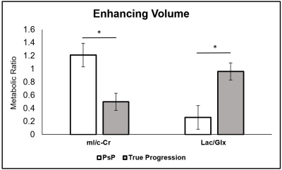 |
1 | Predictive Value of Multimodal MRI using spectroscopy and perfusion can distinguish between pseudoprogression and disease progression in GBM
Mohamed Ehab El-Abtah1, Pratik Talati 2, Melanie Fu1, Benjamin Chun1, Patrick Clark1, Anna Peters 1, Anthony Ranasinghe1, Julian He 1, Otto Rapalino1,3, Gilberto Gonzalez 1,3, William Curry 2, Jorg Dietrich 3,4, Elizabeth Gerstner4,
and Eva-Maria Ratai 1,3
1Athinoula A. Martinos Center for Biomedical Imaging, Boston, MA, United States, 2Department of Neurosurgery, Massachusetts General Hospital, Boston, MA, United States, 3Harvard Medical School, Boston, MA, United States, 4Cancer Center, Massachusetts General Hospital, Boston, MA, United States
There is a need to determine clinical markers that can distinguish between pseudoprogression (PsP) and true progression after patients with GBM undergo resection followed by chemoradiation. We conducted a retrospective study of collected magnetic resonance spectroscopic imaging (MRSI) and perfusion weighted imaging (PWI) data with the aim of investigating their utility in predicting tumor progression. Within the enhancing region on post-contrast T1 imaging, patients with true progression have decreased myo-Inositol normalized by contralateral creatinine (mI/c-Cr), elevated lactate normalized to glutamate+glutamine (Lac/Glx), and elevated relative cerebral blood flow (rCBF) relative to those with PsP.
|
||
3966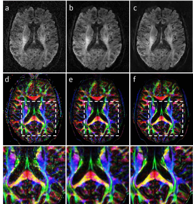 |
2 | High-resolution Diffusion Tensor Imaging at 7T with Multi-band Multi-shot EPI acquisition and Deep Learning Reconstruction
Xinzeng Wang1, Baolian Yang2, Marc R. Label3, Steen Moeller4, and Suchandrima Banerjee5
1GE Healthcare, Houston, TX, United States, 2GE Healthcare, Waukesha, WI, United States, 3GE Healthcare, Calgary, AB, Canada, 4Center for Magnetic Resonance Research, University of Minnesota, Minneapolis, MN, United States, 5GE Healthcare, Menlo Park, CA, United States
Diffusion tensor imaging (DTI) is a well-established tool for providing insights into brain network connectivity and detecting brain microstructure but suffers from artifacts, low SNR, low spatial resolution, and long scan times. High-resolution DTI at 7T with multiband MUSE (MB-MUSE) and noise reduction methods have shown many potentials for mitigating these challenges. In this study, we combine a deep learning reconstruction method with MB-MUSE to overcome the image quality challenges and demonstrate improved quantification of high-resolution DTI at 7T compared with MB-MUSE and MB-MUSE with low-rank denoising.
|
||
3967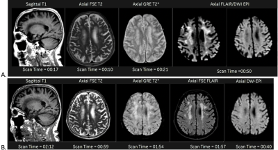 |
3 | Development of a High-Speed MRI Protocol with Deep Learning Reconstruction Method for Brain Imaging in a Clinical Setting Video Not Available
Patrick Quarterman1
1GE Healthcare, Brooklyn, NY, United States
The purpose of this study was to develop a High-Speed MRI (hsMRI) protocol with deep learning reconstruction (DL Recon) for adult and pediatric brain imaging to provide consistent, stout image quality in less than 1/4 the scan time of standard protocols. This was achieved utilizing FSE T1, SS T2w, Gradient Echo (GRE) T2*, and Diffusion Weighted & FLAIR Echo Plane Imaging (EPI) sequences. Evaluation was performed on of 20 patients comparing current protocol to hsMRI with results indicating imaging using hsMRI protocol was of clinically diagnostic quality allowing significant reduction in scan times compared to industry standard routine brain imaging protocols.
|
||
3968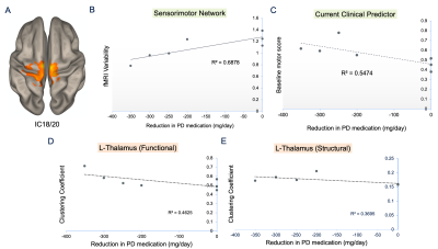 |
4 | Preoperative 7T MRI relates to medication dose reduction after chronic deep brain stimulation for Parkinson’s disease Video Not Available
Sharmaine Khanna1, Amirah Johnson1, Isyss Lyons1, Ben Sipes1, Olga Tymofiyeva1, Janine Lupo1, Jill Ostrem2, Doris Wang3, Philip Starr3, Ian Bledsoe2, and Melanie Morrison1
1Radiology and Biomedical Imaging, University of California San Francisco, San Francisco, CA, United States, 2Neurology, University of California San Francisco, San Francisco, CA, United States, 3Neurological Surgery, University of California San Francisco, San Francisco, CA, United States
Robust objective markers that can predict an individual Parkinson’s disease (PD) patient's potential response to brain stimulation (DBS) are limited. Here we evaluate the relationship between preoperative multimodal 7T MR metrics and a surrogate measure of motor response to DBS: the pre-to-post change in daily dose of dopamine medications. We found that fMRI signal variability in the superior sensorimotor network was more strongly correlated with medication dose reduction after DBS than current clinical prognostic criteria. Brain iron accumulation in the globus pallidus interna furthermore discriminated patients who did versus did not experience a dose change.
|
||
3969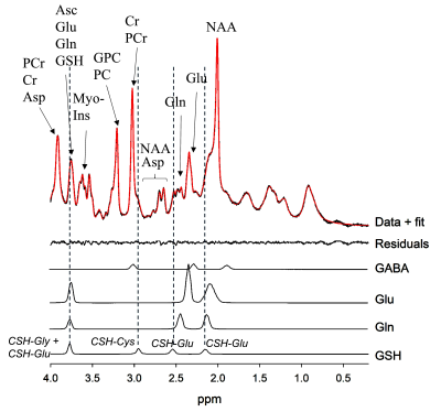 |
5 | Glutathione MRS of the Substantia Nigra in Parkinson Disease
Adil Bashir1, Frank M Skidmore2, and Thomas S Denney1
1Electrical & Computer Engineering, Auburn University, Auburn, AL, United States, 2Department of Neurology, University of Alabama at Birmingham, Birmingham, AL, United States
Glutathione in brain provides protection from oxidative stress. Autopsy studies have shown that glutathione levels are significantly reduced in substantia nigra of patients with Parkinson disease. In this study, we demonstrate the measurement of glutathione with MR spectroscopy in the human substantia nigra in vivo at 7T. Short echo-time STEAM pulse sequence was used to obtain high SNR spectra from Parkinson disease patients and healthy controls. Measured glutathione levels were significantly lower in patients when compared to healthy individuals.
|
||
3970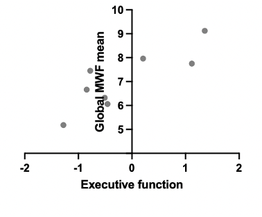 |
6 | Decreased Myelin Content on Myelin Water Imaging and Correlation with Cognitive Performance in Adults with Perinatal HIV Video Permission Withheld
Payal Patel1, Alyssa Vecchio2, Peter Chen3, Jennifer Chiarella4, Shannon Kolind5, Michael Hoff1, Adam Dvorak6, Irene Vavasour5, Serena Spudich4, Christina Marra1, Robert Paul7, and Swati Rane1
1University of Washington, Seattle, WA, United States, 2University of New Mexico, Albuquerque, NM, United States, 3Albert Einstein College of Medicine, Bronx, NY, United States, 4Yale University, New Haven, CT, United States, 5University of British Columbia, Vancouver, BC, Canada, 6University of British Columbia, Vancouver, BC, United States, 7Missouri Institute of Mental Health, St. Louis, MO, United States
Little is known about the pathology of cerebral white matter injury occurring in HIV. Myelin water imaging quantifies myelin content and results are expressed as myelin water fraction (MWF). We defined the cognitive profile of virally suppressed adults with perinatally acquired HIV (pHIV) and demonstrate the association between MWF and cognition. Ten (58%) adults with pHIV were cognitively impaired. Lower global MWF correlated with worse performance in executive function (r: 0.762, p: 0.037). Cognitive impairment is common among our cohort of adults with virally suppressed pHIV. Decreased myelination occurs in pHIV and may be a pathologic substrate of cognitive impairment.
|
||
3971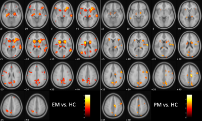 |
7 | Voxel-based assessment of volumetric brain differences in patients with premanifest and early manifest Huntington’s disease
Melanie Morrison1, Angela Jakary1, Andrew Leynes1, Jingwen Yao1, Julia Glueck2, Theresa Driscoll2, Joseph Talkakson2, Alexandra Nelson2, Katherine Possin2, Michael Geschwind2, Christopher Hess1, and Janine Lupo1
1Radiology & Biomedical Imaging, University of California San Francisco, San Francisco, CA, United States, 2Neurology, University of California San Francisco, San Francisco, CA, United States
More robust and accurate methods are needed to characterize disease extent and progression of Huntington's disease in order to develop disease-modifying therapies. Here we used voxel-based morphometry to evaluate patterns of regional brain atrophy in premanifest and early manifest HD patients relative to healthy controls. We found typical wide-spread sub-cortical and cortical gray matter atrophy in early manifest patients and observed similar though less pronounced atrophy in premanifest patients. As the presence of volume loss prior to symptom onset remains controversial, this work contributes to existing evidence of atrophy initiating as early as the premanifest disease stage.
|
||
3972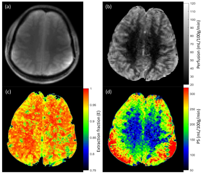 |
8 | Investigation of Blood-Brain Barrier Disruption in Schizophrenia using Magnetization Transfer-ASL at 7T.
Sultan Zaman Mahmud1, Adrienne Lahti2, Nina V. Kraguljac2, Thomas S. Denney1, and Adil Bashir1
1Department of Electrical and Computer Engineering, Auburn University, Auburn, AL, United States, 2Department of Psychiatry, University of Alabama at Birmingham, Birmingham, AL, United States Schizophrenia (SCZ) is a central nervous system (CNS) disease which is characterized by thinking disorder, cognitive impairments and other clinical manifestations that alter daily functioning. SCZ currently does not have satisfactory treatment options to improve cognitive symptoms. Deeper understanding of the underlying mechanism is required for effective treatment of SCZ. Blood-brain barrier (BBB) is the immunological junction between brain and vascular circulation, and its disruption is associated with a number of CNS diseases. This study aims to investigate if the BBB is compromised in SCZ. |
||
3973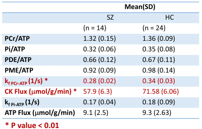 |
9 | Visual Cortex Bioenergetic Abnormalities in Schizophrenia
Adil Bashir1, Adrienne C Lahti2, Nina Kraguljac2, and Thomas S Denney1
1Electrical & Computer Engineering, Auburn University, Auburn, AL, United States, 2Psychiatry and Behavioral Neurobiology, The University of Alabama at Birmingham, Birmingham, AL, United States
This study measured high energy metabolite ratios and CK and ATP metabolic reaction rate in visual cortex of SZ patients and HCs. The forward rate constant hence ATP production flux through CK enzyme was significantly lower in SZ patients when compared to age/sex matched controls. We did not observe abnormalities in metabolite concentration. Impaired CK reaction may underlie abnormal neuro function and information processing in SZ.
|
||
3974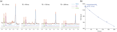 |
10 | T2-relaxation effects on NAA concentration reductions in psychotic disorders Video Not Available
Xi Chen1, Elliot Kuan1, Fei Du1, and Dost Öngür1
1McLean Hospital/Harvard Medical School, Belmont, MA, United States
We used 1H MRS at 4 T to quantify NAA concentrations and apparent T2 relaxation times in 104 psychosis patients compared to 50 matched healthy controls with four TEs (TE = 30, 90, 150 and 200 ms). Even at short TE (30 ms), NAA concentration without T2 correction was significantly lower in chronic psychosis compared to age-matched healthy controls. After T2 correction, no significant differences remained. Thus, it may be neuronal microenvironment indexed by T2 relaxation time, but not neuronal integrity indexed by NAA concentration that underlies the widely reported NAA concentration reductions in psychotic disorders.
|
||
3975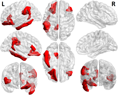 |
11 | Is curvature a biomarker for major depressive disorder? A morphological study using 7T MRI
Tara Lago1, Mariella Reynoso1, Yael Jacob2, Judy Alper3, Bradley N Delman4, James Murrough2, Priti Balchandani3, and Gaurav Verma3
1Staten Island Technical High School, New York, NY, United States, 2Psychiatry, Icahn School of Medicine at Mount Sinai, New York, NY, United States, 3Biomedical Engineering and Imaging Institute, Icahn School of Medicine at Mount Sinai, New York, NY, United States, 4Diagnostic, Molecular and Interventional Radiology, Icahn School of Medicine at Mount Sinai, New York, NY, United States
Numerous studies have analyzed morphological measurements to better understand how depression alters the brain, but few studies have explored curvature as a potential biomarker. By analyzing data from 7T MRI scans, we aimed to determine whether curvature measurements can differentiate between MDD patients and controls. We performed generalized linear models to analyze the relationship between participants’ status and measurements for intrinsic curvature index, Gaussian curvature, mean curvature, and folding index in thirty-four brain regions. Six regions showed significantly higher measures of curvature in MDD patients: the inferior temporal, pars triangularis, supramarginal, lateral orbitofrontal, isthmus cingulate, and entorhinal.
|
||
3976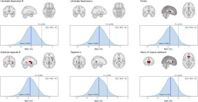 |
12 | Quantitative MRI evaluation of reduced myelin content in white matter tracts in major depressive disorde
Masaya Misaki1, Aki Tsuchiyagaito1, Beni Mulyana1,2, Rayus Kuplicki1, and Martin Paulus1
1Laureate Institute for Brain Research, Tulsa, OK, United States, 2Electrical and Computer Engineering, University of Oklahoma, Tulsa, OK, United States
Quantitative MRI (qMRI) of T1, T2, and proton density (PD) parameters can inform the brain's local microstructure, such as myelin content. We investigated the myelin alteration in white matter tracts for MDD participants compared to healthy controls by qMRI scanning using SyMRI software. MDD group had reduced myelin content in bilateral uncinate fasciculus, fornix, right external capsule, left tapetum, and genu of the corpus callosum regions. These results were consistent with previous reports about the white-matter microstructure alteration in MDD with diffusion tensor imaging (DTI). The results indicate that myelin measurement with qMRI can be an alternative to DTI.
|
||
 Back to Meeting Home
 Back to the Program-at-a-Glance
The International Society for Magnetic Resonance in Medicine is accredited by the Accreditation Council for Continuing Medical Education to provide continuing medical education for physicians.
 Back to Meeting Home
 Back to the Program-at-a-Glance
 View the Presentation