Online Gather.town Pitches
Resting-State fMRI & Novel fMRI Techniques
Joint Annual Meeting ISMRM-ESMRMB & ISMRT 31st Annual Meeting • 07-12 May 2022 • London, UK

Online Gather.town Pitches
Resting-State fMRI & Novel fMRI Techniques
| Booth # | ||||
|---|---|---|---|---|
3321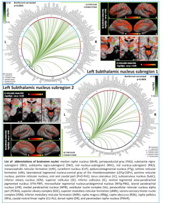 |
1 | Functional and structural connectomes of subthalamic nucleus subregions in living humans using 7 Tesla MRI
Maria Guadalupe Garcia Gomar1,2, Kavita Singh1, Simone Cauzzo1,3,4, and Marta Bianciardi1
1Brainstem Imaging Laboratory, Department of Radiology, Athinoula A. Martinos Center for Biomedical Imaging, Massachusetts General Hospital, Boston, MA, United States, 2Escuela Nacional de Estudios Superiores, Juriquilla, Universidad Nacional Autónoma de México, Querétaro, Mexico, 3Sant'Anna School of Advanced Studies, Institute of Life Sciences, Pisa, Italy, 4Research Center E. Piaggi, University of Pisa, Pisa, Italy
The subthalamic nucleus (STh) plays a crucial role in the basal ganglia movement-related circuit and it is involved in movement disorders such as Parkinson’s disease, dystonia, and chorea, among others. Its study in living humans is challenging due to limited resolution and contrast in conventional MRI. Previous work developed an in-vivo atlas of two STh subregions based on their fractional anisotropy properties. With the goal of characterizing their function and connectivity, in the present study, we evaluated the functional and structural connectivity of these STh subregions using 7 Tesla MRI and tested their preferential connectivity towards associative or motor regions.
|
||
3322 |
2 | The missing third dimension – functional correlations of BOLD signals incorporating white matter
Zhongliang Zu1, Yu Zhao1, Yurui Gao1, Muwei Li1, Zhaohua Ding1, and John C Gore1
1Vanderbilt University Medical Center, Nashville, TN, United States
We describe a novel graph analysis that includes a multigraph with multiple edges between a pair of nodes to model brain functional networks, and introduce a three-way correlation between BOLD signals from a pair of gray matter volumes (nodes) and one white matter bundle to define functional connectivity. Characteristics of inter-nodal communication may be derived from this analysis. By examining selected databases, we show inter-nodal communications vary with ages. By integrating fMRI signals from white matter as a third component in network analysis, more comprehensive descriptions of brain function may be obtained.
|
||
3323 |
3 | Detecting longitudinal dynamic functional connectivity changes using multiband multi-echo fMRI in high school football athletes
Alexander D. Cohen1, Michael D. McCrea2,3, and Yang Wang1
1Radiology, Medical College of Wisconsin, Milwaukee, WI, United States, 2Neurosurgery, Medical College of Wisconsin, Milwaukee, WI, United States, 3Neurology, Medical College of Wisconsin, Milwaukee, WI, United States
Repetitive head impact exposure (RHIE) during contact sports may have damaging neurocognitive effects. In this study, we investigated longitudinal changes in dynamic functional connectivity (dFNC) using multiband multi-echo (MBME) fMRI in high school football players over the course of two seasons. Four recurring connectivity states were identified. A greater total number of dFNC changes were observed over two seasons compared to over the course of one season. Changes across all four states were only observed in the two-season comparison. These preliminary results suggest that RHIE may lead to accumulated effects in functional connectivity patterns.
|
||
3324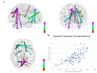 |
4 | Resting state functional connectivity subnetwork relates to prosocial behavior and compassion in adolescents
Benjamin Sipes1, Angela Jakary1, Melanie Morrison1, Tony T Yang2, and Olga Tymofiyeva1
1Radiology, University of California, San Francisco, San Francisco, CA, United States, 2Psychiatry and Behavioral Sciences, University of California, San Francisco, San Francisco, CA, United States In this study, we found that adolescent resting state functional brain networks contains a subnetwork significantly related to self-reported prosocial behavior and compassion. The subnetwork regions indicated connects many previously identified brain regions associated with prosocial behavioral tasks in adolescents, including the bilateral precuneus, left lateral prefrontal cortex, medial prefrontal cortex, inferior frontal gyri, temporal poles, left amygdala, right posterior cingulate cortex, occipital cortex, left supplementary motor area, and cerebellum. |
||
3325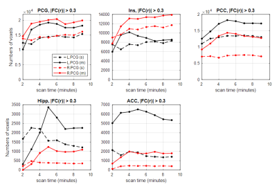 |
5 | Improved Fidelity for Resting-State Connectivity Measurements in High-Spatial-Resolution ME-fMRI on a Compact 3T Scanner
Daehun Kang1, Myung-Ho In1, Maria A Halverson1, Nolan K Meyer1, Erin M Gray1, Thomas K Foo2, Radhika Madhavan2, Zaki Ahmed1, Hang Joon Jo3, Brice Fernandez4, David F Black1, Kirk M Welker1, Joshua D Trzasko1, John Huston, III1,
Matt A Bernstein1, and Yunhong Shu1
1Department of Radiology, Mayo Clinic, Rochester, MN, United States, 2GE Global Research, Niskayuna, NY, United States, 3Department of Physiology, Hanyang University, Seoul, Korea, Republic of, 4GE Healthcare, Buc, France
Through echo-combination, multi-echo echo-planar-imaging (ME-EPI) has shown improved performance for fMRI over single-echo (SE)-EPI. The high-performance gradients on a compact 3T scanner enable whole-brain high-spatial-resolution ME-EPI with reasonable echo times and comparable TR to SE-EPI. To evaluate the fidelity of the ME-EPI sequence in resting state fMRI and compare with that of SE-EPI sequence, seed-based and ICA-based functional connectivity (FC) analyses were applied to evaluate scan-time-dependency and accuracy improvement. ME-EPI extracted more and stronger FCs in seed-based connectivity analysis and produced higher sensitivity and accuracy in ICA-based method, compared to SE-EPI.
|
||
3326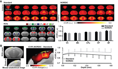 |
6 | NORDIC PCA increases tSNR in both human and mouse resting-state fMRI for potential improvements in cerebrovascular reactivity mapping
Emily L. Tse1, Russell W. Chan1,2, Sarah Y. Wu1, Yixi Xue1, Peiying Liu3, Steen Moeller4, and Kevin C. Chan1,5
1Department of Ophthalmology, New York University Grossman School of Medicine, New York, NY, United States, 2Neuroscience Institute, New York University Grossman School of Medicine, New York, NY, United States, 3Department of Diagnostic Radiology and Nuclear Medicine, University of Maryland School of Medicine, Baltimore, MD, United States, 4Center for Magnetic Resonance Research (CMRR), University of Minnesota, Minneapolis, MN, United States, 5Department of Radiology, New York University Grossman School of Medicine, New York, NY, United States
Cerebrovascular reactivity (CVR) reflects the response of cerebral blood vessels to vasoactive stimuli. Whole-brain relative CVR (rCVR) mapping is achieved using task-free resting-state fMRI, which resembles conventional CVR mapping using hypercapnia challenge. To optimize rCVR mapping, we apply NOise Reduction with DIstribution Corrected (NORDIC) PCA which removes thermal noise originating from the scanner and/or the subject. Results show that NORDIC-correction increases the temporal signal-to-noise ratio in both humans and mice, lowers mouse rCVR variance, and potentially improves mouse rCVR along cortical layers. Future rCVR studies investigating cerebrovascular diseases may incorporate NORDIC-correction for higher sensitivity.
|
||
3327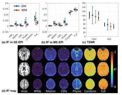 |
7 | Benefits of echo combination for high-spatial-resolution ME resting state FMRI on a compact 3T scanner
Daehun Kang1, Myung-Ho In1, Maria A Halverson1, Nolan K Meyer1, Erin M Gray1, Thomas K Foo2, Radhika Madhavan2, Zaki Ahmed1, Hang Joon Jo3, Brice Fernandez4, David F Black1, Kirk M Welker1, Joshua D Trzasko1, John Huston, III1,
Matt A Bernstein1, and Yunhong Shu1
1Department of Radiology, Mayo Clinic, Rochester, MN, United States, 2GE Global Research, Niskayuna, NY, United States, 3Department of Physiology, Hanyang University, Seoul, Korea, Republic of, 4GE Healthcare, Buc, France
For multi-echo EPI, the T2*-weighted echo combination (T2EC) is a commonly used pre-processing step to boost the data quality. We assessed the benefit of T2EC in high-spatial-resolution multi-echo fMRI with explained variances and functional connectivity detectability. The performance was evaluated and compared with single-echo EPI for resting state fMRI on a compact 3T scanner with high-performance gradients. It was shown that a large portion of unwanted signal variance was globally suppressed through T2EC. Multi-echo acquisition shows improved image intensity and enhanced functional sensitivity in brain regions affected by signal-dropout.
|
||
3328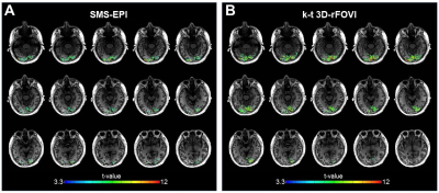 |
8 | Fast 3D fMRI Acquisition over a Small Field-of-View with (k, t)-Space Undersampling
Qingfei Luo1, Kaibao Sun1, Alessandro Scotti1, Guangyu Dan1,2, Muge Karaman1,2, and Xiaohong Joe Zhou1,2,3
1Center for Magnetic Resonance Research, University of Illinois at Chicago, Chicago, IL, United States, 2Department of Biomedical Engineering, University of Illinois at Chicago, Chicago, IL, United States, 3Departments of Radiology and Neurosurgery, University of Illinois at Chicago, Chicago, IL, United States
The simultaneous multi-slice EPI (SMS-EPI) technique is widely used in fast fMRI studies to achieve a short TR (e.g., < 1 s), but its image quality can be degraded when the slices span only a small brain area. In this study we develop a fast fMRI acquisition technique for imaging a small area by employing (k, t)-space undersampling and three-dimension reduced field-of-view imaging (k-t 3D-rFOVI). Our human fMRI experiments covering the visual cortex demonstrate that k-t 3D-rFOVI can provide higher detection sensitivity of brain activations than SMS-EPI.
|
||
3329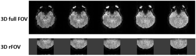 |
9 | Functional MRI with three-dimensional reduced field-of-view
Kaibao Sun1, Guangyu Dan1,2, Alessandro Scotti1, Muge Karaman1,2, Qingfei Luo1, and Xiaohong Joe Zhou1,2,3
1Center for MR Research, University of Illinois at Chicago, Chicago, IL, United States, 2Department of Biomedical Engineering, University of Illinois at Chicago, Chicago, IL, United States, 3Departments of Radiology and Neurosurgery, University of Illinois at Chicago, Chicago, IL, United States
Functional MRI (fMRI) is typically performed with whole brain coverage, even when only a small brain area is of interest. Although zoomed fMRI has been demonstrated using 2D multi-slice acquisitions, extending this capability to 3D focused volume has not been well explored. We have implemented a 3D reduced field-of-view technique for fMRI by using a 2D RF pulse and applied this technique to a visual fMRI study on ten subjects. Compared to a conventional fMRI sequence with a full field-of-view, our technique produced high isotropic spatial resolution and reduced image distortion, despite a moderate reduction in temporal signal-to-noise ratio.
|
||
3330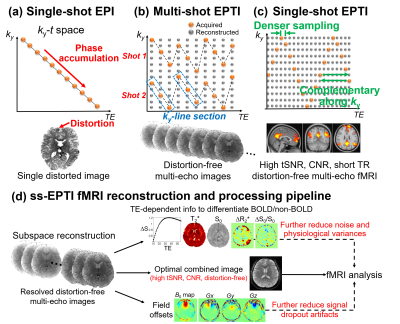 |
10 | Improving fMRI acquisition using single-shot EPTI with distortion-free high-SNR high-CNR multi-echo imaging
Fuyixue Wang1,2, Zijing Dong1,2, Jingyuan Chen1,2, Kawin Setsompop3,4, Jonathan R. Polimeni1,2, and Lawrence L. Wald1,2
1Athinoula A. Martinos Center for Biomedical Imaging, Massachusetts General Hospital, Charlestown, MA, United States, 2Department of Radiology, Harvard Medical School, Boston, MA, United States, 3Department of Radiology, Stanford University, Stanford, CA, United States, 4Department of Electrical Engineering, Stanford University, Stanford, CA, United States
This work aims to address the limitations of EPI and improve fMRI acquisition by developing a single-shot EPTI method, to provide fast distortion-free imaging, with significantly improved tSNR, CNR, robustness to motion, reduced signal dropout, and multi-echo capability for denoising/analysis. The single-shot acquisition is enabled by the newly-designed k-t encoding to provide stronger correlation for improved reconstruction. Results show ss-EPTI achieves: i) 30% higher tSNR efficiency than EPI and 70% higher than 3-shot EPTI; ii) 50% higher CNR than EPI (visual-task); iii) distortion-free fMRI with improved robustness to motion; iv) effective signal dropout reduction; and v) effective non-BOLD variation separation.
|
||
3331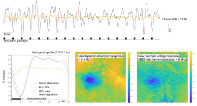 |
11 | Optimization of wide-field optical imaging method towards fMRI integration in mice
Wen-Ju Pan1, Yunmiao Wang2, Harrison Watters1, Lisa Meyer-Baese1, Alaina Corrie Smith1, Dieter Jaeger2, and Shella Keilholz1
1Biomedical Engineering, Emory University/Georgia Institute of Technology, Atlanta, GA, United States, 2Biology, Emory University, Atlanta, GA, United States
We present an optimized wide-field optical imaging method with GEVI in mice to probe the relationship between slow changes in neuronal membrane potential and the BOLD signal in hindpaw stimulation, demonstrate neuronal sub-threshold activity of slow fluctuations could be possibly isolated from hemodynamic signals.
|
||
3332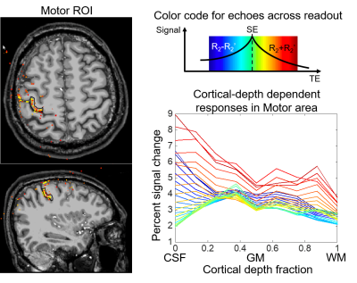 |
12 | Mapping cortical-depth dependent responses in human motor cortex using Spin-echo Echo Planar Time-resolved Imaging (SE-EPTI)
Fuyixue Wang1,2, Zijing Dong1,2, Lawrence L. Wald1,2, Jonathan R. Polimeni1,2, and Kawin Setsompop3,4
1Athinoula A. Martinos Center for Biomedical Imaging, Massachusetts General Hospital, Charlestown, MA, United States, 2Department of Radiology, Harvard Medical School, Boston, MA, United States, 3Department of Radiology, Stanford University, Stanford, CA, United States, 4Department of Electrical Engineering, Stanford University, Stanford, CA, United States
This work utilizes our recently developed spin-echo EPTI technique with minimal T2’-contamination and high specificity to map the layer-dependent responses in primary motor cortex at 7T and further investigate the impact of large vessel bias from T2’ contamination. EPTI resolves multi-contrast images across the readout that are free from distortion and blurring, and simultaneously obtains a SE image with pure T2-weighting and multiple asymmetric SE images with various T2’-weightings. The activation of finger-tapping task was studied using SE-EPTI at 0.9-mm isotropic resolution. Improved specificity of layer-dependent responses was observed using pure SE images.
|
||
3333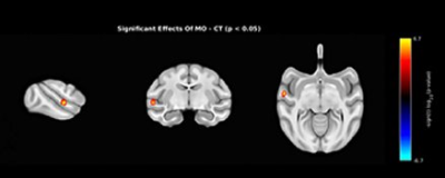 |
13 | Maternal Obesity Alters Neuro-Connectivity in Baboon Offspring
Wei D Zhang1, Peter T Fox 1, Hillary F Huber2, Peter William Nathanielsz3, and Geoffrey D Clarke4
1Research Imaging Institute, UT Health San Antonio, San Antonio, TX, United States, 2Texas Pregnancy & Life-Course Health Research Center, Texas Biomedical Research Institute, San Antonio, TX, United States, 3Animal Science, University of Wyoming, Laramie, WY, United States, 4Research Imaging Insitute, UT Health San Antonio, San Antonio, TX, United States
Maternal obesity status was found to affect neural functional connectivity of adult offspring in in a baboon model. Significant differences in connectivity between offspring of obese mothers and age/sex matched controls were found in regions of the left temporal lobe and frontal lobe using rs-fMRI.
|
||
3334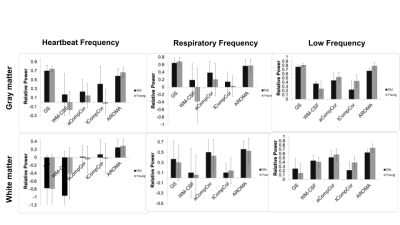 |
14 | Comparing the efficiency of data-driven noise regression in removing cardiac and respiratory signals from rs-fMRI: Difference across age groups
Ali Golestani1 and Jean Chen2,3
1University of Toronto, Toronto, ON, Canada, 2Rotman Research Institute at Baycrest, Toronto, ON, Canada, 3Department of Biophysics, University of Toronto, Toronto, ON, Canada
Data-driven methods have been suggested to remove heartbeat and respiration noises from fMRI signals. We compared the effectiveness of these methods (global-signal regression (GS), white matter and CSF (cerebrospinal fluid) regression, anatomical and temporal CompCor, ICA AROMA) in removing the noise. GS, AROMA, and aCompCor removed the most physiological fluctuation, but GS and AROMA also removed most signals under 0.1 Hz. We also observed that all methods removed less noise power and more low-frequency power from young adult data compared to older adults.
|
||
3335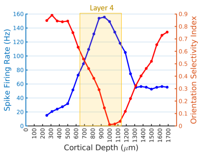 |
15 | Hemodynamic responses based on CBV weighted fMRI strongly correlate with single-unit neuronal action potentials across cortical layers
Wei Zhu1, Corey Cruttenden1, Shinho Cho1, Xiao-Hong Zhu1, Kamil Ugurbil1, and Wei Chen1
1University of Minnesota, Minneapolis, MN, United States
The specificity of hemodynamic signals detected by functional magnetic resonance imaging (fMRI) to neuronal activity is complicated by the neurovascular coupling. To investigate the fidelity of fMRI in differentiating the neuronal activity across cortical layers, we mapped the laminar orientation preference maps in the cat visual cortex by using high-resolution cerebral blood volume weighted (wCBV) fMRI and single-unit neuronal action potential recording, respectively. The strong correlation between wCBV percent change and spike firing rate indicates that wCBV fMRI with sufficient spatial resolution could distinguish the differences of neural activities across layers but with a reduced sharpness.
|
||
3336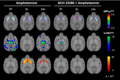 |
16 | Differential D1 and D2 receptor internalization and recycling induced by amphetamine in vivo: a PET/fMRI study
Hanne D Hansen1,2, Martin Schain2, Helen P Deng1, Joseph B Mandeville1,3, Bruce R Rosen1,3, and Christin Y Sander1,3
1A. A. Martinos Center for Biomedical Imaging, Department of Radiology, Massachusetts General Hospital, Charlestown, MA, United States, 2Neurobiology Research Unit and Center for Integrated Molecular Brain Imaging, Copenhagen University Hospital, Rigshospitalet, Copenhagen, Denmark, 3Harvard Medical School, Boston, MA, United States
In this study, we imaged the effect of repeated amphetamine injections over >24h using simultaneous PET/fMRI in non-human primates and demonstrate that both internalization and recycling of receptors take place during this timeframe. We provide novel insight into D1 vs. D2 receptor contributions by using a D1 blocking agent in combination with amphetamine injections. These results shed a new light into the timelines of dopamine receptor subtype adaptations and not only provide a link to existing in vitro results but also a foundation for linking receptor-specific mechanisms to initial stages of psychostimulant drug use and abuse.
|
||
3337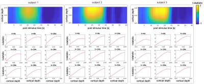 |
17 | Temporal evolution of the cortical-depth profiles of BOLD fMRI response
Anna Izabella Blazejewska1,2, Daniel Gomez1,2,3, and Jonathan R Polimeni1,2,4
1Athinoula A. Martinos Center for Biomedical Imaging, Massachusetts General Hospital, Charlestown, MA, United States, 2Department of Radiology, Harvard Medical School, Charlestown, MA, United States, 3Department of Biomedical Engineering, Boston University, Boston, MA, United States, 4Division of Health Sciences and Technology, Massachusetts Institute of Technology, Cambridge, MA, United States
The gradient-echo BOLD fMRI signal demonstrates greatest changes with activation near large draining veins which results in cortical-depth activation profiles that increase linearly with depth from the white to pial surface. However, due to the sparsity of pial vessels these profiles are likely to vary across cortical locations depending on proximity to vessels. We investigated the temporal evolution and spatial variability of the cortical-depth profiles of the BOLD response within human visual cortex and demonstrated that the response is initially strongest in the mid-cortical depths and in time becomes dominated by the large veins at the pial-surface.
|
||
3338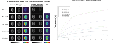 |
18 | RF shielding designs for birdcage coils for high-resolution animal fMRI at 9.4T
Zhangyan Yang1,2, Ming Lu3, Gary Drake2,4, Feng Wang2,4, Pai-Feng Yang2,4, Li Min Chen2,4, John C. Gore1,2,4,5,6, and Xinqiang Yan2,4
1Department of Biomedical Engineering, Vanderbilt University, Nashville, TN, United States, 2Vanderbilt University Institute of Imaging Science, Vanderbilt University Medical Center, Nashville, TN, United States, 3College of Nuclear Equipment and Nuclear Engineering, Yantai University, Shandong, China, 4Department of Radiology and Radiological Sciences, Vanderbilt University Medical Center, Nashville, TN, United States, 5Department of Physics and Astronomy, Vanderbilt University, Nashville, TN, United States, 6Department of Molecular Physiology and Biophysics, Vanderbilt University, Nashville, TN, United States
RF shields are key components of volume coils, but they may cause imaging distortions and the significant heating. The effects of different shielding strategies were evaluated for a birdcage coil used to image animals at 9.4T. Three shields with various thicknesses and two shields with different slot patterns were comparatively investigated ed in terms of performance and temperature rise during fMRI acquisitions.
|
||
3339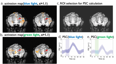 |
19 | BOLD response in squirrel monkey brains evoked by green light without exogenous opsin
zhangyan yang1,2, Pai-Feng Yang2,3, Huiwen Luo1,2, Feng Wang2,3, Jamie L Reed2,3, William A. Grissom1,2,3, Li Min Chen2,3, and John C. Gore1,2,3,4,5
1Department of Biomedical Engineering, Vanderbilt University, Nashville, TN, United States, 2Vanderbilt University Institute of Imaging Science, Vanderbilt University Medical Center, Nashville, TN, United States, 3Department of Radiology and Radiological Sciences, Vanderbilt University Medical Center, Nashville, TN, United States, 4Department of Physics and Astronomy, Vanderbilt University, Nashville, TN, United States, 5Department of Molecular Physiology and Biophysics, Vanderbilt University, Nashville, TN, United States
Optogenetic stimulation combined with MRI detection of blood oxygenation level dependent (BOLD) effects has been developed as a powerful tool to delineate brain circuits. Opsin targeting specific cell types induce neural activity when irradiated with light at a specific wavelength. We detected robust BOLD effects in secondary somatosensory cortex (S2) and downstream interconnected caudate putamen (CPu) of monkey brains from irradiation with low-intensity green light without exogenous green light specific opsin, suggesting hemodynamic changes can be stimulated without any associated neural activity.
|
||
3340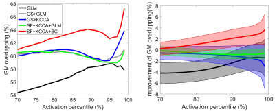 |
20 | A global spatially-adaptive method for task fMRI activation analysis with the advantage of alleviated spatial blurring Video Permission Withheld
Zhengshi Yang1,2, Xiaowei Zhuang1,2, Mark Lowe3, and Dietmar Cordes1,2,4
1Cleveland Clinic Lou Ruvo Center for Brain Health, Las Vegas, NV, United States, 2University of Nevada Las Vegas, Las Vegas, NV, United States, 3Cleveland Clinic, Cleveland, OH, United States, 4University of Colorado, Boulder, CO, United States
Accurately localizing brain activation is of great importance for promoting basic science and clinical application of task fMRI data. In this study, we proposed a global and time-efficient way to conduct brain activation analysis, with the property of spatially-adaptive smoothing and determining individual voxel’s activation status with wholebrain fMRI data considered simultaneously. We have demonstrated its advantage over traditional methods in alleviating spatial blurring artifacts.
|
||
 Back to Meeting Home
 Back to the Program-at-a-Glance
The International Society for Magnetic Resonance in Medicine is accredited by the Accreditation Council for Continuing Medical Education to provide continuing medical education for physicians.
 Back to Meeting Home
 Back to the Program-at-a-Glance
 View the Presentation