Online Gather.town Pitches
Advanced Imaging in Neurology: Psychiatry & Epilepsy
Joint Annual Meeting ISMRM-ESMRMB & ISMRT 31st Annual Meeting • 07-12 May 2022 • London, UK

Online Gather.town Pitches
Advanced Imaging in Neurology: Psychiatry & Epilepsy
| Booth # | ||||
|---|---|---|---|---|
2995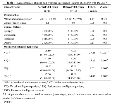 |
1 | Use lesion network localization to locate related brain lesions in incidental white matter lesions children with verbal comprehension delay
Xiaoyu Wang1, Mengxuan Li1, Miaomiao Wang1, Congcong Liu1, Xianjun Li1, and Jian Yang1
1Radiology, The first afflicted hospital of Xi'an Jiaotong University, Xi'an, China
Incidental white matter lesions (IWMLs) is a common disease in children with an incidence rate between 1.9% - 20%. Locating the brain lesions that might be related to the decline in verbal comprehension by overlapping lesion-associated networks can help define the possible response lesion. Our finding would help pediatricians to recognize the significant IWMLs that could potentially cause language delay in children. Thus, an early intervention can be applied to prevent the occurrence and progression of verbal comprehension decline in children.
|
||
2996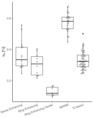 |
2 | Lesion assessment in multiple sclerosis using a two-compartment spherical mean technique
Stefan Eggenreich1, Lukas Pirpamer1, Michael Khalil1, Christian Enzinger1, and Stefan Ropele1
1Department of Neurology, Medical University of Graz, Graz, Austria
Various diffusion-based approaches were suggested for the characterization of lesional tissue in MS-patients. The multi-compartment spherical mean technique (SMT) has been proposed as a way to investigate microscopic diffusion properties in multiple water compartments. However, the potential of SMT for lesion characterization remains unclear. We investigated diffusion-based SMT properties in active and chronic lesions in 58 MS patients and related them to conventional diffusion tensor imaging (DTI). We found that SMT allows a more specific characterization of MS-lesions than DTI and provide evidence that SMT can be considered as a sensitive and robust measure for myelin content in MS patients.
|
||
2997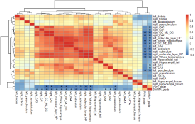 |
3 | Correlation between hippocampal subfields volume and white matter hyperintensity in patients with type 2 diabetes mellitus
Yangyingqiu Liu1, Weiwei Wang1, Yuhan Jiang1, Ailian Liu1, and Yanwei Miao1
1Department of Radiology, First Affiliated Hospital of Dalian Medical University, Dalian, China
Volume of hippocampal subfields in patients with type 2 diabetes mellitus(T2DM) was quantitatively assessed, and its’correlation to white matter hyperintensity (WMH) grade was also analyzed. The results show that the severity of WMH in T2DM patients may affect the atrophy of hippocampal subfields.
|
||
2998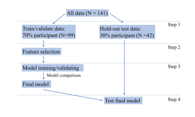 |
4 | Machine Learning based Structural and Diffusion MRI to Identify Suicide Risk in Depressed Patients
Huiru Li1, Huawei Zhang1, Li Yin2, Zhiyun Jia1, and Qiyong Gong1
1Huaxi MR Research Center (HMRRC), Department of Radiology, West China Hospital of Sichuan Universit, Chengdu, China, 2Department of Psychiatry, West China Hospital of Sichuan University, Chengdu, China
In this study, we used 5 machine learning algorithms with structural and diffusion MRI to classify suicidal risk in depressed patients and the best performance was acquired by support vector machine. In addition, the most important 10 features were most located in fronto-temporal–parietal regions. This study found a classification model using MRI data can help diagnosis and assess suicidal risk in depressed patients.
|
||
2999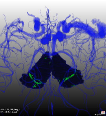 |
5 | Thalamus regulates emotional performance in different pathways with CSVD involved
Bei Wang1, Cen Guo2, He Wang1,3, Yan Han2, and Ying-Hua Chu4
1Institute of Science and Technology for Brain-Inspired Intelligence, Fudan University, Shanghai, China, 2Department of Neurology, Yueyang Hospital of Integrated Traditional Chinese and Western Medicine, Shanghai University of Traditional Chinese Medicine, Shanghai, China, 3Human Phenome Institute, Fudan University, Shanghai, China, 4MR Collaboration, Siemens Healthineers Ltd., Shanghai, China
With the help of 7T Imaging, we investigated the association between the thalamic anterior vascularization patterns and emotions, including depression and anxiety. Cerebral small vessel diseases were considered as a risk factor for emotional performance. The thalamic anterior vascularization pattern was found association with depression and anxiety in subjects without CSVD burden. In the CSVD group, the thalamic volume and cortex thickness in pars triangularis, rostral middle frontal and superior frontal were found significantly correlated with both HAMD and HAMA test scores. It suggests the pathway of the thalamus regulating emotion changes with the development of CSVD.
|
||
3000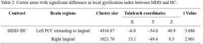 |
6 | Aberrant cortical gyrification in adolescent major depressive disorder
Weijie Bao1, Lingxiao Cao1, Ruohan Feng1,2, Yingxue Gao1, Hailong Li1, Xinyue Hu1, Hui Qiu1, Kaili Liang1, Zilin Zhou1, and Xiaoqi Huang1,3
1Huaxi MR Research Center (HMRRC), Department of Radiology, West China Hospital of Sichuan University, Chengdu, China, 2Department of radiology, the Thrid Hospital of Mianyang, Sichuan Mental Health Center, Mianyang, China, Mianyang, China, 3Psychoradiology Research Unit of the Chinese Academy of Medical Sciences (2018RU011), West China Hospital of Sichuan University, Chengdu, Sichuan, China, Chengdu, China
Evidence has emerged to suggest the important role of cortical gyrification morphology in adult major depressive disorder (MDD). However, little is known about how the altered cortical gyrification contribute to adolescent patients who is at a critical period of brain development. We address this gap by examining the difference of local gyrification index (LGI) between adolescent MDD patients and HCs. We found that MDD patients showed significant increased LGI in the left PCC and bilateral lingual gyrus, which characterize the neurobiological mechanisms of adolescent MDD and may be important to understand the developmental effect of this disorder.
|
||
3001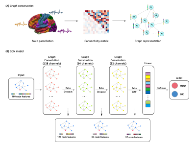 |
7 | Using graph convolutional network modeling to characterize functional network disruption in individuals with major depressive disorder Video Permission Withheld
Kun Qin1, Du Lei2, Ziyu Zhu2, and Qiyong Gong1
1West China Hospital of Sichuan University, Chengdu, China, 2University of Cincinnati College of Medicine, Cincinnati, OH, United States
Few previous studies considered brain network architecture when establishing machine learning models to identify major depressive disorder (MDD). Based on a large, multi-site dataset including 1586 participants, this study aimed to use novel graph convolution network (GCN) to distinguish MDD patients from controls, identify MDD subtypes and characterize related network disruption. We found that GCN enabled excellent classification performance of over 80% accuracy. Besides, shared and distinct disrupted network patterns were identified in first-episode drug-naive and recurrent patients. These findings support the feasibility and effectiveness of network-based GCN classier, illustrating the utility of GCN for detecting disrupted network topology.
|
||
3002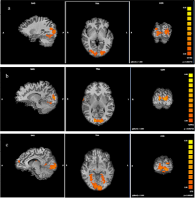 |
8 | Functional Imaging of Body Image Distortion In Anorexia Nervosa: Overweight vs. Underweight
Yagmur Karakus Aydos1, Dicle Dovencioglu2, Kader Karlı Oguz3, Pınar Ozdemir4, Melis Pehlivanturk Kızılkan5, Nuray Kanbur5, Dilek Unal1, Kevser Nalbant1, Fusun Cetin Cuhadaroglu1, and Devrim Akdemir1
1Child and Adolescent Psychiatry, Hacettepe University, Ankara, Turkey, 2Psychology, Middle East Technical University, Ankara, Turkey, 3Radiology, Hacettepe University& Bilkent University UMRAM, Ankara, Turkey, 4Bioistatistics, Hacettepe University, Ankara, Turkey, 5Adolescent Health, Hacettepe University, Ankara, Turkey
Body image disorder is the core symptom of Anorexia Nervosa (AN). Our research focuses on a group of adolescent patients diagnosed with AN to investigate neurobiological aspects of body image distortion, comparing the results with healthy control (HC) and major depression (MD) groups. We planned a block-design task in fMRI by using participants' original and distorted overweight and underweight images. We applied General Linear Models (GLM) for whole-brain analyses and examined correlations of the findings with behavioral data. As a result, AN patients differentiated when evaluating their underweight images. However, all adolescents had similar activations for overweight conditions.
|
||
3003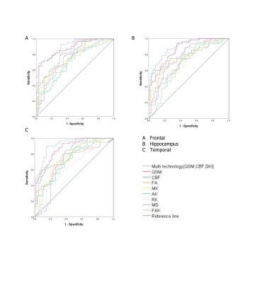 |
9 | Application of quantitative magnetic resonance imaging in the diagnosis of autism in children Video Not Available
Shilong Tang1 and Lisa Nie2
1Children's Hospital of Chongqing Medical University, Chongqing, China, 2GE Healthcare, MR Research China, Beijing, Beijing, China
Purpose: To explore the application of quantitative magnetic resonance imaging in the diagnosis of autism in children. Methods: All the children were scanned using head MRI conventional sequences, 3D-T1,DKI, ESWAN and 3D-pcASL sequences. The QSM, CBF and brain microstructure of each brain area were compared between the groups, and correlations were analyzed. Results: The frontal lobe, temporal lobe and hippocampal regions may be the first areas to show microstructural changes in autistic children . Conclusion: Quantitative magnetic resonance imaging can shows the abnormal changes of brain microstructural in children with autism .
|
||
3004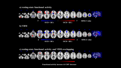 |
10 | A multimodal meta-analysis of regional functional and structural brain abnormalities in obsessive-compulsive disorder
Zibin Yang1, Shu Xiao1, Long Qian2, and Ying Wang1
1Medical Imaging Center, First Affiliated Hospital of Jinan, Guangzhou, China, 2GE Healthcare, Beijing, China, Beijing, China
Numerous neuroimaging studies have revealed abnormalities in specific brain regions in obsessive-compulsive disorder (OCD), but results have been inconsistent. We conducted a whole-brain voxel-wise meta-analysis on resting-state functional imaging and VBM studies between OCD patients and HCs by using Seed-based d Mapping (SDM) software. The meta-analysis demonstrated that OCD exhibits similar abnormalities in both function and structure in the prefrontal and insula. Few regions exhibited only functional or only structural abnormalities in OCD. These results expand the current understanding of functional and structural brain abnormalities in OCD patients, which would provide additional potential targets for therapeutic intervention.
|
||
3005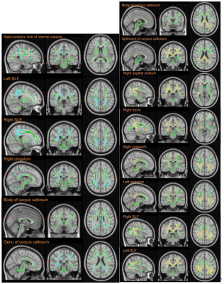 |
11 | Alteration in White Matter Microstructural Integrity and Network Differentiate Drug-naïve Obsessive compulsive disorder and Healthy controls
Suming Zhang 1, Xinyu Hu2, Xuan Bu2, Hailong Li1, LingXiao Cao2, Jing Liu2, KaiLi Liang2, Xue Li3, and Xiaoqi Huang2
1Huaxi MR Research Center (HMRRC), Department of Radiology, West China Hospital of Sichuan University, Chengdu City, China, 2Huaxi MR Research Center (HMRRC), Department of Radiology, West China Hospital of Sichuan University, ChengDu City, China, 3College of Physics, Sichuan University,Chengdu, China, Chengdu City, China
We investigated the alteration of white matter in obsessive compulsive disorder from voxel-level, tractography-level and connectome-level by combining analysis of TBSS, AFQ and graph theory. TBSS and AFQ revealed widespread changes in WM. Network-based statistic (NBS) analysis demonstrated decreased structural connectivity in patients mainly comprising frontal, temporal, limbic areas, which belong to DMN, FPN, limbic, SMN and VAN network. Along with regions commonly described in the CSTC model of pathophysiology, our results indicate an involvement of white matter in frontal-temporal and limbic regions to differentiate OCD patients and healthy controls, supporting their importance for neurobiological alterations in OCD.
|
||
3006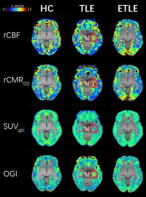 |
12 | Simultaneous PET-MRI reveals differences in aerobic glycolysis in temporal lobe versus extra temporal lobe epilepsy
Qikai Qin1,2, Miao Zhang3, Biao Li3,4, and Garth John Thompson1
1iHuman Institute, ShanghaiTech University, Shanghai, China, 2School of Life Science and Technology, ShanghaiTech University, Shanghai, China, 3Department of Nuclear Medicine, Ruijin Hospital, Shanghai Jiao Tong University School of Medicine, Shanghai, China, 4Collaborative Innovation Center for Molecular Imaging of Precision Medicine, Ruijin Center, Shanghai, China
Locating epileptic foci is critical for surgery in drug-resistant epilepsy. However, conventional methods (MRI, PET, SEEG) often cannot provide sufficient information alone. We have developed a new method, relative oxygen-glucose index (OGI), based on simultaneous PET and calibrated fMRI, which we used as a biomarker to locate epileptic foci during inter-ictal period. Relative OGI reflects aerobic glycolysis relative to global levels, which is useful as many diseases are characterized by metabolic pathway alterations. In this study, we not only demonstrate its potential in foci localization in epilepsy, but also distinguish temporal-lobe and extra-temporal-lobe epilepsy based on their different pathogenesis.
|
||
3007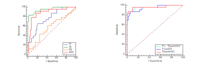 |
13 | Clinical Value of Synthetic MRI and DWI in the diagnosis of Hippocampal Sclerosis in medial temporal lobe Epilepsy
Yucai Bai1, Jian LI1, Xiaocheng Wei2, Yaoxing Ma1, and Bing Chen1
1General Hospital of Ningxia Medical University, Ningxia yinchuan, China, 2GE Healthcare, Beijing, China
The purpose of this study is to explore the value of different quantitative parameters derived from Synthetic MRI and combined with DWI in the diagnosis of hippocampal sclerosis. It was concluded that the values of T1, T2 and ADC in the hippocampal sclerosis were significantly higher than those in the normal control group .In addition, the AUC of the combined model of T2 and ADC was 0.96, which was higher than that of ADC (AUC 0.884) or T2 alone (AUC 0.952). Its sensitivity, specificity, positive predictive value and negative predictive value were 0.95,0.91,0.81,0.98 respectively.
|
||
3008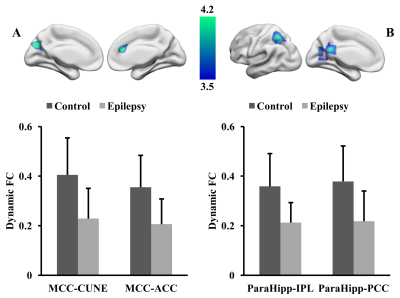 |
14 | Abnormal dynamic functional connectivity in new-onset, drug-naïve adolescent idiopathic generalized epilepsy
Yizhi Yuan1,2, Yuting Wang2,3, Jingping Mou3, Lan Mei2,3, Chunyang Liu3, and Lihua Qiu1,2,3
1Department of Radiology, Chengdu Medical College, Chengdu, China, 2Department of Radiology, The Second People’s Hospital of Yibin, Yibin, China, 3Department of Radiology, Southwest Medical University, Luzhou, China
Dynamic functional connectivity (dFC) is a newly developed analysis strategies which could provide novel understandings of the pathophysiological mechanisms of neuropsychiatric disease. 45 adolescents diagnosed with IGE and 32 well-matched healthy controls completed this study using dynamic functional connectivity (dFC). The decreased dFC between two seed regions(MCC and ParaHipp) and PCC and IPL were part of default mode network(DMN). The impaired DMN in adolescent IGE patients suggest dFC might be more sensitive to reflect the changes of brain function in the early stage of disease and dFC analysis was a promising avenue to deepen our understanding of this disease.
|
||
3009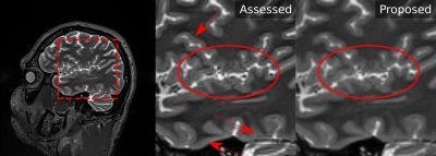 |
15 | Correction of Gibbs ringing artifact improves visualization of epileptogenic pathology
Robert Elton Smith1,2, Eric Pierre1, Graeme Jackson1,2,3, and David Vaughan1,2,4
1Florey Institute of Neuroscience & Mental Health, Heidelberg, Australia, 2Florey Department of Neuroscience and Mental Health, The University of Melbourne, Melbourne, Australia, 3Department of Medicine, Austin Health, Heidelberg, Australia, 4Department of Neurology, Austin Health, Heidelberg, Australia
We evaluate the utility of a recently-proposed method tailored for the removal of Gibbs ringing artifacts in 3D MRI data, by applying it to high-resolution isotropic T2-weighted data acquired during the Australian Epilepsy Project (AEP) pilot. The filter is highly effective at removing this known artifact whilst preserving pathological detail. We therefore encourage its uptake in not only the radiological assessment of the Epilepsies, but any 3D imaging context where the manifestation of Gibbs ringing may be detrimental.
|
||
3010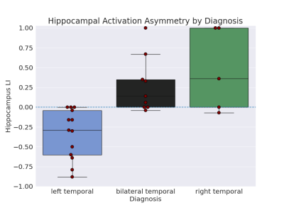 |
16 | Lateralization of Memory Function in Temporal Lobe Epilepsy using Scene Memory fMRI
William Tackett1, Dawn Mechanic-Hamilton1, Sandhitsu Das2, Kathryn Davis1, and John Detre1,2
1Neurology, Perelman School of Medicine, University of Pennsylvania, Philadelphia, PA, United States, 2Radiology, Perelman School of Medicine, University of Pennsylvania, Philadelphia, PA, United States
Surgery to treat mesial temporal lobe epilepsy (TLE) offers freedom from seizures but risks postsurgical memory decline. Lateralizing memory function is essential for mitigating the risk of resecting “eloquent” cortex. Memory laterality has traditionally been assessed using the Wada test, but it is invasive and poses its own risks. The noninvasive method of functional magnetic resonance imaging (fMRI) has become a possible alternative. However, questions remain about fMRI’s reliability in lateralizing memory. Our group has shown that a scene memory task is capable of activating hippocampus and lateralizing memory function. Here, we present an improved version of that experimental paradigm.
|
||
 Back to Meeting Home
 Back to the Program-at-a-Glance
The International Society for Magnetic Resonance in Medicine is accredited by the Accreditation Council for Continuing Medical Education to provide continuing medical education for physicians.
 Back to Meeting Home
 Back to the Program-at-a-Glance
 View the Presentation