Online Gather.town Pitches
Pediatrics: Miscellaneous
Joint Annual Meeting ISMRM-ESMRMB & ISMRT 31st Annual Meeting • 07-12 May 2022 • London, UK

Online Gather.town Pitches
Pediatrics: Miscellaneous
| Booth # | ||||
|---|---|---|---|---|
4022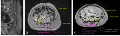 |
1 | Quantifying Infant Lean Body Mass using Free-Breathing 3D Stack-of-Radial MRI
Hannah Parish1, Shu-Fu Shih2,3, Shahnaz Ghahremani3, Sevgi Gokce Kafali2,3, Kara Calkins4, and Holden Wu2,3
1David Geffen School of Medicine, University of California Los Angeles, Los Angeles, CA, United States, 2Department of Bioengineering, University of California Los Angeles, Los Angeles, CA, United States, 3Department of Radiological Sciences, University of California Los Angeles, Los Angeles, CA, United States, 4Department of Pediatrics, University of California Los Angeles, Los Angeles, CA, United States Lean body mass (LBM) is an important measure of metabolic ability. Decreased LBM during infancy may be associated with impaired growth and increased risk for future metabolic disease. This study measured skeletal muscle area as a surrogate for LBM in infants using free-breathing MRI. Psoas and paraspinal muscle areas were measured in 14 infants at the L4-L5 intervertebral disc level. Cross-sectional area of the thigh muscles was measured at the right midthigh in 8 infants. Infants with higher birth weights and those born large for gestational age had larger muscle areas than infants born lighter and/or small for gestational age. |
||
4023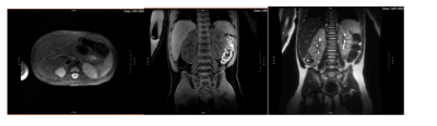 |
2 | Clinical Evaluation of a Lightweight, Flexible Pediatric RF Coil Video Not Available
Ricardo Flores1, Fei Tan1, Michael Ohliger1, and Peder Larson1
1Radiology and Biomedical Imaging, University of California, San Francisco, San Francisco, CA, United States
In this abstract, we conducted a clinical evaluation of a prototype lightweight, flexible pediatric RF Coil. We evaluated the image quality on phantoms, an adult volunteer, and four pediatric clinical patients. For the phantom scans, identical 2D GRE protocols were prescribed between arrays and scanners for study reproducibility and accurate image comparisons. For the pediatric clinical patients, different anatomical studies were clinically ordered. Each study was reviewed clinically and found to achieve diagnostic quality with excellent tissue contrast, coverage, and SNR.
|
||
 |
4024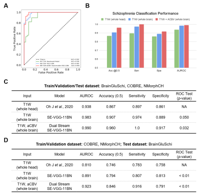 |
3 | Detecting schizophrenia using structural and synthetic cerebral blood volume MRI with deep learning
Junhao Zhang1, Nicolas Acosta1, Vishwanatha Mitnala Rao1, Pin-Yu Lee1, Sabrina Gjerswold-Selleck1, Zihan Wan1, Xinyang Feng1, Jeffrey A. Lieberman2, Scott A. Small3, and Jia Guo2
1Department of Biomedical Engineering, Columbia University, New York, NY, United States, 2Department of Psychiatry, Columbia University, New York, NY, United States, 3Department of Neurology, Columbia University, New York, NY, United States |
|
4025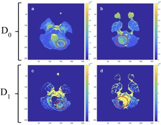 |
4 | Differentiation of Pediatric Brain Tumor Grades Using High b-Value Diffusion MRI with a Varying Diffusion Curvature Model
Jay Fu1, William Hou2, Muge Karaman3,4, and Xiaohong Joe Zhou3,4,5
1Watchung Hills Regional High School, Warren, NJ, United States, 2Montgomery High School, Skillman, NJ, United States, 3Center for MR Research, University of Illinois at Chicago, Chicago, IL, United States, 4Department of Bioengineering, University of Illinois at Chicago, Chicago, IL, United States, 5Departments of Radiology and Neurosurgery, University of Illinois at Chicago, Chicago, IL, United States
Imaging-based determination of tumor grade is highly desirable, particularly for pediatric brain tumors where surgical biopsy carries considerable risks. We have applied a novel diffusion model – varying diffusion curvature (VDC) model, with two b-value ranges (0-2000 sec/mm2 and 0-4000 sec/mm2), to differentiate between low- and high-grade pediatric brain tumors on 70 patients. Our results showed that the combination of the VDC parameters substantially outperformed the ADC for differentiating low- from -high grade pediatric brain tumors. Increasing the maximal b-value from 2000 to 4000 sec/mm2 did not noticeably improve the performance.
|
||
4026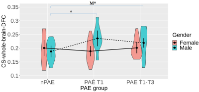 |
5 | Disrupted structural-functional coupling in children with prenatal alcohol exposure Video Not Available
Xiaoyun Liang1,2, Chun-Hung Yeh3,4, and Peter J. Anderson1,5
1Victorian Infant Brain Study (VIBeS), Murdoch Children's Research Institute, Melbourne, Australia, 2Florey Department of Neuroscience and Mental Health, University of Melbourne, Melbourne, Australia, 3Institute for Radiological Research, Chang Gung University and Chang Gung Memorial Hospital, Taoyuan, Taiwan, 4Department of Psychiatry, Chang Gung Memorial Hospital, Taoyuan, Taiwan, 5Turner Institute for Brain and Mental Health, Monash University, Clayton, Australia
In this study, we investigated whether prenatal alcohol exposure (PAE) disrupts the coupling strength between SC and static FC or dynamic FC using multimodal connectomes. Greater coupling strengths were identified in the PAE T1 and PAE T1-T3 groups within males, indicative of brain structural and functional changes in children with PAE. Distinct age effects on coupling strength were also revealed. Further, the reduction of assortative coefficients indicated that PAE could compromise the robustness of brain networks. These findings suggest that multimodal connectomes, especially integrating network dynamics, could be advantageous to revealing brain structural and functional changes.
|
||
4027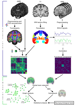 |
6 | Combined structural and functional connectivity changes in Fabry disease
Ilaria Gabusi1,2, Giuseppe Pontillo2,3, Simona Schiavi1,4, Sara Bosticardo1,5, Maria Petracca6, Matteo Battocchio1,7, Antonio Pisani8, Arturo Brunetti2, Alessandro Daducci1, and Sirio Cocozza2
1Department of Computer Science, University of Verona, Verona, Italy, 2Department of Advanced Biomedical Sciences, University of Naples "Federico II", Naples, Italy, 3Department of Electrical Engineering and Information Technology, University of Naples "Federico II", Naples, Italy, 4Department of Neuroscience, Rehabilitation, Ophthalmology, Genetics, Maternal and Child Health (DINOGMI), University of Genoa, Genoa, Italy, 5Department of Biomedical Engineering, University Hospital Basel and University of Basel, Basel, Switzerland, 6Department of Human Neurosciences, Sapienza University of Rome, Rome, Italy, 7University of Sherbrooke, Sherbrooke, QC, Canada, 8Department of Public Health, University of Naples "Federico II", Naples, Italy
Central nervous system involvement in Fabry disease (FD) patients is known, but the effects on brain connectivity have never been explored at macro-scale level. Here, we investigate both structural (SC) and functional connectivity (FC) by analyzing diffusion and resting-state functional MRI. We applied a graph theory approach to compare connectomes of patients and controls and we explored how altered global network metrics are related to patients’ outcome in neuropsychological tests. Our results show that FD patients present a loss of axonal integrity which leads to a widespread reorganization of brain structural and functional architecture, related to their clinical performances.
|
||
4028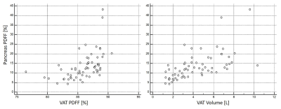 |
7 | Pancreatic PDFF in persons with obesity: change during weight loss and correlations with adipose tissue PDFF
Daniela Junker1, Selina Rupp1, Jessie Han1, Mingming Wu1, Anna Reik2, Meike Wiechert2, Hans Hauner2,3, Marcus M. Makowski1, Christina Holzapfel2, and Dimitrios C. Karampinos1
1Department of Diagnostic and Interventional Radiology, School of Medicine, Technical University of Munich, Munich, Germany, 2Institute for Nutritional Medicine, School of Medicine, Technical University of Munich, Munich, Germany, 3Else Kroener-Fresenius-Center of Nutritional Medicine, School of Life Sciences, Technical University of Munich, Freising, Germany
Risk stratification strategies in patients with metabolic diseases are urgently needed for the selection of appropriate lifestyle interventions. Pancreatic fat content has been shown to be associated with the metabolic syndrome and type 2 diabetes mellitus. MRI-based proton density fat fraction (PDFF) mapping based on a multi-echo gradient echo acquisition is the method of choice for non-invasive pancreatic fat quantification. This study evaluates how the PDFF of the pancreas correlates to adipose tissue PDFF and evolves after an 8-week weight loss intervention.
|
||
 Back to Meeting Home
 Back to the Program-at-a-Glance
The International Society for Magnetic Resonance in Medicine is accredited by the Accreditation Council for Continuing Medical Education to provide continuing medical education for physicians.
 Back to Meeting Home
 Back to the Program-at-a-Glance
 View the Presentation