Online Gather.town Pitches
Pediatric Imaging
Joint Annual Meeting ISMRM-ESMRMB & ISMRT 31st Annual Meeting • 07-12 May 2022 • London, UK

| Booth # | ||||
|---|---|---|---|---|
 |
3714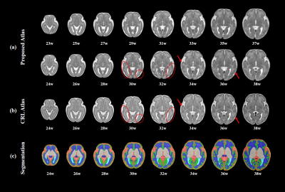 |
1 | Spatiotemporal structural atlas of the fetal brain depicts cortical developmental patterns in a Chinese population
Xinyi Xu1, Jiwei Sun1, Wen Shi1,2, Yao Shen1, Ruoke Zhao1, Wanrong Luo1, Cong Sun3, Ruike Chen1, Mingyang Li1, Yi-Cheng Hsu4, Yi Sun4, Yi Zhang1, Guangbin Wang3,5, and Dan Wu1
1Key Laboratory for Biomedical Engineering of Ministry of Education, Department of Biodical Engineering, College of Biomedical Engineering & Instrument Science, Zhejiang University, HangZhou, China, 2Department of Biomedical Engineering, Johns Hopkins University School of Medicine, Baltimore, MD, United States, 3Department of Radiology, Cheeloo College of Medicine, Shandong Provincial Hospital, Shandong University, Jinan, China, 4MR Collaboration, Siemens Healthineers Ltd, Shanghai, China, 5Department of Radiology, Shandong Provincial Hospital Affiliated to Shandong First Medical University, Jinan, China We constructed a 4D spatiotemporal atlas of the normal fetal brain development from 23 to 38 gestational weeks in a Chinese population. We depicted the developmental trajectories of morphological indices of the cerebral cortex, which showed a characteristic regional variations and indicated the developmental order and rates in the different cortical parcels. The fetal brain atlas and fetal cerebral cortex trajectory offer a better understanding of fetal brain development and can be used as an analytic reference for clinicians in diagnostic or research settings. |
|
3715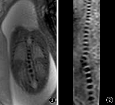 |
2 | Evaluation of fetal spinal anatomy and vertebral malformation using three-dimensional T2-star FFE sequence at 3T: clinical experience
Shuang He1, Qiang Lei1, Xiaoli Zhao1, Ya Yang1, Zhigang Wang1, Hua Lai1, Xiaoyong Zhang2, and Xilin Wen1
1Chengdu Women's and Children's Central Hospital, School of Medicine, University of Electronic Science and Technology of China, Chengdu, China, 2Philips Healthcare, Chengdu, China Magnetic resonance image (MRI) has been increasingly used to evaluate fetal malformations. Currently, the commonly used fetal spine scanning sequences include conventional 2D Single-Shot TSE (2D SSH TSE) and 2D balanced fast field echo sequence (2D BTFE), However, 2D MR sequences have disadvantages such as unclear display of tiny structures especially the fetal cervical vertebra, furthermore the image quality is strongly affected by fetal postures. In this study we compared 3D T2-star-weighted FFE sequence with conventional 2D SSH TSE sequence and 2D BTFE sequence. The 3D T2-star FFE sequence has high prenatal diagnosis value for fetal spine malformation, and can be used as an important supplementary sequence for fetal spine diagnosis in clinical practice. |
||
|
3716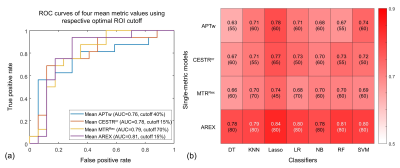 |
3 | Radiomic Analysis of APT MRI for Preoperative Risk Stratification of Pediatric Neuroblastoma
Wenqi Wang1, Jia Xuan2, Jiawei Liang2, Xiaohui Ma2, Weibo Chen3, Dan Wu1, Hongxi Zhang2, Can Lai2, and Yi Zhang1
1Key Laboratory for Biomedical Engineering of Ministry of Education, Department of Biomedical Engineering, College of Biomedical Engineering & Instrument Science, Zhejiang University, Hangzhou, China, 2Department of Radiology, Children’s Hospital, Zhejiang University School of Medicine, Hangzhou, China, 3Philips Healthcare, Shanghai, China
Neuroblastoma (NB) is most often diagnosed in young children, and high-risk NB indicates a poor prognosis and requires aggressive treatment. Here, we assessed the utility of radiomics for amide proton transfer (APT) imaging in the preoperative classification of high-risk and low-risk abdominal NBs. Thirty-three pediatric patients underwent APT scans, with 16 confirmed high-risk NBs and 17 confirmed low-risk NBs. Radiomic models were constructed from four APT-related metric maps using 7 classifiers, with their prediction performances evaluated by associated AUCs. The optimal model achieved an AUC of 0.86, demonstrating the potential of APT MRI-based radiomics for risk stratification of NB.
|
|
3717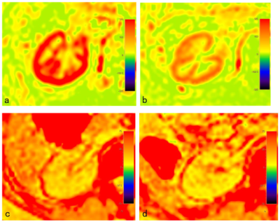 |
4 | Imaging evaluation of pediatric IgAN and HSPN based on ASL and mDIXON-Quant
Yupeng Zhu1, Yun Peng1, Xiaorong Liu2, Jianxiu Lian3, Zhiwei Shen3, and Jiazheng Wang3
1Radiology, Beijing Children's Hospital, Capital Medical University, National Center for Children's Health, Beijing, China, 2Beijing Children's Hospital, Capital Medical University, National Center for Children's Health, Beijing, China, 3Philips Healthcare, Beijing, China
It is difficult to identify IgA nephropathy (IgAN) and Henoch Schonlein purpura nephritis (HSPN) due to their similar image features on conversional MR anatomic images. Arterial spin labeling (ASL) and mDIXON-Quant MR imaging can provide more data about perfusion and relaxation time to evaluate the functional state of kidney. In this study, we explore to different IgAN from HSPN in pediatric patients with nephropathy using ASL and mDIXON-Quant MR sequences, and hope to improve accuracy in differential diagnosis of IgAN and HSPN.
|
||
3718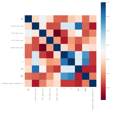 |
5 | Machine learning and the prediction of cerebral ventricular changes in fetuses with ventriculomegaly: a fetal MRI study Video Permission Withheld
Xue Chen1, Zhou Huang2, Yonggang Li2,3,4, Jibin Zhang1, Xiaowen Gu1, and Zhisen Li1
1Department of Radiology, The Affiliated Suzhou Hospital of Nanjing Medical University, Suzhou Municipal Hospital, Suzhou city, Jiangsu province, 215002, China, 2Department of Radiology, The First Affiliated Hospital of Soochow University, Suzhou city, Jiangsu province, 215000, China, 3Institute of Medical Imaging, Soochow University, Suzhou city, Jiangsu province, 215000, China, 4National Clinical Research Center for Hematologic Diseases, the First Affiliated Hospital of Soochow University, Suzhou city, Jiangsu province, 215000, China
Fetal ventriculomegaly (FV) is one of the central nervous system (CNS) major malformations. Prenatal clinical research has seen only a few applications of machine learning. To our knowledge, radiomic machine learning for predicting the change of cerebral ventricular in fetuses with ventriculomegaly has not been reported. We discovered that a combination of clinical characteristics and fetal MRI features could accurately predict postnatal ventricular changes in fetuses with ventriculomegaly. The occipital lobe white matter on the dilated lateral ventricle side may play an important role in the pathophysiological process in FV.
|
||
3719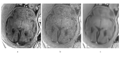 |
6 | Fetal brain T1-weighted imaging with a deep learning constrained compressed SENSE reconstruction
Jing Wang1, Zhuo Wang1, Yi Zhu2, and Ke Jiang2
1Radiology, the First Hospital of Jilin University, Changchun, China, 2Philips Healthcare, Beijing, China
The quality of commonly used fetal T1-weighed inversion recovery(IR) images is relatively poor. Compressed SENSE(CS) technique allows shortening of scan time, but the overall image quality has not been significantly improved. In this study, Compressed-SENSE Artificial intelligence(CS-AI) framework was applied to reduce the scan time and increase the spatial resolution. This study aims at acquiring high-resolution fetal brain T1-weighted image with reduced scan time and compare the image quality among images reconstructed with CS-AI, CS and conventional SENSE.
|
||
|
3720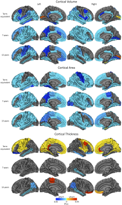 |
7 | Cortical development over the first 13 years of life after very preterm birth
Claire Kelly1,2,3, Deanne K Thompson2,3,4, Chris Adamson3, Gareth Ball3,4, Thijs Dhollander3, Richard Beare3, Bonnie Alexander3,5, Jeanie LY Cheong2,6,7, Marc L Seal3,4, Terrie E Inder8, Lex W Doyle2,4,6,7, and Peter J Anderson1,2
1Turner Institute for Brain and Mental Health, School of Psychological Sciences, Monash University, Melbourne, Australia, 2Victorian Infant Brain Studies (VIBeS), Murdoch Children's Research Institute, Melbourne, Australia, 3Developmental Imaging, Murdoch Children's Research Institute, Melbourne, Australia, 4Department of Paediatrics, The University of Melbourne, Melbourne, Australia, 5Department of Neurosurgery, The Royal Children's Hospital, Melbourne, Australia, 6The Royal Women's Hospital, Melbourne, Australia, 7Department of Obstetrics and Gynaecology, The University of Melbourne, Melbourne, Australia, 8Department of Pediatric Newborn Medicine, Brigham and Women’s Hospital, Harvard Medical School, Boston, MA, United States
Longitudinal cortical development following very preterm (VP) birth has been infrequently studied. We undertook a unique longitudinal MRI study of 202 VP infants and 66 term-born infants from shortly after birth to age 13 years. Longitudinal changes in surface-based volume, area and thickness measurements for 62 cortical regions were modelled using generalised additive models. We reveal cortical alterations in the neonatal period and altered cortical development in the early years of life following VP birth. Cortical alterations persisted to age 13, with several regions of reduced volume and area, thickness reductions in temporal regions, and thickness increases in frontal regions.
|
|
3721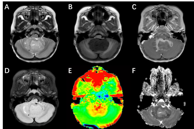 |
8 | Distinguishing sonic hedgehog from Group 4 medulloblastoma using amide proton transfer–weighted MR Imaging Video Permission Withheld
AnDong Ma1, YaoMing Qu1, MingJun Lu1, Xia Zou1, XinZi Liu1, Chen Zhao2, and ZhiBo Wen1
1Radiology, ZhuJiang Hospital of Southern Medical University, GuangZhou, China, 2Philips Healthcare, GuangZhou, China
Preoperative prediction of molecular subgroup based on magnetic resonance imaging (MRI) remains a challenge. The objective of this study was to assess amide proton transfer-weighted (APTw) method in medulloblastoma (MB). Preliminary results showed that the mean value of APT value in sonic hedgehog (SHH) -activated MB tended to be higher than that of Group 4 MB.
|
||
| 3722 | 9 | Relationship between fetal brainstem growth and corresponding gestational weeks: an MRI study
jie LI1, xiao Ling1, songhong Yue1, haoyuan Li1, tao Wen1, jing Zhang1, and kai Ai2
1Lanzhou University Second Hospital, Lanzhou, China, 2Philips Healthcare, Xi'an, China
We use midsagittal T2WI-based structural MRI images to investigate the relationship between fetal brainstem growth and corresponding gestational weeks. The changes of fetal brainstem substructure in of 153 pregnant women in the middle and late stages were quantitatively evaluated. We found that all measurement indices (height, anteroposterior diameter and cross-sectional area) of normal fetal brainstem were significantly correlated with gestational age. Our study demonstrates that MRI can clearly show the fetal brainstem structure, and the changes of brainstem substructure follow certain rules in fetus.
|
||
3723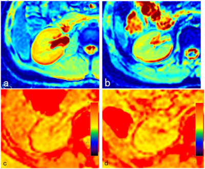 |
10 | The value of IVIM and mDIXON-Quant MR imaging in the identification of pediatric IgAN and HSPN
Yupeng Zhu1, Yun Peng1, Xiaorong Liu2, Jianxiu Lian3, Zhiwei Shen3, and Geli Hu3
1Radiology, Beijing Children's Hospital, Capital Medical University, National Center for Children's Health, Beijing, China, 2Beijing Children's Hospital, Capital Medical University, National Center for Children's Health, Beijing, China, 3Philips Healthcare, Beijing, China
It is difficult to identify IgA nephropathy (IgAN) and Henoch Schonlein purpura nephritis (HSPN) due to their similar clinical features. Functional magnetic resonance imaging (fMRI) can simultaneously obtain tissue structure and function imaging, and evaluate the functional state of kidney. In this study, intravoxel incoherent motion (IVIM) and mDIXON-Quant were performed in pediatric patients with nephropathy, and it was found that IgAN and HSPN were remarkably different in renal function. The values of medulla’s D and cortex’s R2* obtained by IVIM and mDIXON-Quant had the ability in differential diagnosis of IgAN and HSPN.
|
||
3724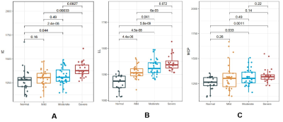 |
11 | The value of synthetic MRI in detecting the brain change and hearing impairment of children with sensorineural hearing loss
Penghua Zhang1, Xiaoan Zhang1, Xin Zhao1, Lin Lu1, Jinxia Guo2, Qingna Xing1, Meiying Cheng1, and Lingsong Meng1
1The Third Affiliated Hospital of Zhengzhou University, Zhengzhou, China, 2MR Research,GE Healthcare,Beijing,China, Beijing, China Study explored the brain development of children with sensorineural hearing loss (SNHL),which may cause children's language discrimination ability and voice localization ability worse.The auditory pathway was regarded impaired and the related brain structure may change in SNHL,especially in early development stage for children age less than 3 months.Studies with Diffusion tensor imaging,diffusion kurtosis imaging and fMRImainly investigate the patient age over 2 years old,when it’s a timechildren's brain is myelinated[1] . Synthetic MRI was used in this study to quantitatively evaluate the differences among normal and SNHL with varying degree of hearing loss. The quantitative value of T1 in inferior colliculus,lateral lemniscus and middle cerebellar peduncle were found help early diagnosis of hearing loss of SNHL infants. |
||
 |
3725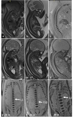 |
12 | Susceptibility-Weighted Imaging for Evaluation of Fetal Vertebrae and Vertebral Anomalies Video Permission Withheld
Xianyun Cai1,2, Jinxia Zhu3, and Guangbin Wang1
1Cheeloo College of Medicine, Shandong University, Jinan, Shandong, China; Department of Radiology, Shandong Provincial Hospital Affiliated to Shandong First Medical University, Jinan, Shandong, China., Jinan, China, China, 2Department of Radiology, Shandong Provincial Hospital Affiliated to Shandong First Medical University, Jinan, Shandong, China., Jinan, China, China, 3MR Collaboration, Siemens Healthineers Ltd, Beijing, China. Email: jinxia.zhu@siemens-healthineers.com, beijing, China
This study explored the feasibility and clinical value of susceptibility weighted imaging (SWI) in depicting fetal vertebral growth and related anomalies. The results showed that vertebral development in fetuses on SWI was remarkably linearly correlated with gestational ages. In addition, SWI demonstrated superior image quality and higher diagnostic accuracy compared to conventional Half-Fourier acquisition single-shot turbo spin-echo (HASTE) or true fast imaging with steady-state precession (TrueFISP) sequences. In conclusion, SWI is a reliable choice when imaging fetal vertebrae and vertebral anomalies.
|
|
|
3726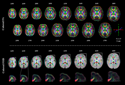 |
13 | Reconstructing a four-dimensional diffusion magnetic resonance imaging atlas of the fetal brain
Ruike Chen1, Cong Sun2, Yuhao Liao1, Tingting Liu1, Junyan Wang3, Yi-Cheng Hsu4, Yi Sun4, Yi Zhang1, Guangbin Wang2, and Dan Wu1
1Key Laboratory for Biomedical Engineering of Ministry of Education, Department of Biomedical Engineering, College of Biomedical Engineering & Instrument Science, Zhejiang University, Hangzhou, China, 2Department of Radiology, Shandong Provincial Hospital, Cheeloo College of Medicine, Shandong University, Jinan, Shandong, China, 3Zhejiang Lab, Hangzhou, China, 4MR Collaboration, Siemens Healthineers Ltd., Shanghai, China
Diffusion MRI (dMRI) is a powerful tool to characterize the developing brain, e.g., to map the spatiotemporal development of early white matter (WM) in the fetal brain and to quantify microstructural changes for diagnosis of prenatal disorders. We proposed a pipeline for dMRI atlas generation with fiber orientation distribution-based registration and shape update, and constructed the first spatiotemporal dMRI atlas of the fetal brain in a Chinese population of 90 normal fetuses from 24 to 38 gestational weeks. The atlas provided information for deciphering the morphological and microstructural development of WM tracts during the second to third trimester.
|
|
3727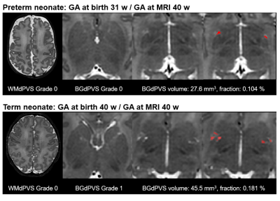 |
14 | Dilated perivascular spaces in neonates
Hyun Gi Kim1, Sumin Kim2, JeeYoung Kim1, Na Young Shin1, and Yoonho Nam2
1The Catholic University of Korea, Seoul, Korea, Republic of, 2Division of Biomedical Engineering, Hankuk University of Foreign Studies, Yongin, Korea, Republic of
Perivascular spaces (PVS) are fluid-filled cavities around the blood vessels in the brain. In adults, dilated PVS (dPVS) is known to increase with age but dPVS in neonates is poorly understood. In this study, we performed both visual and volumetric evaluation of dPVS in neonates using T2-weighted images from the Developing Human Connectome project. We found that gender did not affect dPVS extent in neonates, preterm neonates had lower dPVS extent than term neonates, and dPVS fraction decreased with gestational age (GA) at MRI scan.
|
||
3728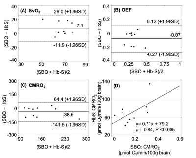 |
15 | Whole-brain oxygen metabolism in adults with sickle-cell disease using susceptometry and T2-based method
Pei-Hsin Wu1, Felix W Wehrli2, and Seyed A Nabavizadeh2
1Department of Electrical Engineering, National Sun Yat-sen University, Kaohsiung, Taiwan, 2Department of Radiology, University of Pennsylvania, Philadelphia, PA, United States SvO2 and CMRO2 in patients with sickle cell anemia (SCA) were evaluated by MR susceptibility-based oximetry and compared with that derived from T2-based methods. The results show that (1) SvO2 trends lower and OEF tends to be higher in T2-based methods, (2) there is high correlation of CMRO2 between SBO and HbS model, (3) CMRO2 derived from SBO indicates tCBF was elevated to maintain oxygen delivery and oxygen metabolism in the brain given that CMRO2 is comparable with the healthy subjects reported previously. The preliminary results demonstrate the feasibility of OEF and CMRO2 estimation via SBO in subjects with SCA. |
||
|
3729 |
16 | Hemodynamics, brain volume and cortical development in fetuses with complex congenital heart disease Video Permission Withheld
Cong Sun1, Xinyi Xu2, Jiaguang Song3, Yufan Chen1, Chao Zhang1, Yuhao Liao2, Wanrong Luo2, Jinxia Zhu4, Dan Wu2, and Guangbin Wang1,3
1Radiology, Shandong Provincial Hospital Affiliated to Shandong University, Jinan, China, 2Key Laboratory for Biomedical Engineering of Ministry of Education, Department of Biomedical Engineering, College of Biomedical Engineering & Instrument Science, Zhejiang University, Hangzhou, China, 3Shandong Provincial Hospital Affiliated to Shandong First Medical University, Jinan, China, 4Siemens Healthineers Ltd, Beijing, China
Fetuses with complex congenital heart disease (CHD, n=23) and healthy gestational age-matched controls (n=25) were investigated. Hemodynamics were assessed by Doppler examinations. Fetal brain MR images were segmented to get global and regional morphological measurements. Results showed that fetuses with CHD have abnormally higher umbilical artery pulsation index and smaller global and regional brain volumes as early as 20-30 weeks; however, thickness, mean curvature, and sulcal depth of different brain lobes showed no statistical difference from the healthy controls. These results add to growing evidence of antenatal brain abnormalities of the CHD fetuses during the second and early third trimesters.
|
|
3730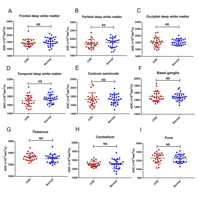 |
17 | Regional brain ADC value changes in fetuses with complex congenital heart diseases in the second and early third trimesters
Cong Sun1, Jiaguang Song2, Yufan Chen1, Jinxia Zhu3, and Guangbin Wang1,4
1Shandong Provincial Hospital Affiliated to Shandong University, Jinan, China, 2Ultrasound, Shandong Provincial Hospital Affiliated to Shandong First Medical University, Jinan, China, 3Siemens Healthineers Ltd, Beijing, China, 4Shandong Provincial Hospital Affiliated to Shandong First Medical University, Jinan, China, Jinan, China
We measured apparent diffusion coefficient (ADC) values in the brains of 64 normal fetuses at 20-40 gestational weeks and 23 fetuses with complex CHD during second and early third trimesters. We also compared ADC values between the fetuses with CHD and the 27 gestational age-matched normal controls using covariance analyses. Our results showed normal variations in ADC values for fetal brains during maturation in utero. Moreover, the ADC values in the CHD group were not significantly different than the GA-matched normal fetuses, indicating that myelin development was not delayed in the CHD fetuses during the second and early third trimesters.
|
||
 Back to Meeting Home
 Back to the Program-at-a-Glance
The International Society for Magnetic Resonance in Medicine is accredited by the Accreditation Council for Continuing Medical Education to provide continuing medical education for physicians.
 Back to Meeting Home
 Back to the Program-at-a-Glance
 View the Presentation