Online Gather.town Pitches
Multiparametric MR: Breast Cancer
Joint Annual Meeting ISMRM-ESMRMB & ISMRT 31st Annual Meeting • 07-12 May 2022 • London, UK

Online Gather.town Pitches
Multiparametric MR: Breast Cancer
| Booth # | ||||
|---|---|---|---|---|
 |
4263 |
1 | Can combined DWI, IVIM and DKI better differentiate malignant from benign breast lesions with TIC type II?
Wang Hongjie1, Wang Weiwei2, Dou Weiqiang3, Lv Siqiang1, Zhu Laimin2, Chen Yueqin2, and Sun Zhanguo2
1Clinical Medical College of Jining Medical University, Jining, China, 2Department of Medical Imaging, Affiliated Hospital of Jining Medical University, Jining, China, 3MR Research China, GE Healthcare, Beijing, China Accurate preoperative prediction of benign and malignant breast lesions is essential in clinical practice. Currently, the diagnostic specificity of DCE-MRI is relatively low for breast lesions with time-intensity-curve (TIC) type-II. Different diffusion-weighted models, including mono-exponential diffusion-weighted imaging (DWI), intravoxel-incoherent-motion diffusion-weighted imaging (IVIM) and diffusion kurtosis imaging (DKI), have been previously reported effective in the differential diagnosis of overall breast lesions. In this study, we found that, compared with DWI alone, combined DWI, IVIM and DKI can better differentiate malignant from benign breast TIC type-II lesions, with IVIM parameter of D and DKI parameter of MK as demonstrated independent influencing factors. |
|
4264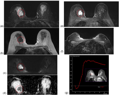 |
2 | stratification of axillary lymph node metastasis burden with standard breast MRI in breast cancer Video Permission Withheld
Jieying Chen1, Xiaolian Su1, Tingting Xu1, Qifeng Luo2, Jilei Zhang3, Lin Zhang1, and Guangyu Tang1
1Department of Radiology, Shanghai Tenth People’s Hospital, Tongji University School of Medicine, Shanghai, China, 2Department of General Surgery, Shanghai Tenth People’s Hospital, Tongji University School of Medicine, Shanghai, China, 3Philips Healthcare, Shanghai, China Assessment of axillary lymph node metastasis burden before surgery in breast cancer patients is warranted for axillary management. This study developed a simple model based on the standard breast MRI features of breast tumor and axillary lymph node to differentiate patients with no, low or heavy axillary metastasis burden. With the help of this model, patients and clinicians would make more rational decision when to choose omitting surgery, sentinel lymph node biopsy or axillary lymph node dissection for axilla management. |
||
4265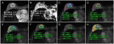 |
3 | A risk stratification scoring system for predicting axillary lymph node metastasis in breast cancer using multiparametric MRI Video Not Available
Xiaofeng Chen1, Jiada Yang1, Nina Fan1, Zhiqi Yang1, Ruibin Huang2, Yue Li1, Cuixia Wan1, Junhao Gong1, Yuting Liao3, Guijin Li4, Mengzhu Wang5, Fengyan Cheng1, Xiangguang Chen1, Zhuozhi Dai6,
and Weixiong Fan1
1Meizhou People's Hospital, Meizhou, China, 2First Affiliated Hospital of Shantou University Medical College, Shantou, China, 3GE Healthcare, Guangzhou, China, 4Siemens Healthineers, Shanghai, China, 5Siemens Healthineers, Guangzhou, China, 6Shantou Central Hospital, Shantou, China
This study developed and validated a point-based scoring system (PSS) for stratifying the axillary lymph node (ALN) metastasis risk of breast cancer (BC) based on preoperative clinicopathological and MRI features compared with MRI-reported ALN status model and to explore its prognostic significance. The PSS in our study provides an easy tool for surgeons to assess breast cancer patients' risk for ALN metastasis prior to surgery. And the results showed that PSS had a relatively better performance than MRI-reported ALN status model (P=0.045),with a higher AUC, accuracy, and specificity, suggesting that PSS can be routinely used in axillary nodal staging.
|
||
4266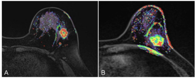 |
4 | Quantitative analysis of DCE-MRI for predicting lymphovascular invasion in breast cancer Video Permission Withheld
Zhiqi Yang1, Tianfu Lai2, Xiaofeng Chen1, Ruibin Huang3, Weichao Yang1, Yuting Liao4, Mengzhu Wang5, Guijin Li6, Zhuozhi Dai7, and Xiangguang Chen1
1Meizhou People’s Hospital, Meizhou, China, 2Department of Radiology, Meizhou People’s Hospital, Meizhou, China, 3First Affiliated Hospital of Shantou University Medical College, Shantou, China, 4GE Healthcare, Guangzhou, China, 5Siemens Healthineers, Guangzhou, China, 6Siemens Healthineers, Shanghai, China, 7Shantou Central Hospital, Shantou, China
The optimal biomarkers for the diagnosis of breast lymphovascular invasion have not yet been found. DCE-MRI features can evaluate the tumor microenvironment which is related to LVI. This study explored the independent predictor of LVI, as well as developed and validated the parametric combined prediction model for the diagnosis of lymphovascular invasion in breast cancer using quantitative analysis of DCE-MRI. The prediction model based on Kep and N stage could improve the performance of LVI prediction, compared to clinicopathological or magnetic resonance parameters.
|
||
4267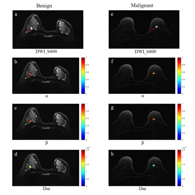 |
5 | Classification of Benign and Malignant Breast Lesions Using high b-value Diffusion MRI with a Continuous-time Random-walk (CTRW) Model
Yuan Jiang1, Ke Xue2, Yongming Dai2, Dongmei Wu3, Junzhe Yang1, Naishan Qin1, and Jianxing Qiu1
1Peking university first hospital, Beijing, China, 2Central Research Institute, United Imaging Healthcare, Shanghai, China, 3Shanghai Key Laboratory of Magnetic Resonance, School of Physics and Electronics Science, East China Normal University, Shanghai, China
The high b-value DWI with a CTRW Model is useful in classification of benign and malignant proliferative breast lesions. The identification model using the combinations of CTRW parameters, Dm and β, has a moderate efficiency.
|
||
4268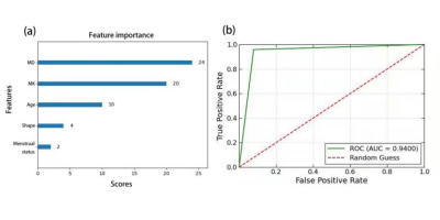 |
6 | Constructing XGboost prediction model based on 3.0T diffusion kurtosis imaging improves the diagnostic performance for breast cancer Video Permission Withheld
Han Zhou1, Wan Tang1,2, Tianhong Quan3, Xiaoyan Chen1, Huanian Zhang1, Zijie Fu1, Renhua Wu1, and Yan Lin1
1Radiology Department, Second Affiliated Hospital of Shantou University Medical College, Shantou, China, 2Institute of Health Monitoring,Inspection and Protection,Hubei Provincial Center for Disease Control and Prevention,Hubei Provincial Key Laboratory for Applied Toxicology, Wuhan, China, 3Shantou University,College Of Engineering, Shantou, China
This study demonstrated that MK derived from DKI was performed better than MD, ADC, Ve, Kep and Ktrans for differentiating between benign and malignant BLs. Also, MK has great potentialities in predict histological grades, lymph node status and Ki-67 expression of BCs. Finally, a XGboost model was constructed by combining MD, MK, age, shape and menstrual status, which exhibited superior diagnostic performance for BC characterization and an improved assessment of BLs. The findings of current study will aid the development of a novel noninvasive approach for BC screening and clinical diagnosis, therefore reducing unnecessary biopsies and patient`s anxiety.
|
||
 |
4269 |
7 | Improved Differentiation of Benign and Malignant Breast Lesions with Synthetic Relaxometry and Kaiser Score
Lingsong Meng1, Xin Zhao1, Jinxia Guo2, Lin Lu1, Qingna Xing1, Yafei Guo1, Honglei Shang1, Penghua Zhang1, Yongbing Sun1, and Xiaoan Zhang1
1The Third Affiliated Hospital of Zhengzhou University, Zhengzhou, China, 2MR Research, GE Healthcare, Beijing, China
Early detection and accurate characterization can reduce significantly the death rates of breast cancer patients. Kaiser score (KS) demonstrates robust and wonderful performance in the assessment of breast lesions. Nevertheless, this scoring system lacks quantitative parameters, which may lead to false negatives, especially for breast cancers exhibiting atypical morphological features. In this study, the quantitative value from Synthetic MRI was added to the KS assessment to improve the differentiation of the malignant and benign breast lesions.
|
|
4270 |
8 | Multiband Accelerated IMPULSED Protocol for Breast Tumor Imaging
Jie Ding1, Zhen Zhang1, Yishi Wang2, Xiuzheng Yue2, Rongrong Zhu1, and Ruoshui Ha1
1Medical Imaging Center, People's Hospital of Ningxia Hui Autonomous Region, Yinchuan, China, 2Philips Healthcare, Beijing, China
The investigation of time dependent diffusion imaging techniques such as IMPULSED is emerging for clinical research since recently made available on 3T scanners. IMPULSED requires the acquisition of diffusion images at different diffusion times and diffusion weightings. In this study, we proposed to use Multiband to accelerate the data acquisition for IMPULSED and compared the ADC spectrum for two protocols. Our results showed the consistency in ADC calculation for the Multiband accelerated protocol with the result without using Multiband.
|
||
4271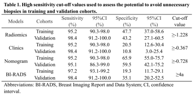 |
9 | Potential of a Nomogram Based on DCE MRI and Clinical Features to Reduce Unnecessary Biopsies of Breast Lesions: Comparison with BI-RADS Video Permission Withheld
Youfan Zhao1, Zhongwei Chen1, Jiejie Zhou1, Haiwei Miao1, Shuxin Ye1, Huiru Liu1, Meihao Wang1, and Min-Ying Su2
1Department of Radiology, The First Affiliated Hospital of Wenzhou Medical University, Wenzhou, China, 2Department of Radiological Sciences, University of California, Irvine, CA, United States
318 patients (331 breast lesions) with DCE-MRI and clinical features were analyzed to establish a nomogram for diagnosis of breast cancer. The dataset was split to 233 (145 malignant 88 benign) for training, and 98 (61 malignant 37 benign) for testing. Radiomics features were extracted from DCE-MRI and selected to calculate the radiomics score. The clinical features were analyzed by univariate and multivariate analyses. Then a nomogram was established based on clinical features and rad-score. When applying cut-off values with ≥95% sensitivity, the nomogram can reduce 59.5% to 65.9% unnecessary biopsies, which was higher than that of BI-RADS.
|
||
4272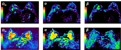 |
10 | Differentiating malignant and benign breast tumors using a continuous-time random-walk diffusion model
Mu Du1, Da Zou2, Yanzhen Hou1, Liyun Zheng2, Dongmei Wu3, Yongming Dai2, and Yubao Liu1
1Medical Imaging Center, Shenzhen Hospital, Southern Medical University, Shenzhen, China, 2United Imaging Healthcare, Shanghai, China, 3Shanghai Key Laboratory of Magnetic Resonance, Department of Physics, East China Normal University, Shanghai, China
Magnetic resonance imaging (MRI) is an important diagnostic method for the breast cancer. However, for conventional MRI techniques such as diffusion-weighted imaging (DWI), it ignores the non-Gaussian behaviors of the water diffusion caused by the restriction of tissue microstructure, thus the conventional method is insensitive to microstructural and heterogeneity changes in breast tumor. This study explores the feasibility of applying a continuous-time random-walk (CTRW) non-Gaussian diffusion model to the differentiation and assessment of malignant and benign breast tumors. The correlations between CTRW parameters and breast tumor immunohistochemical features such as oestrogen receptor (ER), progesterone receptor (PR) and Ki67 expression status are studied. The CTRW model has potential in future applications of breast tumor malignancy and therapeutic efficacy evaluation.
|
||
4273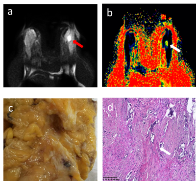 |
11 | Diffusion and IVIM MR imaging-based virtual elastography for the differentiation between invasive ductal carcinoma and fibroadenoma Video Not Available
Ting Liang1, Xianjun Li1, and Jian Yang1
1Department of Radiology, the First Affiliated Hospital of Xi'an Jiaotong University, Xi'an, China
Preoperative accurate differential diagnosis of benign and malignant breast tumors is very important to avoid unnecessary surgical treatment. This study aims to evaluate the potential efficacy of the virtual elastography (vMRE) method in the differential diagnosis of IDC and FA, and help guide clinical management. Our results suggested that mean virtual stiffness obtained by this vMRE method can accurately distinguish the hardness difference between FA and IDC.
|
||
4274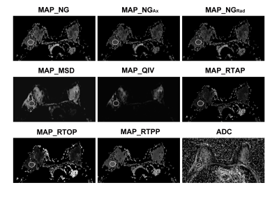 |
12 | Mean Apparent Propagator-MRI: A Novel Diffusion-Based Approach for Quantitative Assessment of Tumor-Stromal Ratio in Breast Carcinoma Video Not Available
Xiang Zhang1, Wei Jiang1, Mengzhu Wang2, Zehong Yang1, Xu Yan2, Guang Yang3, Chunping Mao1, and Jun Shen1
1Sun Yat-Sen Memorial Hospital, Sun Yat-Sen University, Guangzhou, China, 2MR Scientific Marketing, Siemens Healthineers, Guangzhou, China, 3Shanghai Key Laboratory of Magnetic Resonance, School of Physics and Electronic Science, East China Normal University, Shanghai, China
Mean apparent propagator (MAP)-MRI establishes a robust analytical framework based on the random motion distribution of natural water molecules. This study investigated the application of quantitative parameters derived from MAP-MRI in patients with breast cancer and determined whether MAP-MRI can be adopted as a better method than apparent diffusion coefficient (ADC) from conventional DWI to preoperative diagnosis tumor-stromal ratio. The results suggested that quantitative assessment with MAP-MRI showed superior predictive performance than conventional DWI for preoperative diagnosis of tumor-stromal ratio in patients with breast cancer.
|
||
|
4275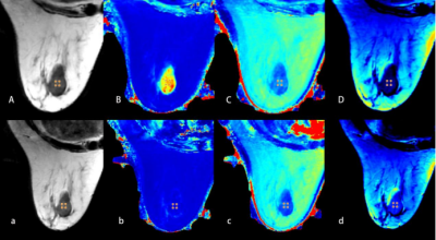 |
13 | Clinical Value of Synthetic MRI and DCE-MRI in the Differentiation of Benign and Malignant Breast Lesions
jinrui liu1, mengyin xu1, zhihao li2, jialiang ren3, and zhaohui an1
1General Hospital of Ningxia Medical University, Yinchuan, China, 2GE Healthcare China, Xi’an, China, 3GE Healthcare China, Beijing, China
In this study, an evaluation was proposed based on the clinical value of synthetic MRI parameters in distinguishing benign and malignant breast lesions. From conclusion, the T2-pre values and PD-pre values were statistically significant different between benign and malignant lesion. The combined T2-pre, PD-pre with BI-RADS outperformed BI-RADS only in discriminating malignant and benign lesions, which has potential clinical value for improving diagnostic accuracy of breast lesions.
|
|
4276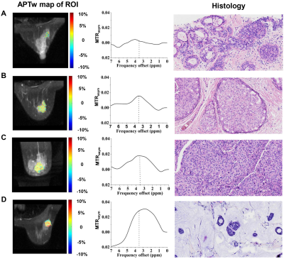 |
14 | Breast amide proton transfer imaging at 3T: diagnostic performance and correlation with pathologic characteristics
Zhou Liu1, Jie Wen1, Meng Wang1, Ya Ren1, Qian Yang1, Long Qian2, Honghong Luo1, Cuiju He1, Yin Wu3, and Dehong Luo1
1National Cancer Center/National Clinical Research Center for Cancer/Cancer Hospital & Shenzhen Hospital, Chinese Academy of Medical Sciences and Peking Union Medical College, Shenzhen, China, 2MR Research, GE Healthcare, Beijing, China, 3Paul C. Lauterbur Research Center for Biomedical Imaging, Shenzhen Institute of Advanced Technology, Chinese Academy of Sciences, Shenzhen, China
To date, great efforts have been made to investigate the performance of APT imaging in breast cancer diagnosis and prognosis, but with discrepant findings. Hence, this study aims to investigate the diagnostic performance and association of APT imaging with clinical-pathologic characteristics of breast cancer in a relatively large cohort of patients at 3T. Results showed significantly higher APTw signals in malignant lesions than that in benign ones, and high histologic grade, T stage and proliferation Ki-67 index in breast cancer patients. The results confirm the usefulness of APT imaging in the differentiation of lesion malignancy and the prediction of prognosis.
|
||
4277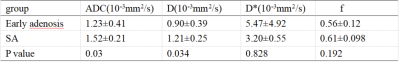 |
15 | Intravoxel incoherent motion imaging for the differentiating early breast adenosis from sclerosing adenosis Video Permission Withheld
Qi Wu1, lina zhang2, qingwei song2, ailian liu2, liangjie lin3, xiaoxiao zhang3, and xiaofang xu3
1The First Affiliated Hospital of Dalian Medical University, daLian, China, 2The First Affiliated Hospital of Dalian Medical University, dalian, China, 3philips Healthcare, Beijing, China
Sclerosing adenosis (SA) of the breast is the late development stage of breast adenosis which is difficult to diagnose. Because its imaging manifestations are similar to malignant lesions of the breast, which often needs pathological diagnosis. Intravoxel incoherent motion(IVIM ) imaging is an MR method to separate diffusion and perfusion effects by fitting a double exponential model of signal attenuation and can accurately display the diffusion movement of water molecules. This study confirmed that some parameters of IVIM can be used in the identification of early breast adenosis and SA. Therefore, IVIM is of great value in the diagnosis of breast adenosis.
|
||
 Back to Meeting Home
 Back to the Program-at-a-Glance
The International Society for Magnetic Resonance in Medicine is accredited by the Accreditation Council for Continuing Medical Education to provide continuing medical education for physicians.
 Back to Meeting Home
 Back to the Program-at-a-Glance
 View the Presentation