Online Gather.town Pitches
Multiparameric MRI: Prostate & Rectal Cancer
Joint Annual Meeting ISMRM-ESMRMB & ISMRT 31st Annual Meeting • 07-12 May 2022 • London, UK

Online Gather.town Pitches
Multiparameric MRI: Prostate & Rectal Cancer
| Booth # | ||||
|---|---|---|---|---|
3731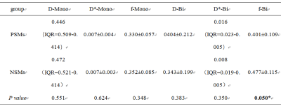 |
1 | The efficacy of Intravoxel incoherent motion in predicting positive margins of radical prostatectomy
Wanting Gan1, Shuang Meng1, Lihua Chen1, Nan Wang1, Yunsong Liu1, and Ailian Liu1
1Department of Radiology,the First Affiliated Hospital of Dalian Medical University, Dalian, China
Positive surgical margins(PSMs) are associated with local recurrence and distant metastasis of prostate cancer which affect the long-term survival rate of patients1-3. IVIM can simultaneously observe microcirculatory perfusion of blood and intracellular and extracellular water molecules diffusion4. The results of our study show that f-Bi can be used to predict positive margins of radical prostatectomy.
|
||
3732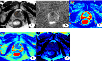 |
2 | Research on the Application of Diagnosis and Invasion Assessment of Transition Zone Prostate Cancer by Relaxation Maps From Synthetic MRI
Na Song1, Zhiqiang Chen1, Xiaohua Chen1, Zhuo Wang1, Shaoru Zhang1, Xiaocheng Wei2, Dan Zhang1, and Kai Zhu1
1Radiology, The Department of Radiology, General Hospital of Ningxia Medical University, Yinchuan, China, 2GE Healthcare, MR Research China, Beijing, Beijing, China In this retrospective study, we aim to investigate the application value of quantitative relaxation time obtained from synthetic MRI in the diagnosis and assessment of aggressiveness of transition zone prostate cancer. It was concluded that quantitative relaxation T1 and T2 values of synthetic MRI are useful parameters for discriminate TZ PCa from BPH, and T1 value demonstrated the similar diagnostic performance as ADC value. Gleason staging could be preliminarily assessed by T2 value of SyMRI in patients who cannot be punctured.
|
||
3733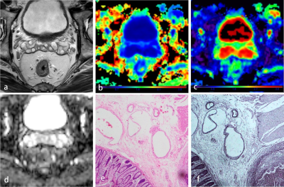 |
3 | Combining diffusion kurtosis imaging and clinical data for predicting the extramural venous invasion of rectal adenocarcinoma Video Not Available
QiuRong Wei1, WeiCui Chen2, LiTing Mao2, Kan Deng3, WeiKang Huang2, Bo Liu2, Xian Liu2, and ZhaoXian Yan2
1The Second Clinical College of Guangzhou University of Chinese Medicine, Guangzhou, China, 2the Second Affiliated Hospital of Guangzhou University of Chinese Medicine, Guangzhou, China, 3Philips Healthcare, Guangzhou, China
The extramural venous invasion (EMVI) is considered as the potential prognostic factor for patients with rectal carcinoma. We tried to investigate the diagnostic ability of diffusion kurtosis imaging (DKI) - derived parameters as risk factors for EMVI’s involvement status.Our results showed that the Kmean values, lymph node involvement, and an advanced tumor stage (T3) were independent risk factors for the prediction of EMVI status, which may suggest that the DKI as a potential biomarker for evaluating the EMVI status of rectal cancer is feasible.
|
||
3734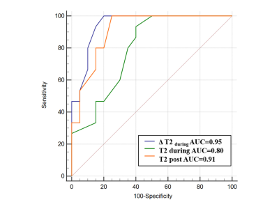 |
4 | Quantitative T2 mapping in early prediction of treatment response to chemoradiation for patients with local advanced rectal cancer Video Permission Withheld
Yuxi Ge1,2 and Weiqiang Dou3
1Affiliated Hospital of Jiangnan University, Wuxi, China, 2MR Research, GE Healthcare, Nanjing, China, 3GE Healthcare, Nanjing, China
This study aimed to investigate the feasibility of T2 mapping in predicting the response to chemoradiation (CRT) for patients with local advanced rectal cancer (LARC). Thirty-five LARC patients were measured with T2 mapping before-, during- and after-CRT. Resultant T2 values during- and after-CRT, and the T2 decrease ratio of before- to during-CRT were correlated significantly with tumor regression grade. Compared to poor responders, good responders showed lower T2 during- and after-CRT and higher T2 decrease ratio of before- to during-CRT. Therefore, quantitative T2 mapping may be useful for predicting the response to CRT for LARC patients.
|
||
3735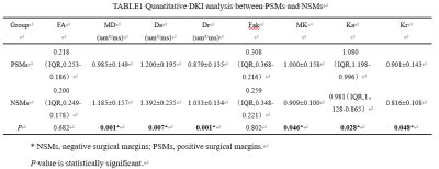 |
5 | Diffusion kurtosis imaging predicts positive margins after radical resection of localized prostate cancer
Shuang Meng1, Lihua Chen2, Nan Wang2, Yunsong Liu2, and Ailian Liu2
1Radiology, The First Affiliated Hospital of Dalian Medical University, Dalian, China, 2The First Affiliated Hospital of Dalian Medical University, Dalian, China
PSMs are an independent risk factor for biochemical recurrence in patients after radical prostatectomy. DKI quantifies the non-Gaussian nature of the real water molecule diffusion, and accurately reveals the changes in the microstructure of the tissue. Results of this study indicate that MD, Da , Dr, MK, Ka and Kr can be used to predict predicts positive margins after radical resection of localized prostate cancer.
|
||
3736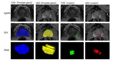 |
6 | MRI-derived radiomics to guide initial prostate biopsy for initial biopsy patients
Cheng Xueqing1, Li Haixia2, and Chen Yuntian1
1Department of radiology, West China Hospital, Chengdu, China, 2Philips Healthcare, Guangzhou, China
In this study, we applied the radiomics features of both suspected lesion and prostate gland on bi-parametric MRI (bpMRI) (T2W and ADC) to predict whether targeted biopsy alone is enough in detecting clinically significant prostate cancer (csPCa) in biopsy naïve men and compare their value with PI-RADS category.
|
||
3737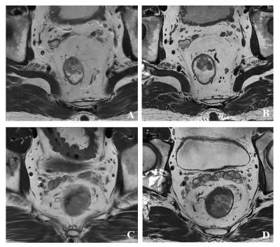 |
7 | Added Value of Isotropic High-Resolution 3D T2-weighted Imaging in Differentiation of Tis–T1 from T2 Stage in Rectal Cancers
Yu Guo1, Yu Fu1, Jiansen Li2, Lei Zhang1, Zhuo Wang1, and Huimao Zhang1
1The First Hospital of Jilin University, Changchun, China, 2United Imaging Healthcare, Shanghai, China
2D T2WI obtained in multi plane is recommend in preoperative assessment of rectal cancer, but 2D acquisition suffers from heavy partial volume effects. As a result, 2D T2WI cannot assess early rectal cancer stages accurately. Comparing with 2D T2WI, 3D T2WI could provide higher spatial resolution, especially the thin slice thickness, with highly reduced partial volume effects in rectal MRI. The purpose of the study was to compare the local-regional staging accuracy of the conventional 2D T2WI protocol and the 3D T2WI protocol for preoperative MR imaging in early rectal cancer patients.
|
||
3738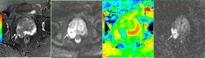 |
8 | Detection of prostatic peripheral zone cancer based on quantitative analysis of 3.0T MR Diffusion Tensor Imaging Video Permission Withheld
Zhuo Wang1, Zhiqiang Chen1, Shaoru Zhang1, Xiaohua Chen1, Dan Zhang1, Na Song1, Yuhui Xiong2, Lei Cai3, and Bing Chen3
1The Department of Radiology, General Hospital of Ningxia Medical University,, YinChuan, China, 2GE Healthcare, Beijing, China, 3The Department of Radiology, General Hospital of Ningxia Medical University,, Yinchuan, China
In this study, we aim to evaluate the diagnostic performance of diffusion tensor imaging (DTI) in detecting prostatic peripheral zone cancer and to deduce its clinical utility. The area under curve (AUC) of the receiver operating characteristic (ROC) curves was used to evaluate the diagnostic efficiency of ADC value, FA value and their combination. The diagnostic threshold values of ADC and FA were also determined. It was concluded that DTI quantitative indexes has moderately high diagnostic accuracy in detecting prostate cancer , while the ADC value had higher diagnostic efficiency than the FA value.
|
||
3739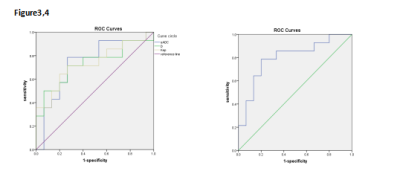 |
9 | The combination of DCE-MRI and IVIM to differentiate rectal mucinous adenocarcinoma and tubular adenocarcinoma Video Not Available
yuhui liu1,2, anliang chen1, Wan Dong1, and Ailian Liu1
1The First Affiliated Hospital of Dalian Medical University, Dalian, China, 2Dalian Medical University, Dalian, China
The purpose of this study is to use IVIM(intravoxel incoherent motion)and DCE(dynamic contrast-enhanced)imaging techniques to identify the value of rectal mucinous adenocarcinoma and tubular adenocarcinoma, and to provide rich information and help for clinical decision-making.
|
||
3740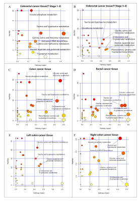 |
10 | NMR-based metabonomic fingerprinting reveals temporal and spatial heterogeneities of metabolic characterization in colorectal cancer tissue
Yan Lin1, Huanian Zhang1, Ting Ouyang1, Rongzhi Cai1, Peie Zheng1, yao Fu1, and Renhua Wu1
1Radiology Department, Second Affiliated Hospital of Shantou University Medical College, Shantou City, China
This study aimed to profile the metabolic differences of colorectal cancer tissues (CCT) in different stages and sites, as compared with their adjacent noncancerous tissues (ANT), to investigate the temporal and spatial heterogeneities of metabolic characterization. Our NMR-based metabonomics fingerprinting revealed that many of the metabolite levels were significantly altered in CCT as compared with ANT, indicating deregulations of glucose metabolism, one-carbon metabolism, glutamine metabolism, amino acid metabolism, fatty acid metabolism, TCA cycle, choline metabolism, ect. Significant metabolic differences were found in CRC tissues at different pathological stages and sites, suggesting temporal and spatial heterogeneities of metabolic characterization in CCT.
|
||
3741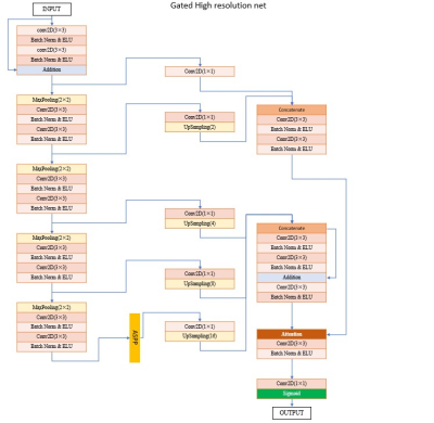 |
11 | Deep slice-crossed network with local weighted loss for brain metastases detection and segmentation Video Permission Withheld
Jiao Qu1, Xin Shu2, Wenjing Zhang1, Mengyuan Xu1, Ying Wang1, Lituan Wang2, Lei Zhang2, and Su Lui1
1Huaxi MR Research Center (HMRRC), Department of Radiology, West China Hospital of Sichuan University, Chengdu, China, 2College of Computer Science, Sichuan University, Chengdu, China
Brain metastases detection and segmentation on magnetic resonance images is laborious, error-prone and often irreproducible for radiologists and radiation oncologist. We present a specific deep slice-crossed network with local weighted loss to automatically detect and segment brain metastases on contrast-enhanced T1WI images. The results demonstrated the good performance, high robustness and generalizability of the model. In addition, compared with radiologists, the model showed higher sensitivity and increased efficiency in identifying and segmenting brain metastases. The results jointly suggested that the proposed model is a promising tool to assist the workflow in the clinical practice.
|
||
3742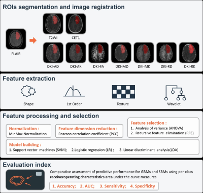 |
12 | A preliminary investigation of radiomics-based diffusion kurtosis imaging differences between glioblastoma and solitary brain metastasis
Eryuan Gao1, Guohua Zhao1, Huiting Zhang2, Ankang Gao1, Xiaoyue Ma1, Jie Bai1, Xu Yan2, Yusong Lin3, Guang Yang4, and Jingliang Cheng1
1department of magnetic resonance imaging, the First Affiliated Hospital of Zhengzhou University, Zhengzhou, China, 2MR Scientific Marketing, Siemens Healthcare, Shanghai, China, 3the School of Software, Zhengzhou University, Zhengzhou, China, 4Shanghai Key Laboratory of Magnetic Resonance, East China Normal University, Shanghai, China
The differentiation of glioblastomas from solitary brain metastases by using routine magnetic resonance imaging (MRI) alone presents a challenge. As a representative of diffusion MRI technology, diffusion kurtosis imaging (DKI) can closely reflect the real situation of water molecule movement in tumor tissues. We speculate that DKI radiomics is superior to routine MRI radiomics in distinguishing GBMs from SBMs. We developed a series of radiomics models of DKI parameter maps and routine MRI to compare their performance. Finally, several independent parameters models perform well in distinguishing two tumors. The combined DKI radiomics model obtained the best performance.
|
||
3743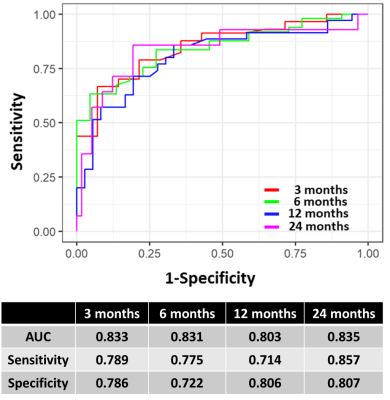 |
13 | A Deep Survival Model based on MR Radiomics and EGFR status to Predict Overall Survival after Radiosurgery in Brain Metastases
Chien-Yi Liao1, Cheng-Chia Lee2,3,4, Huai-Che Yang2,3, Wen-Yuh Chung2,3, Hsiu-Mei Wu3,5, Wan-Yuo Guo3,5, Ren-Shyan Liu6, and Chia-Feng Lu1,7
1Department of Biomedical Imaging and Radiological Sciences, National Yang Ming Chiao Tung University, Taipei, Taiwan, 2Department of Neurosurgery, Neurological Institute, Taipei Veteran General Hospital, Taipei, Taiwan, 3School of Medicine, National Yang Ming Chiao Tung University, Taipei, Taiwan, 4Brain Research Center, National Yang Ming Chiao Tung University, Taipei, Taiwan, 5Department of Radiology, Taipei Veteran General Hospital, Taipei, Taiwan, 6Department of Medical Imaging, Cheng-Hsin General Hospital, Taipei, Taiwan, 7Institute of Biophotonics, National Yang Ming Chiao Tung University, Taipei, Taiwan
About 30% of Non-small cell lung cancer (NSCLC) patients develop brain metastases (BMs) during the course of the disease. MR radiomics and EGFR mutation status were reported with the potential to predict the local tumor control of Gamma Knife stereotactic radiosurgery (GKRS). The prediction of overall survival after GKRS can further benefit the management of BM patients. We proposed a deep learning-based model using the clinical information, EGFR mutation status, and MR radiomic features to predict the overall survival after GKRS. We suggested that pre-GKRS MRI characteristics combined with gene and clinical information can improve the prediction of overall survival.
|
||
3744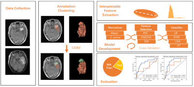 |
14 | Habitat Analysis of Multi-parametric MRI for Predicting Postoperative Recurrence of Meningioma
Chengxiu Zhang1, Yang Song2, Lin Lin3, Rufei Zhang3, Minxiong Zhou4, Xu Yan2, and Guang Yang1
1Shanghai Key Laboratory of Magnetic Resonance, East China Normal University, Shanghai, China, 2MR Scientific Marketing, Siemens Healthineers, Shanghai, China, 3Department of Radiology, Fujian Medical University Union Hospital, Fuzhou, China, 4Shanghai University of Medicine & Health Sciences, Shanghai, China
We used multi-parametric MRI to predict the postoperative recurrence of meningioma. Compared to the usual radiomics on analyzing the whole tumor, we used an unsupervised clustering method to explore the tumor habitats. Interpretable features were extracted from subregions of the lesion and used to build a habitat radiomic model. The habitat model achieved an AUC of 0.711 compared with 0.569 achieved by whole tumor analysis. The split subregions of the tumor also have clear biological meanings to the radiologists.
|
||
3745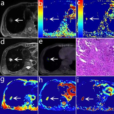 |
15 | Comparative analysis of IVIM and PET imaging versus APTw in assessing Ki-67 expression of non-small cell cancer Video Not Available
Ting Fang1,2, Nan Meng1, Zhun Huang2, Pengyang Feng2, Ziqiang Li2, Yang Yang3, Jianmin Yuan4, and Meiyun Wang2
1Zhengzhou University People's hospital, Zhengzhou, China, 2Henan Provincial People's Hospital, Zhengzhou, China, 3Beijing United Imaging Research Institute of Intelligent Imaging, Beijing, China, 4Central Research Institute, Shanghai, China
Intra-Voxel Incoherent Motion (IVIM), PET imaging, and Amide Proton Transfer weighted imaging (APTw) are all effective tools for distinguishing the pathological types of solitary pulmonary lesions. The difference between their ability to distinguish the Ki-67 high expression (HE) and low expression (LE) group is still unclear. Our study shows IVIM and PET imaging are better methods than APTw for distinguishing Ki-67 HE and LE groups of non-small cell cancer.
|
||
 Back to Meeting Home
 Back to the Program-at-a-Glance
The International Society for Magnetic Resonance in Medicine is accredited by the Accreditation Council for Continuing Medical Education to provide continuing medical education for physicians.
 Back to Meeting Home
 Back to the Program-at-a-Glance
 View the Presentation