Online Gather.town Pitches
Preclinical Imaging III
Joint Annual Meeting ISMRM-ESMRMB & ISMRT 31st Annual Meeting • 07-12 May 2022 • London, UK

Online Gather.town Pitches
Preclinical Imaging III
| Booth # | ||||
|---|---|---|---|---|
4639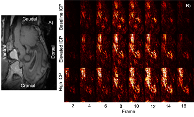 |
1 | Correlating Raised Intracranial Pressure with Increased Brain Motion in a Ovine Model
Alice Little1, Eryn Kwon1,2,3, Soroush Safaei2, Gonzalo Maso Talou2, David Dubowitz1, Miriam Scadeng1,3, Sarah-Jane Guild2,4, and Samantha Holdsworth1,3
1Department of Anatomy and Medical Imaging, Faculty of Medical and Health Sciences & Centre for Brain Research, University of Auckland, Auckland, New Zealand, 2Auckland Bioengineering Institute, Auckland, New Zealand, 3Mātai Medical Research Institute, Gisborne, New Zealand, 4Department of Physiology, Faculty of Medical and Health Sciences, University of Auckland, Auckland, New Zealand
Heartbeat-driven brain motion has historically remained limited to the domain of research. Such motion may provide a window into conditions that alter the brain pressure where direct, invasive measurements are difficult to justify. Amplified MRI (aMRI) is a recently developed method which amplifies subtle brain motion, providing high temporal resolution and high contrast ‘videos’ of brain motion patterns. In an ovine model of elevated intracranial pressure we show preliminary data that aMRI can be used to detect changes in brain motion associated with pressure, indicating potential to provide non-invasive insight into the mechanical changes in patients with altered intracranial pressure.
|
||
4640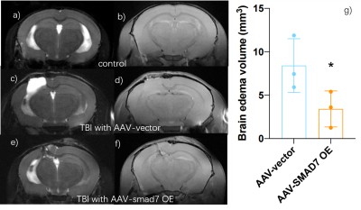 |
2 | T2/T1-weighted MRI revealed the therapeutic role of microglial SMAD7 in a traumatic brain injury (TBI) mouse model Video Permission Withheld
Yang Ruan1, Yimin Huang2, Xuejun He2, Huaqiu Zhang2, and Hongxia Lei1
1Wuhan United Imaging Life Science Instrument Co., Ltd., China, Wuhan, China, 2Department of Neurosurgery, Tongji Hospital, Tongji Medical College,, Huazhong University of Science and Technology, Wuhan, China
We applied T2/T1-weighted MRI together with behavior, Hematoxylin&Eosin(HE)and immunohistochemistry fluorescence staining (IHC) to evaluate the role of microglial SMAD7, a novel therapeutic approach, in a TBI mouse model. The consistency between MRI results and behavior and myelin and neurnal marker outcomes suggest that the protective role of over-expression of microglial SMAD7 in TBI.
|
||
4641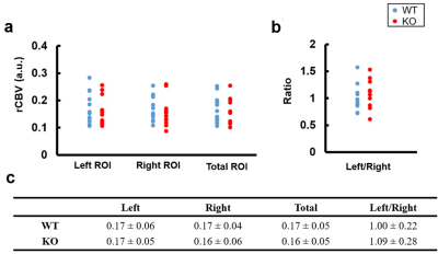 |
3 | Quantifications of Relative Cerebral Blood Volume and MRS metabolites in Aging Collapsin Response Mediator Protein 1 Gene Knockout Mice
Tzu-Ming Hung1, Sheng-Min Huang2, Yun-Chieh Tsai3, Ting-Yu Chin4, and Hsu-Hsia Peng1
1Department of Biomedical Engineering and Environmental Sciences, National Tsing Hua University, Hsinchu, Taiwan, Hsinchu, Taiwan, 2Institute of biomedical engineering and nanomedicine, National Health Research Institutes, Miaoli, Taiwan, Miaoli, Taiwan, 3Graduate Institute of Life Sciences, National Defense Medical Center, Taipei City 114, Taiwan, Taipei, Taiwan, 4Department of Bioscience Technology, Chung Yuan Christian University, Taoyuan City 320, Taiwan, Taoyuan, Taiwan
Aging and collapsin response mediator protein 1 (CRMP-1) gene knockout (KO) mice may exhibit neural disorganization in hippocampus and demonstrate memory and spatial learning dysfunction. The purpose of this study was to quantify relative cerebral blood volume (rCBV) and MRS metabolites in hippocampus in wild type and aging CRMP-1 KO mice. There was no significant difference of rCBV value between the two groups. In comparison with wild type (WT) mice, KO mice possessed significant lower Glx/t-Cr level in hippocampus, illustrating the altered neural transmission function in aging CRMP-1 KO mice.
|
||
4642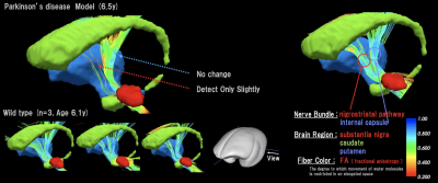 |
4 | Detection of cranial nerve degeneration in a transgenic Parkinson’s disease model marmoset using multi-contrast mechanism MRI Video Permission Withheld
Junichi Hata1,2,3, Reona Kobayashi2, Yawara Haga2, Mai Mizumura1, Hinako Oshiro1, Kanako Muta1, Naoya Hayashi2, Daisuke Yoshimaru3, Kei Hagiya2, Hirotaka James Okano3, and Hideyuki Okano2
1Tokyo Metropolitan University, Tokyo, Japan, 2RIKEN Center for Brain Science, Saitama, Japan, 3The Jikei University, School of Medicine, Tokyo, Japan In this study, we evaluated the characteristics of the brain in a genetically modified marmoset model of Parkinson’s disease. Various contrast mechanism images were acquired using magnetic resonance imaging (MRI), and the whole brain underwent explorative investigation with each contrast. This study was evaluated for Parkinson’s disease marmoset by anatomical MRI, neural fiber tractography and awakening state functional MRI. Malti-contrast MRI showed diseases characteristics in the thalamus, the nigral striatum, motor circuit and anymore. The findings suggest that the marmoset is useful as a model animal to study human diseases. |
||
4643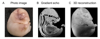 |
5 | High-resolution multimodal imaging of the embryonic and neonatal mouse
Tomokazu Tsurugizawa1,2,3, Takuma Kumamoto4, Daisuke Yoshimaru5, and Yoshichika Yoshioka6
1Human Informatics and Interaction Research Institute, National Institute of Advanced Industrial Science and Technology (AIST), Tsukuba, Japan, 2Faculty of Engineering, University of Tsukuba, Tsukuba, Japan, 3Center for Information and Neural Networks (CiNet), Osaka University and National Institute of Information and Communications Technology (NICT), Osaka, Japan, 4Developmental Neuroscience Project, Department of Brain & Neurosciences, Tokyo Metropolitan Institute of Medical Science, Tokyo, Japan, 5Division of Regenerative Medicine, Jikei University school of Medicine, Tokyo, Japan, 6Graduate School of Frontier Biosciences, Osaka University, Osaka, Japan
The potential of magnetic resonance microscopy is to image the multimodal function of the brain in small brain, such as embryo. In this study, we developed high resolution imaging of T1 image, T2 mapping, mean diffusivity, and neurite orientation dispersion by diffusion MRI in embryo and early postnatal (P07) in mouse. These values were different between embryo and early postnatal. Immunohistochemistry revealed distinct proliferation and maturation of neurons and astrocytes in these stages. These results indicate the possibility of high-resolution MR microscopy to detect the cellular maturation in development.
|
||
4644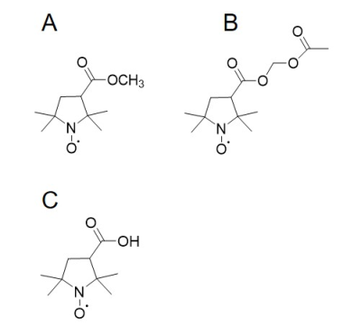 |
6 | Brain redox imaging using the blood–brain barrier-permeable nitroxides with different resistances to esterase Video Permission Withheld
Miho C EMOTO1, Shingo Sato2, Hideo Sato-Akaba3, and Hirotada G Fujii4
1Department of Clinical Laboratory Science, Health Sciences University of Hokkaido, Sapporo, Japan, 2Yamagata University, Yamagata, Japan, 3Osaka University, Toyonaka, Japan, 4Health Sciences University of Hokkaido, Ishikari, Japan
Lipophilic 3-methoxycarbonyl-2,2,5,5-tetramethylpiperidine-1-oxyl (MCP) has been used as a redox-sensitive nitroxide probe in MRI and EPR brain redox studies. Despite its success for this purpose, MCP cannot evaluate the redox status at both hydrophobic and hydrophilic brain regions. Accordingly, it is necessary to identify a lipophilic nitroxide that can penetrate the BBB and is hydrolyzed to hydrophilic nitroxide by neuronal esterase in the brain. For this purpose, lipophilic 3-acetoxymethoxycarbonyl-2,2,5,5-tetramethylpyrrolidine-1-oxyl was prepared and compared with MCP. Brain redox EPR imaging revealed that these two probes each detect a different redox status according to the antioxidant levels in the brain.
|
||
4645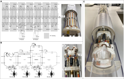 |
7 | Design and construction of a volume Tx coil and conformal Rx array for simultaneous squirrel monkey brain and spinal cord imaging at 9.4 T
Ming Lu1, Zhangyan Yang2,3, Feng Wang2,4, Gary Drake2,4, Li Min Chen2,4, John Gore2,3,4, and Xinqiang Yan2,4
1College of nuclear equipment and nuclear engineering, Yantai University, Yantai, China, 2Vanderbilt University Institute of Imaging Science, Vanderbilt University Medical Center, Nashville, TN, United States, 3Department of Biomedical Engineering, Vanderbilt University, Nashville, TN, United States, 4Department of Radiology and Radiological Sciences, Vanderbilt University Medical Center, Nashville, TN, United States
Simultaneous imaging of brain and spinal cord regions potentially provides valuable information about how they work together and interact. However, to date, almost all studies have investigated these two highly interconnected systems separately, mainly because of a lack of adequate imaging coils. We designed a volume Tx and conformal receive array dedicated to simultaneous imaging of squirrel monkey brain and spinal cord at 9.4 T. The coil exhibits excellent SNR in both brain and spinal cord areas and can be used for studies investigating how the brain and spinal cord work together.
|
||
4646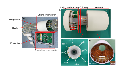 |
8 | Design and Construction of an 8-channel transceiver coil array for Rat imaging at 9.4T
Feng Du1,2, Nan Li1,2, Xing Yang1,2, Baogui Zhang3, Xiaoliang Zhang4, Xin Liu1,2, Hairong Zheng1,2, and Ye Li1,2
1Lauterbur Research Center for Biomedical Imaging, Shenzhen Institutes of Advanced Technology, Chinese Academy of Sciences, Shenzhen, China, 2Key Laboratory for Magnetic Resonance and Multimodality Imaging of Guangdong Province, Shenzhen, China, 3Brainnetome Center, Institute of Automation, Chinese Academy of Sciences, Beijing, China, 4Department of Biomedical Engineering, State University of New York, Buffalo, NY, United States
The development of RF coils for the ultrahigh-field(UHF) MRI system is greatly significant for biomedical research as it determines imaging performance. In this work,an 8-channel transceiver array was designed and constructed for UHF MRI at 9.4 T to take full advantage of the benefits provided by higher field strengths. The phantom and in vivo studies were performed on preclinical 9.4 T MRI system to verify the proposed tansceiver performance. The results proved the ability of the proposed 8-channel transceiver array to obtain high signal-to-noise ratio (SNR) and high-spatial resolution MR images at 9.4T and indicated the potentiality for UHF MRIapplications.
|
||
4647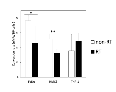 |
9 | Glycolytic Flux Alterations Following Radiotherapy in Cancer and Immune Cells: Hyperpolarized Carbon-13 Magnetic Resonance Imaging Study
Ying-Chieh Lai1,2, Ching-Yi Hsieh2,3,4, Kuan-Ying Lu1,2, Albert P Chen5, Shu-Hang Ng1,4, Fang-Hsin Chen4,6, and Gigin Lin1,2,3,4
1Department of Medical Imaging and Intervention, Chang Gung Memorial Hospital at Linkou and Chang Gung University, Taoyuan, Taiwan, Taoyuan, Taiwan, 2Clinical Metabolomics Core Laboratory, Chang Gung Memorial Hospital at Linkou, Taoyuan, Taiwan, Taoyuan, Taiwan, 3Medical Imaging Research Center, Institute for Radiological Research, Chang Gung University, Taoyuan, Taiwan, Taoyuan, Taiwan, 4Department of Medical Imaging and Radiological Sciences, Chang Gung University, Taoyuan, Taiwan, Taoyuan, Taiwan, 5GE Healthcare, Toronto, ON, Canada, Toronto, ON, Canada, 6Radiation Biology Research Center, Institute for Radiological Research, Chang Gung University, Taoyuan, Taiwan, Taoyuan, Taiwan
Alterations in metabolism following radiotherapy affect therapeutic efficacy, although the mechanism underlying such alterations is unclear. The present study demonstrates the potential for monitoring early metabolic alterations in both cancer and immune cells following radiation treatment by using hyperpolarized 13C-MRI. We demonstrated that cancer cells (human FaDu squamous carcinoma) and immune cells (HMC3 microglial cells and THP-1 monocytes) had distinct metabolic responses to ionizing radiation. Western blot analysis confirmed the similar trends in LDHA and LDHB expression levels. We furthered the knowledge of metabolic alterations resulting from not only cancer cells but also immune cells in the tumor microenvironment.
|
||
4648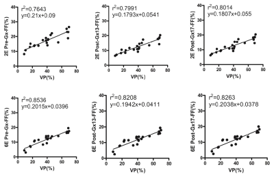 |
10 | Influence of gadoxetate disodium to the liver fat fraction quantified with Dixon sequences in rabbits
Zhe Huang1, Yu Jiang#1, Xia Wang#1, Sheng Zhang#1, Heng Li#1, and Dong Yue Han*1
1Department of Radiology, Xi’an GaoXin Hospital, Xi’an Jiao Tong University, Xi’an, China
A study was carried out on the effect of Gadoxetate disodium (Gx) on the accuracy of liver fat content determined by 2-echos and 6-echos VIBE Dixon in rabbits with nonalcoholic fatty liver disease in hepatobiliary specific phase. Proton density fat-fraction (PDFF) before and 13min. and 17min. after Gx enhancement had strong correlation with that of histopathology (r22E= 0.7643-0.8014 vs. r26E= 0.8208-0.8536). PDFF measured by 6E-VIBE Dixon was stable and reliable, especially for that before Gx enhancement(r26E= 0.8536). PDFF measured by 2E-VIBE Dixon fluctuated to some extent, however, it could be effectively corrected in the specific hepatobiliary phase after Gx enhancement.
|
||
4649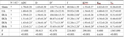 |
11 | IVIM and DCE MRI to early detect hepatic injury and microcirculation alteration induced by intestinal ischemia reperfusion in a rat model Video Permission Withheld
jiaxing yang1, Weiqiang Dou2, changjie pan3, and haifeng shi3
1Radiology, The Affiliated ChangzhouNo.2 People's Hospital of Nanjing Medical University, Changzhou, China, 2MR Research China, Beijing, China, 3The Affiliated ChangzhouNo.2 People's Hospital of Nanjing Medical University, Changzhou, China
This study demonstrated that intravoxel incoherent motion (IVIM) and dynamic contrast enhanced (DCE) magnetic resonance imaging (MRI) can effectively estimate hepatic pathophysiological processes in a rat model with intestinal ischemia reperfusion (IIR) induced hepatic injury. The changes in microcirculation and perfusion were considered the major factors of IIR-induced hepatic injury. IVIM derived parameters of f and D* as well as DCE related parameter of Ve and Ktrans revealed high sensitivity to histological change, implying that IVIM and DCE-MRI might be an effective tool to evaluate the process of IIR-induced hepatic injury.
|
||
4650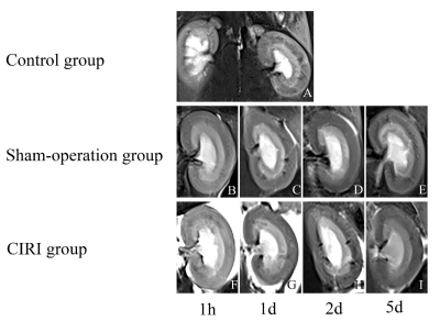 |
12 | Intravoxel incoherent motion magnetic resonance imaging for the early detection of renal cold ischemia–reperfusion injury in a rat model Video Permission Withheld
Yan Ren1 and Wen Shen1
1Tianjin First Central Hospital, Tianjin, China
The study evaluated the use of Intravoxel Incoherent Motion (IVIM) imaging to detect dynamic changes in renal microvascular characteristics during cold ischemia-reperfusion injuries (CIRIs). As previous studies have investigated warm ischemia-reperfusion injuries, we aimed to assess MR diffusion imaging in a renal CIRI Sprague Dawley rat model. Results showed that IVIM imaging is a sensitive tool to monitor changes in renal functional characteristics.
|
||
4651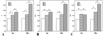 |
13 | Detection of early metastatic lymph nodes using inflow-based vascular-space-occupancy (iVASO) MR imaging
Yuankui Wu1, Liuji Guo1, Jun Hua2,3, Xiaomin Liu1, Yikai Xu1, and Kan Deng4
1Department of Medical Imaging, Nanfang Hospital, Southern Medical University, Guangzhou, China, 2Neurosection, Division of MRI Research, Department of Radiology,, Johns Hopkins University School of Medicine, Baltimore, MD, United States, 3F.M. Kirby Research Center for Functional Brain Imaging, Kennedy Krieger Institute, Baltimore, MD, United States, 4Philips Healthcare, Guangzhou, China
Accurate preoperative evaluation of lymph nodes (LNs) involvement is of vital importance for clinical decisions and optimizing individual treatment regimens. Related studies showed elevated perfusion at the level of capillary in metastatic LNs. Inflow-based vascular space occupancy (iVASO) is a novel and noninvasive perfusion technology that can provide absolute blood volume of precapillary arterioles (arteriolar blood volume, BVa). In this study, the potential value of BVa in detecting metastatic LNs was investigated. The results showed that the BVa might have the potential to detect early metastases within lymph nodes.
|
||
 |
4652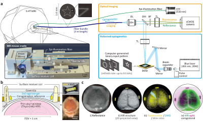 |
14 | Whole-brain mouse fMRI combined with cortex-wide patterned optogenetic stimulation
Hyun Seok Moon1,2,3, Seonghoon Kim1,4, Thanh Tan Vo1,2,3, Geun Ho Im1, Myunghwan Choi1,4, and Seong-Gi Kim1,2,3
1Center for Neuroscience Imaging Research, Institute for Basic Science (IBS), Suwon, Korea, Republic of, 2Department of Biomedical Engineering, Sungkyunkwan University, Suwon, Korea, Republic of, 3Department of Intelligent Precision Healthcare Convergence, Sungkyunkwan University, Suwon, Korea, Republic of, 4School of Biological Sciences, Seoul National University, Seoul, Korea, Republic of
Cortex-wide, spatial-patterned optogenetic mouse fMRI with online stimulation planning in situ.
|
|
4653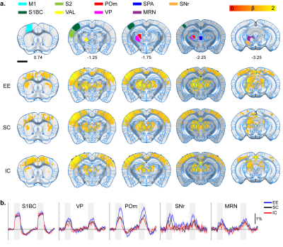 |
15 | Tales of Two Environments: Enriched Mice show Stronger Sensory Evoked BOLD fMRI Responses, while Socially Isolated Ones Respond Less
Taekwan Lee1, Taeyi You2,3, Geun Ho Im3, Seong-Gi Kim2,3,4, Sungkwon Chung5, and Jung Hee Lee2,3,6
1Korea Brain Research Institute, Daegu, Korea, Republic of, 2Biomedical Engineering, Sungkyungkwan University, Suwon, Korea, Republic of, 3Center for Neuroscience Imaging Research, Institute for Basic Science, Suwon, Korea, Republic of, 4Intelligent Precision Healthcare Convergence, Sungkyungkwan University, Suwon, Korea, Republic of, 5Physiology, Sungkyunkwan University, Suwon, Korea, Republic of, 6Radiology, Sungkyungkwan University, Suwon, Korea, Republic of
It is well known environmental factors can affect brain plasticity in humans, yet finding strong correlative factors is difficult due to the long development and complexity of human research. Mouse enrichment studies allows for better controlled research and by combining it with fMRI, makes mapping brain-wide plasticity changes possible. Here, we treated mice into three groups of enrichment, standard caging, and isolated caging to see how their brain responds to multiple-sensory stimulations. We found the enrichment group responded stronger in multimodal midbrain and thalamic areas. The isolated group responded less suggesting mouse fMRI is viable in detecting plasticity changes.
|
||
 Back to Meeting Home
 Back to the Program-at-a-Glance
The International Society for Magnetic Resonance in Medicine is accredited by the Accreditation Council for Continuing Medical Education to provide continuing medical education for physicians.
 Back to Meeting Home
 Back to the Program-at-a-Glance
 View the Presentation