Online Gather.town Pitches
Preclinical Imaging I
Joint Annual Meeting ISMRM-ESMRMB & ISMRT 31st Annual Meeting • 07-12 May 2022 • London, UK

Online Gather.town Pitches
Preclinical Imaging I
| Booth # | ||||
|---|---|---|---|---|
3267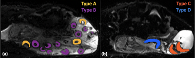 |
1 | Measuring changes in placental morphology using magnetic resonance imaging in a preclinical model of human pregnancy
Dimitra Flouri1,2, Jack RT Darby3, Stacey L Holman3, Sunthara R Perumal4, Sebastien Ourselin1, Anna L David5,6, Andrew Melbourne1,2, and Janna L Morrison3
1School of Biomedical Engineering & Imaging Sciences, King's College London, London, United Kingdom, 2Department of Medical Physics & Biomedical Engineering, University College London, London, United Kingdom, 3Early Origins of Adult Health Research Group, University of South Australia, Adelaide, Australia, 4Preclinical Imaging and Research Laboratories, South Australian Health and Medical Research Institute, Adelaide, Australia, 5Institute for Women's Health, University College London, London, United Kingdom, 6NIHR Biomedical Research Center, University College London Hospitals, London, United Kingdom
Abnormalities of placental development and function underlie many pathologies of pregnancy including preeclampsia and fetal growth restriction (FGR). Advances in MRI techniques provide capacity to obtain additional placental in vivo information to support clinical decision-making. Animal models are used in invasive validation studies are possible in animal models and allow for controlled experiments during pregnancy. Here, we investigated whether MRI technology could be used to study the anatomical morphology of placentae in vivo in sheep; and we characterise diffusion and perfusion properties in normal pregnancies and those complicated by induced FGR.
|
||
3268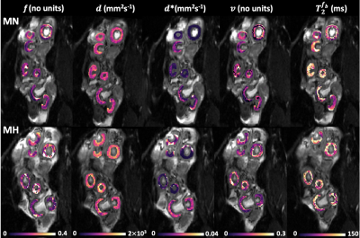 |
2 | MRI assessment of placental oxygenation changes in response to maternal hyperoxygenation in a sheep model of human pregnancy
Dimitra Flouri1,2, Jack RT Darby3, Stacey L Holman3, Georgia K Williams4, Sebastien Ourselin1, Anna L David5,6, Janna L Morrison3, and Andrew Melbourne1,2
1School of Biomedical Engineering & Imaging Sciences, King's College London, London, United Kingdom, 2Department of Medical Physics & Biomedical Engineering, University College London, London, United Kingdom, 3Early Origins of Adult Health Research Group, University of South Australia, Adelaide, Australia, 4Preclinical Imaging and Research Laboratories, South Australian Health and Medical Research Institute, Adelaide, Australia, 5Institute for Women's Health, University College London, London, United Kingdom, 6NIHR Biomedical Research Center, University College London Hospitals, London, United Kingdom
Abnormalities of placenta development and function may result in fetal growth restriction (FGR). Advances in MRI technology enable estimation of quantitative indices that reflect tissue diffusivity and oxygenation. We investigated the physiological impact of maternal hyperoxygenation on the placenta pregnant sheep. We applied a multi-compartment MRI signal model to measure the changes in oxygenation caused by changes in maternal blood oxygen level. The expected increase in feto-placental blood relaxation and feto-placental oxygen saturation with maternal hyperoxygenation was observed. Results suggested that diffusion and relaxation-based MRI is sensitive to acute changes in maternal and feto-placental oxygen level.
|
||
3269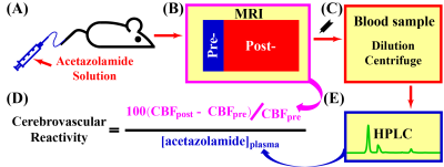 |
3 | Quantitative cerebrovascular reactivity MRI in mice using acetazolamide challenge
Zhiliang Wei1,2, Yuguo Li1,2, Xirui Hou3, Zheng Han1,2, Jiadi Xu1,2, Michael T. McMahon1,2, Wenzhen Duan4,5, Guanshu Liu1,2, and Hanzhang Lu1,2,3
1Russell H. Morgan Department of Radiology and Radiological Science, Johns Hopkins University School of Medicine, Baltimore, MD, United States, 2F. M. Kirby Research Center for Functional Brain Imaging, Kennedy Krieger Research Institute, Baltimore, MD, United States, 3Department of Biomedical Engineering, Johns Hopkins University School of Medicine, Baltimore, MD, United States, 4Department of Psychiatry and Behavioral Sciences, Johns Hopkins University School of Medicine, Baltimore, MD, United States, 5The Solomon H. Snyder Department of Neuroscience, Johns Hopkins University School of Medicine, Baltimore, MD, United States
Cerebrovascular reactivity (CVR), which denotes brain’s vasodilatory capacity, is broadly utilized in cerebrovascular and neurodegenerative diseases. However, the most popular hypercapnia method in human studies is unsuitable for small animals due to the difficulty in measuring end-tidal CO2. Here, we took a different approach using a pharmacological vasodilatory stimulus, acetazolamide. Plasma level of acetazolamide and vascular responses were quantified by high-performance liquid chromatography and perfusion MRI, respectively. Evidences of feasibility, safety, temporal characteristics, and dose-dependence have been demonstrated. This new CVR technique may open several avenues for preclinical research on cerebrovascular diseases and therapeutic testing in different animal models.
|
||
3270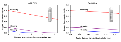 |
4 | A Radial Flow Cell Perfusion System for Hyperpolarized 13C NMR Metabolic Studies at Low Oxygen Levels
Anthony Mancuso1, Mehrdad Pourfathi2, Ryan M. Kiefer2, Michael C Noji2, Sarmad Siddiqui2, Enri Profka2, Stephen J Kadlecek2, Rahim Rizi2, and Terence PF Gade1,2
1Cancer Biology, University of Pennsylvania, Philadelphia, PA, United States, 2Radiology, University of Pennsylvania, Philadelphia, PA, United States
We have developed new methods for studying cultured cancer cell metabolism with hyperpolarized 13C magnetic resonance spectroscopy (HP 13C MRS) at low oxygen concentrations. Cells grown on the surfaces of microcarriers inside the spectrometer were perfused radially (rather than axially) in a modified NMR tube at controlled oxygen levels. Computational and experimental results demonstrated that cell mass oxygen profiles with radial flow were much more uniform than with conventional axial flow. The metabolism of HP [1-13C]pyruvate was markedly different between the two flow configurations, demonstrating the importance of avoiding large oxygen gradients in cell perfusion experiments.
|
||
3271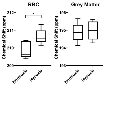 |
5 | Effect of Inhaled Oxygen Concentration on 129Xe Chemical Shift of Red Blood Cells in Rat Brain
Yonni Friedlander1,2, Brandon Zanette1, Andras Lindenmaier1,2, Daniel Li1, Stephen Kadlecek3, Giles Santyr1,2, and Andrea Kassner1,2
1Hospital for Sick Children, Toronto, ON, Canada, 2University of Toronto, Toronto, ON, Canada, 3University of Pennsylvania, Philadelphia, PA, United States
Hyperpolarized 129Xe RBC chemical shift in the rat brain was measured to be higher during hypoxic ventilation than during normoxic ventilation.
|
||
3272 |
6 | Longitudinal biochemical and behavioral alterations in a gyrencephalic model of blast related mild traumatic brain injury
Shiyu Tang1,2, Su Xu1,2, Li Jiang1,2, Donna Wilder3, Joseph Long3, Alexandre E. Medina4, Xin Li1,2, Gary Fiskum5,6, Venkata Siva Sai Sujith Sajja3, and Rao P. Gullapalli1,2
1Department of Diagnostic Radiology and Nuclear Medicine, University of Maryland School of Medicine, Baltimore, MD, United States, 2Center for Advanced Imaging Research (CAIR), University of Maryland School of Medicine, Baltimore, MD, United States, 3Blast Induced Neurotrauma Branch, Walter Reed Army Institute of Research, Silver Spring, MD, United States, 4Department of Pediatrics, University of Maryland School of Medicine, Baltimore, MD, United States, 5Department of Anesthesiology, University of Maryland School of Medicine, Baltimore, MD, United States, 6Shock, Trauma, and Anesthesiology Research Center, University of Maryland School of Medicine, Baltimore, MD, United States Compared to rodents, the ferret model has greater similarities to humans in terms of developmental process, brain structure and sophisticated behavior. In this study, we assessed the longitudinal changes in brain metabolism and impulsivity behavior in a ferret model that closely mimics the blast exposure conditions encountered by Warfighters. Ferrets demonstrated concomitantly increased behavioral impulsivity and metabolite alterations in prefrontal cortex following blast exposure. Our findings agree with clinical observations in patients, suggesting that this model is a good gyrencephalic animal model to study brain biochemical profile changes and neuropsychiatric alterations associated with blast exposure. |
||
3273 |
7 | Early sex dependent changes in the default mode network of the TgF344-AD rat model of Alzheimer’s disease
Raúl Tudela1, Emma Muñoz-Moreno2, Xavier López-Gil2, Federico Varriano3, and Guadalupe Soria1,3
1Biomedical Imaging Group, Consorcio Centro de Investigación Biomédica en Red (CIBER) de Bioingeniería, Biomateriales y Nanomedicina (CIBER-BBN), Barcelona, Spain, 2Experimental 7T MRI Unit, Magnetic Resonance Imaging Core Facility, Institut d’Investigacions Biomèdiques August Pi I Sunyer (IDIBAPS), Barcelona, Spain, 3Laboratory of Surgical Neuroanatomy, Faculty of Medicine and Health Sciences, Institute of Neurosciences, University of Barcelona, Barcelona, Spain
TgF344-AD rat model has been revealed as an interesting animal model for investigating Alzheimer's disease. About two thirds of persons diagnosed with AD dementia are women, suggesting important sex and gender differences in risk factors for the development of AD. Therefore, it is of high interest to also investigate this issue in the AD animal models. In this study we present preliminary results regarding the effect of sex difference in the functional default mode network between young TgF344-AD rats and Wild-type.
|
||
3274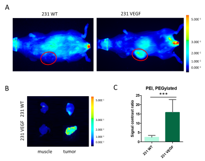 |
8 | VEGF overexpression in breast cancer xenograft significantly increases nanoparticle-mediated siRNA delivery and target gene downregulation
Shanshan Tan1, Zhihang Chen1, Yelena Mironchik1, Noriko Mori1, Marie-France Penet1, Balaji Krishnamachary1, and Zaver M. Bhujwalla1,2,3
1The Russell H Morgan Department of Radiology and Radiological Science, Johns Hopkins University, Baltimore, MD, United States, 2Sidney Kimmel Comprehensive Cancer Center, Baltimore, MD, United States, 3Department of Radiation Oncology and Molecular Radiation Science, Johns Hopkins University School of Medicine, Baltimore, MD, United States
Effective tumor delivery is a major challenge in achieving small interfering RNA (siRNA) mediated gene silencing. Poor tumor vascularization can limit the delivery of therapeutic siRNA. Here, we used a VEGF overexpression breast cancer murine model and its wildtype counterpart to investigate the influence of increasing tumor vasculature on choline kinase-alpha siRNA nanoparticle delivery.
|
||
3275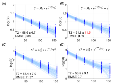 |
9 | Comparison of different T2 fitting models in renal tissue relaxometry Video Permission Withheld
Wan-Ting Zhao1,2,3, Karl-Heinz Herrmann1, Martin Krämer1, Daniel Güllmar1, Jürgen R. Reichenbach1, and Verena Hoerr1,4,5
1Institute of Diagnostic and Interventional Radiology, University Hospital Jena, Jena, Germany, 2Center for Sepsis Control and Care, University Hospital Jena, Jena, Germany, 3Institute of Medical Microbiology, University Hospital Jena, Jena, Germany, 4Clinic for Radiology, University Hospital Muenster, Muenster, Germany, 5Heart Center, University Hospital Bonn, Bonn, Germany
While relaxation rate estimation substantially varies on the chosen fitting method, a tissue-specific T2 fitting scheme for the kidney is lacking. The current study aims to compare multiple T2 fitting methods, including square signal and mono-exponential with constant offsets, in high SNR and low SNR scenarios from identical data points. We compare standard deviations for fitting robustness and RMSE for the goodness of fit. Our finding suggests a mono-exponential with constant offsets is the most suitable method, for it yielded the lowest RMSE both in high and low SNR.
|
||
3276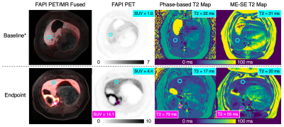 |
10 | Phase-Based T2 Mapping for Assessment of Liver Fibrosis in a Porcine Model
Ruvini Navaratna1,2, Daiki Tamada2, Sarvesh Periyasamy2,3, Jennifer J Meudt4, Paul Laeseke2,3, Dhanansayan Shanmuganayagam4,5,6, Scott B Reeder1,2,3,7,8, and Ali Pirasteh1,2
1Medical Physics, University of Wisconsin-Madison, Madison, WI, United States, 2Radiology, University of Wisconsin-Madison, Madison, WI, United States, 3Biomedical Engineering, University of Wisconsin-Madison, Madison, WI, United States, 4Animal and Dairy Sciences, University of Wisconsin-Madison, Madison, WI, United States, 5Surgery, University of Wisconsin-Madison, Madison, WI, United States, 6Center for Biomedical Swine Research and Innovation, University of Wisconsin-Madison, Madison, WI, United States, 7Medicine, University of Wisconsin-Madison, Madison, WI, United States, 8Emergency Medicine, University of Wisconsin-Madison, Madison, WI, United States
Fibrosis is the sequela of and the most important outcome predictor in chronic liver disease. MRI T2 mapping can provide safe and noninvasive detection/staging of liver fibrosis. However, current T2 mapping techniques suffer from prolonged acquisition times, prohibiting widespread clinical utilization. To address this unmet need, we evaluated the feasibility of a whole-liver, single-breath-hold, phase-based T2 mapping technique to assess liver fibrosis in a human-sized porcine model of liver fibrosis. We demonstrated a strong correlation between our phase-based liver T2 values and liver uptake of the fibroblast activation protein inhibitor on PET as well as the reference spin-echo T2 estimates.
|
||
3277 |
11 | Minimally-Invasive Surgical Treatment of Lung Cancer using Magnetic Resonance Guided Focused Ultrasound in a Porcine Model
Braden Miller1, Lauren Powlovich2, David Moore2, Linda Martin1, and Jaime Mata1
1University of Virginia, Charlottesville, VA, United States, 2Focused Ultrasound Foundation, Charlottesville, VA, United States
This new technique has the potential to increase lung cancer survival rates while decreasing morbidity associated with current practices as it allows a less invasive manner for ablation or tumor debulking.
|
||
3278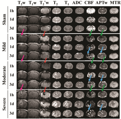 |
12 | Using amide proton transfer-weighted (APTw) MRI to detect severity and predict outcome after traumatic brain injury (TBI) in rats
Yinfeng Dong1, Yanting Gu1, Jianhua Lu2, Jieru Wan1, Shanshan Jiang2, Raymond C. Koehler1, and Jinyuan Zhou2
1Department of Anesthesiology and Critical Care Medicine, Johns Hopkins University, Baltimore, MD, United States, 2Department of Radiology, Johns Hopkins University, Baltimore, MD, United States
After TBI, secondary injury severity is difficult to determine. The objective of this study was to investigate the capacity of noninvasive APTw MRI to assess TBI injury in different brain regions and predict long-term neurobehavior outcomes. Fifty-five male and female rats were subjected to a controlled cortical impact with one of three different impactor depths to produce different degrees of TBI, and scanned on a 4.7 T horizontal bore animal imager. Our results suggest that APTw imaging can be used for detecting the level of inflammation and as a potential predictor of long-term outcomes from TBI.
|
||
3279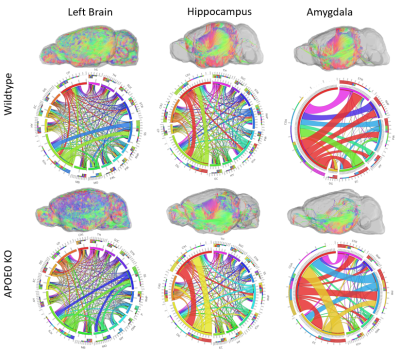 |
13 | Apolipoprotein-E (APOE) Deficiency Leads to Neuronal Network Remodeling Characterized by Diffusion MRI and Graph Theory
Margaret Caroline Stapleton1, Philipp Boehm-Sturm2, Stefan Koch2, Susanne Mueller2, Devin Raine Everaldo Cortes1, and Yijen Wu1
1Developmental Biology, University of Pittsburgh, Pittsburgh, PA, United States, 2Department of Experimental Neurology, Charite University Medicine Berlin, Berlin, Germany
Allelic difference in Apolipoprotein-E (APOE) is a well-known risk factor for the late-onset Alzheimer’s disease (AD) but how APOE affects brain functions and the subsequent cognitive declination remains unclear. Diffusion MRI followed by network topology analysis found that APOE deficiency in knockout mice resulted in altered neuronal network in the brain regions known to be affected by Alzheimer’s Disease. This study suggests the possible role of APOE in neuronal network organization, which might predispose brains to differential AD vulnerability.
|
||
 Back to Meeting Home
 Back to the Program-at-a-Glance
The International Society for Magnetic Resonance in Medicine is accredited by the Accreditation Council for Continuing Medical Education to provide continuing medical education for physicians.
 Back to Meeting Home
 Back to the Program-at-a-Glance
 View the Presentation