Online Gather.town Pitches
Novel Image Reconstruction Techniques II
Joint Annual Meeting ISMRM-ESMRMB & ISMRT 31st Annual Meeting • 07-12 May 2022 • London, UK

Online Gather.town Pitches
Novel Image Reconstruction Techniques II
| Booth # | ||||
|---|---|---|---|---|
3441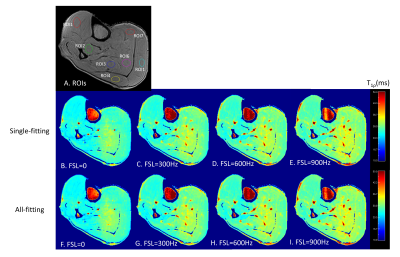 |
1 | Integrated T1rho Dispersion Imaging and Quantification
Qi Peng1, Gregory Peng1, and Can Wu2
1Department of Radiology, Albert Einstein College of Medicine, Bronx, NY, United States, 2Department of Medical Physics, Memorial Sloan Kettering Cancer Center, New York, NY, United States
T1ρ dispersion imaging is an emerging MRI technique for tissue characterization. Multiple independent repetitions of T1ρ experiments at different spin-lock frequencies have to be performed to generate tissue T1ρ dispersion curve. In this work, we demonstrate the feasibility of an integrated imaging and quantification approach for T1ρ dispersion imaging, which allows simultaneous generation of T1ρ maps at multiple spinlock frequencies in one coherent workflow. This opens door for further data undersampling to exploit redundancy at higher dimension data space, further reducing total scan time needed for the time-consuming T1ρ dispersion imaging.
|
||
3442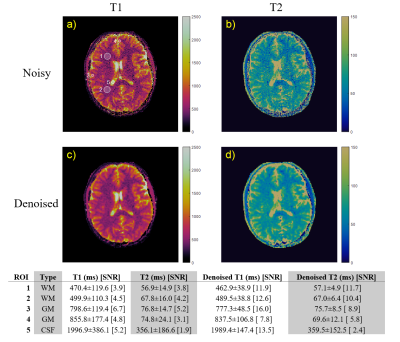 |
2 | Denoising MR Fingerprinting by matching against General Noise Model at 0.55 T
Ruogu Matthew Zhu1, Nicole Seiberlich2, and Yun Jiang2
1Department of Electrical and Computer Engineering, University of Michigan, Ann Arbor, MI, United States, 2Department of Radiology, University of Michigan, Ann Arbor, MI, United States
Low SNR is a challenge for magnetic resonance fingerprinting (MRF) at low-field (0.55 T). In this work, we apply a locally low rank denoising method based on elimination of noise-only principal components according to the Marchenko-Pastur distribution to MRF data. We show that this method is effective at denoising both phantom and in vivo MRF images.
|
||
3443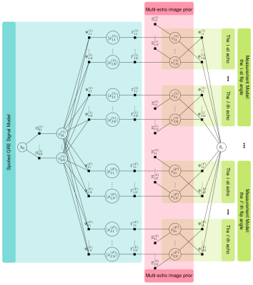 |
3 | Bayesian Quantitative T1 Mapping with Variable-Density and Poisson-Disk sampling
Shuai Huang1, James J. Lah2, Jason W. Allen1, and Deqiang Qiu1
1Radiology and Imaging Sciences, Emory University, Atlanta, GA, United States, 2Neurology, Emory University, Atlanta, GA, United States
We propose a Bayesian approach with built-in parameter estimation to perform T1 mapping from undersampled measurements. Apart from using measurements acquired at multiple flip angles, the Bayesian approach offers a convenient way to synthesize measurements from multiple echoes as well to obtain better image quality. The sparse prior on the image wavelet coefficients could further improve the performance when we perform undersampling in the k-space to reduce scan time. The proposed Bayesian approach automatically and adaptively estimates the induced regularization and other parameters by undersampling, making it a better choice over approaches that require manual regularization parameter tuning.
|
||
3444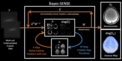 |
4 | Bayesian sensitivity encoding enables parameter-free, highly accelerated joint multi-contrast reconstruction
Alexander Lin1, Demba Ba1, and Berkin Bilgic2,3,4
1Harvard University, Cambridge, MA, United States, 2Harvard-MIT Health Sciences and Technology, Massachusetts Institute of Technology, Cambridge, MA, United States, 3Athinoula A. Martinos Center for Biomedical Imaging, Charlestown, MA, United States, 4Department of Radiology, Harvard Medical School, Boston, MA, United States
To address limitations with classical SENSE algorithms, we propose Bayesian Sensitivity Encoding (Bayes-SENSE), which obviates the need to tune regularization penalties, provides variance maps that can quantify algorithmic uncertainty, and is easily extendable to multi-contrast reconstruction. Bayes-SENSE is a synergistic combination of SENSE and Bayesian CS (BCS). We adapt recent work accelerating BCS to develop an efficient and highly parallelizable inference algorithm for Bayes-SENSE based on the conjugate gradient (CG) method. We evaluate Bayes-SENSE in several undersampling settings with parallel imaging, and demonstrate that it outperforms L2-/L1-SENSE in terms of reconstruction error while also providing the aforementioned benefits.
|
||
3445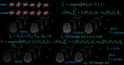 |
5 | Synergistic Combination of Golden-angle Radial Sampling and Dual-Subspace Modeling for Rapid and Robust High Spatiotemporal Resolution MRA
Zhifeng Chen1,2 and Lirong Yan1,2
1USC Stevens Neuroimaging and Informatics Institute, Keck School of Medicine, University of Southern California, Los Angeles, CA, United States, 2Department of Neurology, Keck School of Medicine, University of Southern California, Los Angeles, CA, United States This study proposes a rapid and robust ASL-based time-resolved MRA technique with high spatiotemporal resolution termed Dual-Subspace MRA (DS-MRA), which employs subspace modeling from both temporal and control/label dimensions as sparsity constraints to improve the robustness of image reconstruction with under-sampled golden-angle radial dynamic MRA. The performance of DS-MRA was compared with conventional iGRASP and 5-dimensional GRASP. Our preliminary data suggests that DS-MRA outperforms conventional GRASP reconstructions with less residual streaking artifacts and noise, especially at higher acceleration rates. |
||
 |
3446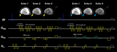 |
6 | 3D-CAIPI-BUDA and Joint Hankel Low-Rank Reconstruction Enable Rapid and Distortion-free High-Resolution T2* Mapping and QSM
Zhifeng Chen1,2,3, Congyu Liao4, Xiaozhi Cao4, Benedikt A Poser5, Zhongbiao Xu6, Wei‐Ching Lo7, Manyi Wen8, Jaejin Cho1, Qiyuan Tian1, Yaohui Wang9, Yanqiu Feng3, Wufan Chen3, Ling Xia10, Feng Liu11, and Berkin Bilgic1,2
1Athinoula A. Martinos Center for Biomedical Imaging, Massachusetts General Hospital, Charlestown, MA, United States, 2Department of Radiology, Harvard Medical School, Charlestown, MA, United States, 3School of Biomedical Engineering, Guangdong Provincial Key Laboratory of Medical Image Processing, Southern Medical University, Guangzhou, China, 4Department of Radiology, Stanford University, Stanford, CA, United States, 5Maastricht Brain Imaging Center, Faculty of Psychology and Neuroscience, University of Maastricht, Maastricht, Netherlands, 6Department of Radiotherapy, Cancer Center, Guangdong Provincial People's Hospital & Guangdong Academy of Medical Science, Guangzhou, China, 7Siemens Medical Solutions, Boston, MA, United States, 8Department of Chemical Pathology, The Chinese University of Hong Kong, Hong Kong, China, 9Division of Superconducting Magnet Science and Technology, Institute of Electrical Engineering, Chinese Academy of Sciences, Beijing, China, 10Department of Biomedical Engineering, Zhejiang University, Hangzhou, China, 11School of Information Technology and Electrical Engineering, The University of Queensland, Brisbane, Australia Quantitative imaging has been very useful in neuroscientific and clinical applications, including glioma, tumor diagnosis and prognosis, brain maturation, and Alzheimer's disease. EPI is a powerful tool for quantitative imaging owing to its extremely fast acquisition. This work aims to develop a distortion-free, blip-up/down acquisition (BUDA) 3D-EPI with controlled aliasing in parallel imaging (CAIPI) sampling and joint Hankel low-rank image reconstruction for fast and robust multi-contrast high-resolution whole-brain imaging. The developed technique could generate distortion-free high-resolution whole-brain T2* mapping and quantitative susceptibility mapping in 47s at 1.1×1.1×1 mm3 resolution. |
|
3447 |
7 | Improved Multi-band Multi-shot Diffusion MRI Reconstruction with Joint Usage of Structured Low-rank Constraints and Explicit Phase Mapping
Erpeng Dai1, Merry Mani 2, and Jennifer A McNab1
1Departmnet of Radiology, Stanford University, Stanford, CA, United States, 2Departmnet of Radiology, University of Iowa, Iowa City, IA, United States Inter-shot phase correction is a critical step in multi-band multi-shot diffusion MRI. The phase correction can be accomplished by first estimating the explicit phase map and then inputting it into the diffusion signal formulation model to recover the diffusion images. Alternatively, the phase information can be used in an indirect manner to determine structured low rank constraints in k-space. The two methods differ in terms of reconstruction accuracy and efficiency. In this study, we propose a new way to combine the two approaches for improved image quality, termed “Joint Usage of structured Low-rank constraints and Explicit Phase mapping” (JULEP). |
||
|
3448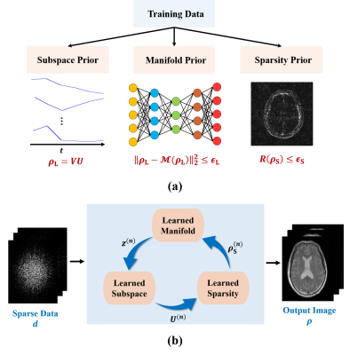 |
8 | Integrating Subspace Learning, Manifold Learning, and Sparsity Learning to Reconstruct Image Sequences
Yudu Li1,2, Yue Guan3, Yibo Zhao1,2, Rong Guo1,2, Yao Li4, and Zhi-Pei Liang1,2
1Department of Electrical and Computer Engineering, University of Illinois at Urbana-Champaign, Urbana, IL, United States, 2Beckman Institute for Advanced Science and Technology, University of Illinois at Urbana-Champaign, Urbana, IL, United States, 3Institute for Medical Imaging Technology, Shanghai Jiao Tong University, Shanghai, China, 4School of Biomedical Engineering, Shanghai Jiao Tong University, Shanghai, China
Many imaging applications, such as dynamic imaging and multi-contrast imaging, involve the acquisition of a sequence of images. This work addresses the underlying image reconstruction problem by incorporating priori information such as partial separability, image sparsity, and manifold structure jointly to enable high-quality image reconstruction from highly sparse data. To this end, we propose a new deep learning-based framework that enforces those constraints effectively and consistently. The proposed method has been validated using multi-contrast imaging data and produced impressive results. The image reconstruction framework can be extended for incorporating additional constraints and/or solving other sequential image reconstruction problems.
|
|
3449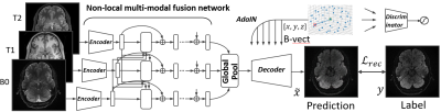 |
9 | Adversarial Non-local Multi-modality MRI Aggregation for Directional DWI Synthesis
Xiaofeng Liu1, Fangxu Xing1, Van Jay Wedeen1, Georges El Fakhri1, and Jonghye Woo1
1Dept. of Radiology, MGH and Harvard Medical School, Boston, MA, United States
Diffusion MRI is sensitive to subject motion, and with prolonged acquisition time, it suffers from motion corruption and artifacts. To address this, we present an adversarial non-local network-based multi-modality MRI fusion framework for directional DWI synthesis. Our framework is based on a generative model conditioned on a specified b-vector sampled in q-space, where it efficiently fuses information from multiple structural MRIs, including T1- and T2-weighted MRI, and B0 image, with an adaptive attention scheme. Experimental results, using a total of ten q-ball data, show its potential to synthesize high-fidelity DWIs at arbitrary q-space coordinates and facilitate quantification of diffusion parameters.
|
||
3450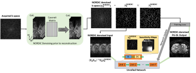 |
10 | High-Quality 0.5mm Isotropic Functional MRI Using a Synergistic Combination of NORDIC Denoising and Deep Learning Reconstruction
Omer Burak Demirel1,2, Steen Moeller2, Luca Vizioli2,3, Burhaneddin Yaman1,2, Logan Dowdle2,3, Essa Yacoub2, Kamil Ugurbil2, and Mehmet Akçakaya1,2
1Electrical and Computer Engineering, University of Minnesota, Minneapolis, MN, United States, 2Center for Magnetic Resonance Research, University of Minnesota, Minneapolis, MN, United States, 3Department of Neurosurgery, University of Minnesota, Minneapolis, MN, United States
Submillimeter fMRI allows studying brain function at the mesoscale level, but scans at such resolutions require trade-offs in SNR and coverage, necessitating better image reconstruction. In this work, we combine NOise Reduction with Distribution Corrected (NORDIC) denoising prior to image reconstruction with self-supervised physics-guided deep learning (PG-DL) for high-quality 0.5mm isotropic fMRI. The former removes components of image series that cannot be distinguished from thermal noise, while the latter enables higher acceleration rates. Results show that the proposed combination of NORDIC and PG-DL improves on NORDIC or PG-DL alone, both visually, and in terms of tSNR and GLM-derived t-maps.
|
||
3451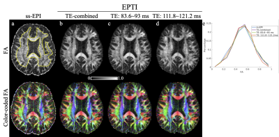 |
11 | A Subspace EPTI Reconstruction with Magnitude-only Bases and Synergistic Phase Bias Updating for Distortion-Free Diffusion-Relaxometry MRI
Erpeng Dai1, Zijing Dong2,3, Kawin Setsompop1,4, and Jennifer A McNab1
1Departmnet of Radiology, Stanford University, Stanford, CA, United States, 2Athinoula A. Martinos Center for Biomedical Imaging, Massachusetts General Hospital, Charlestown, MA, United States, 3Department of Radiology, Harvard Medical School, Boston, MA, United States, 4Departmnet of Electrical Engineering, Stanford University, Stanford, CA, United States
The distortion-free diffusion and relaxometry images provided by echo-planar time resolved imaging (EPTI) ) represent a valuable acquisition strategy for mapping brain tissue microstructure. However, given the large under-sampling factor of EPTI acquisition and the intrinsically low SNR of diffusion MRI, an SNR-efficient reconstruction is vital. Subspace reconstruction can improve SNR efficiency by reducing the number of unknowns. In subspace reconstruction, the selection of bases strongly affects the reconstruction image fidelity, SNR, and computational efficiency. Here, we explore a new subspace-based EPTI reconstruction with magnitude-only bases and synergistic phase bias updating and demonstrate its performance for microstructural mapping.
|
||
3452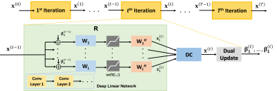 |
12 | Learning Deep Linear Convolutional Transforms For Accelerated MRI
Hongyi Gu1,2, Burhaneddin Yaman1,2, Steen Moeller2, Il Yong Chun3, and Mehmet Akçakaya1,2
1Electrical and Computer Engineering, University of Minnesota, Minneapolis, MN, United States, 2Center for Magnetic Resonance Research, University of Minnesota, Minneapolis, MN, United States, 3Electrical and Computer Engineering, University of Hawai’i at Mānoa, Honolulu, HI, United States
Research shows that deep learning (DL) based MRI reconstruction outperform conventional methods, such as parallel imaging and compressed sensing (CS). Unlike CS with pre-determined linear representations for regularization, DL uses nonlinear representations learned from a large database. Transform learning (TL) is another line of work bridging the gap between these two approaches. In this work, we combine ideas from CS, TL and DL to learn deep linear convolutional transforms, which has comparable performance to DL and supports uniform under-sampling unlike CS, while enabling sparse convex optimization at inference time.
|
||
3453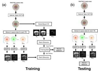 |
13 | The impact of streak-removal on deep learning reconstruction of radial datasets
Brian Patrick Toner1, Zhiyang Fu2, Rohit Philip3, Diego R. Martin4, Maria Altbach3, and Ali Bilgin2,3,5
1Applied Mathematics, University of Arizona, Tucson, AZ, United States, 2Electrical and Computer Engineering, University of Arizona, Tucson, AZ, United States, 3Department of Medical Imaging, University of Arizona, Tucson, AZ, United States, 4Houston Methodist Hospital, Houston, TX, United States, 5Biomedical Engineering, University of Arizona, Tucson, AZ, United States
Recently, deep learning models have been developed for reconstructing data acquired using radial turbo spin echo sequences, yielding images at multiple echo times as well as co-registered T2 maps. In radial imaging, streaking artifacts from anatomical regions where the magnetic field gradients are nonlinear can obscure pathology and impact accuracy of parameter mapping. In this work, we demonstrate that removal of streaking artifacts from data prior to training can provide substantial improvement in the reconstruction performance of deep learning methods.
|
||
3454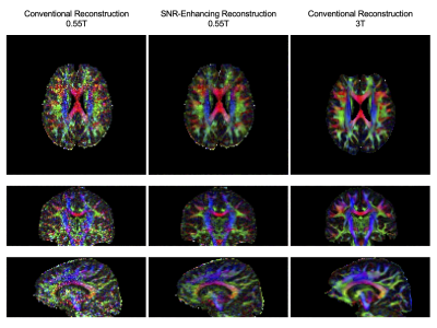 |
14 | Diffusion Tensor Imaging of the Brain on a Prototype 0.55T System using SNR-Enhancing Joint Reconstruction
Hao-Ting Kung1, Sophia X. Cui2, Jonas T. Kaplan3, Anand A. Joshi1, Richard M. Leahy1, Krishna S. Nayak1, Jay Acharya4, and Justin P. Haldar1,3
1Signal and Image Processing Institute, University of Southern California, Los Angeles, CA, United States, 2Siemens Medical Solutions USA, Inc., Los Angeles, CA, United States, 3Brain and Creativity Institute, University of Southern California, Los Angeles, CA, United States, 4Department of Clinical Radiology, University of Southern California, Los Angeles, CA, United States
There has been substantial recent interest in MRI systems with lower $$$B_0$$$ field strengths, which can improve the value and accessibility of MRI. This work investigates the performance of diffusion tensor imaging on a prototype whole-body 0.55T system equipped with high-performance shielded gradients. Although the images suffer from noise contamination when using conventional image reconstruction techniques, we demonstrate that the use of an SNR-enhancing joint reconstruction technique can substantially reduce noise concerns, enabling high quality diffusion tensor imaging results. In addition, compared to diffusion data acquired on a conventional 3T scanner, the 0.55T images demonstrate substantially reduced susceptibility-induced geometric distortions.
|
||
3455 |
15 | Using Low Rank Plus Sparse (L+S) Reconstruction to Accelerate Dynamic Hyperpolarized 13C Spiral Chemical Shift Imaging In Vivo
Minjie Zhu1, Aditya Jhajharia1, Joshua Rogers1, and Mayer Dirk1
1University of Maryland, Baltimore, Baltimore, MD, United States
The goal of this study is to validate the Low Rank Plus Sparse Reconstruction algorithm in in-vivo spectroscopic imaging applications. The proposed method can be used to increase temporal and/or spatial resolution of dynamic hyperpolarized 13C imaging without compromising image quality.
|
||
3456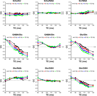 |
16 | Cross-correlation between metabolites due to spectral overlap
Sungtak Hong1, Li An1, and Jun Shen1
1National Institute of Mental Health, National Institutes of Health, Bethesda, MD, United States It is shown that spectral overlap can cause an apparent correlation between metabolites regardless of underlying biological correlations or the lack thereof. Because MRS signals are often used to correlate with other MRS signals or clinical measures a theoretical framework is developed to estimate correlations originating from spectral overlap at long echo time. Monte Carlos simulations were also performed to quantify the correlations. Our results showed that significant correlations may occur even at long TE, when the contribution from macromolecule background becomes negligible. The proposed theoretical framework was proven to be useful for predicting cross-correlation coefficients originating from spectral overlap.
|
||
3457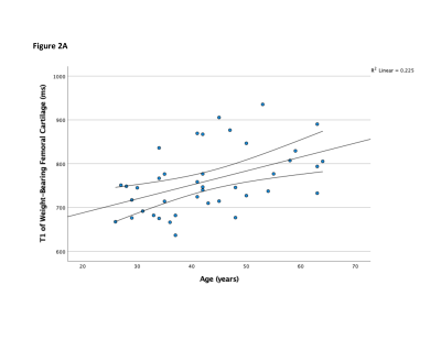 |
17 | Assessment of T1, T2, and T1ρ Values of Knee Cartilage and Menisci in Healthy Subjects using 3T MRI: A Preliminary Study
Ralph Zeitoun1, Chikara Noda1, Bharath Ambale-Venkatesh1, Yoshimori Kassai2, João A C Lima1, and Chia-Ying Liu2
1Division of Cardiology, Johns Hopkins University School of Medicine, Baltimore, MD, United States, 2Canon Medical Systems Corporation, Otawara, Japan Efforts have been focused on early detection of osteoarthritis using MRI. Our study reports T1, T1ρ and T2 values for knee cartilage and menisci in normal subjects using 3T MRI, and evaluates associations with age, gender, and BMI. We found that WB-FC had higher T1 and lower T2 values than LWB-FC, and WB-FC had higher T1 values in females. T1 values of WB-FC and LM were associated with age, and T1ρ values of WB-FC were associated with BMI. Our study presents novel data on T1 mapping, reinforcing the utility of MRI as a potential tool for early detection of osteoarthritis. |
||
 Back to Meeting Home
 Back to the Program-at-a-Glance
The International Society for Magnetic Resonance in Medicine is accredited by the Accreditation Council for Continuing Medical Education to provide continuing medical education for physicians.
 Back to Meeting Home
 Back to the Program-at-a-Glance
 View the Presentation