Online Gather.town Pitches
Brain, Head & Neck Cancer
Joint Annual Meeting ISMRM-ESMRMB & ISMRT 31st Annual Meeting • 07-12 May 2022 • London, UK

Online Gather.town Pitches
Brain, Head & Neck Cancer
| Booth # | ||||
|---|---|---|---|---|
3128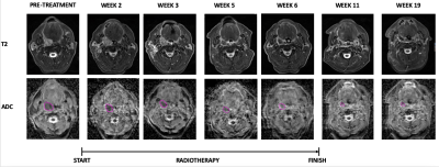 |
1 | Changes in serial DWI MRI imaging during radiation therapy can predict treatment response in patients with head and neck cancer
Yuvnik Trada1,2, Paul Keall2, Michael Jameson3, Daniel Moses4,5, Peter Lin6,7, Phillip Clap5,8, Lois Holloway8,9, Myo Min10,11, Dion Forstner3, Allan Fowler9, and Mark Lee5,9
1Radiation Oncology, Calvary Mater Newcastle, Waratah, Australia, 2The University of Sydney, Sydney, Australia, 3GenesisCare St Vincents Hospital, Sydney, Australia, 4Department of Medical Imaging, Prince of Wales Hospital, Sydney, Australia, 5University of New South Wales, Sydney, Australia, 6Department of Nuclear Medicine and PET, Sydney, Australia, 7Western Sydney University, Sydney, Australia, 8Ingham Institute of Applied Medical Research, Liverpool, Australia, 9Liverpool Hospital, Liverpool, Australia, 10Sunshine Coast University Hospital, Sunshine Coast, Australia, 11University of Sunshine Coast, Britinya, Australia
In head and cancer there is a need for a predictive biomarker for patients undergoing radiotherapy(RT) to allow personalisation of treatment.55 patients with HNSCC had DWI imaging performed at baseline, during (week 2, 3, 5 and 6) and post RT (1 and 3 months). Absolute and change in primary tumour ADCmean was correlated to local recurrence.Week 3 was the optimal timepoint for mid-treatment response assessment. Change in week 3 ADCmean of <24.4% predicted local recurrence; corresponding 2 year LRFS of 97% vs 42%.Changes in mid-treatment DWI imaging could be utilised in design of future adaptive clinical trials.
|
||
| 3129 | 2 | The performance of pretreatment MR-based radiomics in RHES combined chemoradiotherapy response prediction in nasopharyngeal carcinoma.
Lixuan Huang1, Zongxiang Yang1, Hao Ren2, Cheng Tang1, Huiting Zhang3, Yang Song3, and Liling Long1
1The First Affliated Hospital of Guangxi Medical University, Nanning, China, 2Guangxi Medical University Kaiyuan Langdong Hospital, Nanning, China, 3Siemens Healthineers, Shanghai, China This study is to investigate a MRI radiomics model based on pre-treatment texture features to explore its application value in predicting the efficacy of recombinant human endostatin(RHES) combined concurrent chemoradiotherapy for nasopharyngeal carcinoma. The results showed that the AUC and the accuracy of radiomics-clinics combine model and radiomics model for response assessment in nasopharyngeal carcinoma(NPC) was higher than clinics model. These suggest that pretreatment MRI based radiomics could predict RHES combined concurrent chemoradiotherapy response in NPC, its accuracy was better than clinics model. |
||
3130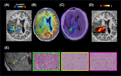 |
3 | Combined 7T CEST/MRS imaging to predict tumor metabolism in glioma
Yifan Yuan1, Qi Yue1, Xiang Zou1, Yu Guo1, Ying-Hua Chu2, Yi-Cheng Hsu2, Patrick Liebig3, Wenwen Yu4, He Wang4,5, Liang Chen1, and Ying Mao1
1Department of Neurosurgery, Huashan Hospital, Fudan University, Shanghai, China, 2Siemens Healthineers Ltd., Shanghai, China, 3Siemens Healthcare GmbH, Erlangen, Germany, 4Institute of Science and Technology for Brain-Inspired Intelligence, Fudan University, Shanghai, China, 5Human Phenome Institute, Fudan University, Shanghai, China
Magnetic resonance imaging has been applied in intraoperative neuro-navigation and irradiation guidance in glioma diagnosis. The contrast-enhanced MRI or peritumor edema on T2w image is not precise enough to delineate tumor infiltration. MR spectroscopy (MRS) and chemical exchange saturation transfer (CEST) have been used to evaluate tumor metabolism. Studies showed that amino acid PET surpassed conventional MR imaging for tumor delineation. Here, we explored the correlation between MRS and CEST at 7T and chose PET as gold standard to investigate multimodal MR imaging for glioma metabolic characteristics and tumor extent. An exemplary case with stereotactic biopsy proof was shown.
|
||
3131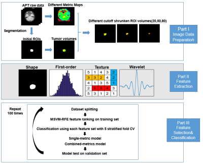 |
4 | Molecular subgrouping of pediatric medulloblastoma by APT radiomics
Junjie Wen1, Hongxi Zhang2, Zhipeng Shen3, Xiaohui Ma2, Xinchun Chen2, Weibo Chen4, Dan Wu1, and Yi Zhang1
1Key Laboratory for Biomedical Engineering of Ministry of Education, Department of Biomedical Engineering, College of Biomedical Engineering & Instrument Science, Zhejiang University, Hang Zhou, Zhejiang, China, 2Department of Radiology, Children’s Hospital, Zhejiang University School of Medicine, Hang Zhou, Zhejiang, China, 3Department of Neurosurgery, Children’s Hospital, Zhejiang University School of Medicine, Hang Zhou, Zhejiang, China, 4Philips Healthcare, Shanghai, China
APT imaging and its derived metric maps were applied to identify molecular subgroups of medulloblastoma for the first time. Thirty-eight newly-diagnosed pediatric patients with medulloblastoma were enrolled in this study and scanned on a 3T scanner. We implemented a radiomic analysis of the APT-related metric maps, with initial regions of interest delineated by an experienced radiologist and then shrunk automatically. After feature extraction and selection, five different classifiers were tested with both single-metric and multi-metric maps. We successfully established predictive models to differentiate the subgroups of medulloblastoma with good accuracy of 0.754.
|
||
3132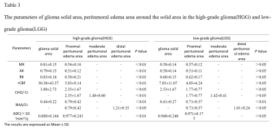 |
5 | The application of multi-mode MR imaging technology in the tumor infiltration boundary of cerebral glioma
Jin Li1, Xiangrong Li1, and Huiting Zhang2
1The First Affiliated Hospital of Guangxi Medical University, Nanning, China, 2MR Scientific Marketing, Siemens Healthineers, Wuhan, China
We explored the applied value of multi-mode MR imaging technology, including diffusion kurtosis imaging (DKI), arterial spin labeling (ASL) and 1H-MRS, in glioma tumor invasion boundary. Results showed mean kurtosis from DKI and apparent diffusion coefficient had the highest diagnostic efficiency between high-grade (HGG) and low-grade gliomas (LGG). There were significant differences in CHO/Cr and NAA/Cr from 1H-MRS between the solid area and the proximal peritumoral edema area (PEA), and CHO/Cr and NAA/Cr in different PEA of LGG or HGG. PEA within 1cm around the solid area is recommended to be resected to decrease recurrence rate.
|
||
3133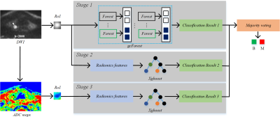 |
6 | Multi-stage ensemble machine learning for predicting the pathology of thyroid micronodules on small-datasets high b-value thyroid DWI
ChengLong Deng1,2, BingChao Wu1,2, QingJun Wang3, QingLei Shi4, Bei Guan1,2, Dacheng Qu5, ChenXi Li1,2, DaoGuang Zan1,2, XiaoLin Chen1,2, and YongJi Wang1,2
1Collaborative Innovation Center, Institute of Software Chinese Academy of Sciences, Beijing, China, 2University of Chinese Academy of Sciences, Beijing, China, 3Department of Radiology, The 6th Medical Center of Chinese PLA General Hospital, Beijing, China, 4MR Scientific Marketing, Siemens Healthcare, Beijing, China, 5School of Computer Science and Technology, Beijing Institute of Technology, Beijing, China
In this paper, a multi-stage ensemble learning based on the majority voting mechanism was designed to leverage the contradiction between an insufficient number of thyroid MRI and well-trained deep learning models that accurately predicted the pathology of thyroid micronodules. And its clinical applicability value was also assessed in terms of micronodule risk stratification and optimal regimen selection on high b-value (2000 s/mm2) diffusion-weighted images. Experimental results proved that our model had the capability of effectively distinguishing benign and malignant micronodules on small-dataset thyroid DWI images.
|
||
3134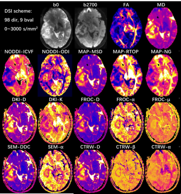 |
7 | Feasibility of Integrating 7 Diffusion Models within a Single Acquisition and Their Performance Comparison in Glioma Grading Video Not Available
Minxiong Zhou1, Huiting Zhang2, Ankang Gao3, Shaoyu Wang2, Jie Bai3, Guang Yang4, Jingliang Cheng3, and Xu Yan2
1Shanghai University of Medicine & Health Sciences, Shanghai, China, 2MR Scientific Marketing, Siemens Healthineers, Shanghai, China, 3Department of MRI, The First Affiliated Hospital of Zhengzhou University, Zhengzhou, China, 4Shanghai Key Laboratory of Magnetic Resonance, School of Physics and Electronic Science, Shanghai Key Laboratory of Magnetic Resonance, School of Physics and Electronic Science,East China Normal University, Shanghai, China
There are many advanced diffusion models available for neural and body researches, while their acquisitions are different and normally difficult to be joint applied and compared in one study. In this study, we tried to calculate 7 diffusion models, including 4 neural and 3 cancer related models, using data from one acquisition, and applied it for glioma. Two diffusion acquisition strategy were evaluated, and the performance of these models in glioma grading was also compared. The result showed that both acquisition strategies could generate high quality quantitative parameters for all models, which showed significant differences between high- and low-grade tumor.
|
||
3135 |
8 | Evaluation of diagnostic and prognostic value of high-resolution 3D 1H-MRSI for patients with newly diagnosed glioma
Bin Bo1, Tianyao Wang2, Ziyu Meng1, Rong Guo3,4, Yudu Li3,4, Yibo Zhao3,4, Xin Yu5, Zhi-Pei Liang3,4, and Yao Li1
1School of Biomedical Engineering, Shanghai Jiao Tong University, Shanghai, China, 2Radiology Department, The Fifth People's Hospital of Shanghai, Fudan University, Shanghai, China, 3Beckman Institute for Advanced Science and Technology, University of Illinois at Urbana-Champaign, Urbana, IL, United States, 4Department of Electrical and Computer Engineering, University of Illinois at Urbana-Champaign, Urbana, IL, United States, 5Department of Biomedical Engineering, Case Western Reserve University, Cleveland, OH, United States
Conventional structural MRI has limited specificity in defining the extent and grade of glioma. MRSI complements MRI in tumor tissue characterization with neurometabolic fingerprints. In this study, we investigated the use of high-resolution 3D MRSI for glioma grading and evaluation of patient overall survival. Our results showed that the neurometabolic biomarkers could differentiate low-grade from high-grade gliomas and provide prognostic value for overall survival of newly diagnosed glioma patients.
|
||
3136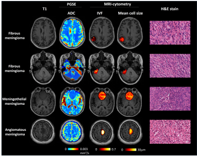 |
9 | Characterizing meningioma subtypes using MRI-cytometry
Ying-Hua Chu1, Yifan Yuan2, Xueying Zhao3, He Wang3,4, Thorsten Feiweier5, and Yi-Cheng Hsu1
1Siemens Healthineers Ltd., Shanghai, China, 2Department of Neurosurgery, Huashan Hospital, Fudan University, Shanghai, China, 3Institute of Science and Technology for Brain-Inspired Intelligence, Fudan University, Shanghai, China, 4Human Phenome Institute, Fudan University, Shanghai, China, 5Siemens Healthcare GmbH, Erlangen, Germany
We used MRI-cytometry to estimate the mean cell diameter and intracellular volume of tumors of the meningioma subtype classification. We found cell size and intracellular volume to be very different between the three meningioma subtypes in this preliminary study. Our results reflected high collagen volume among the extracellular space in fibrous meningioma. We plan to scan more patients in the future to find a useful biomarker using MRI-cytometry.
|
||
3137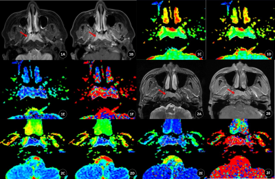 |
10 | Differentiation of T1 stage nasopharyngeal carcinoma and lymphoid hyperplasia by mono-exponential and bi-exponential metrics of TSE DWI Video Not Available
Yifan Liu1, Shasha Bao1, Nan Xu1, Xiaobin Guo1, Chengde Liao1, Zhongping Zhang2, and Tengfei Ke1
1Dept. of Radiology, Yunnan Cancer Hospital, Kunming, China, 2Philips Healthcare China, Guangzhou, China
We aimed to differentiate between T1 stage nasopharyngeal carcinoma (NPC) and nasopharyngeal lymphoid hyperplasia using multiple b-values Turbo-spin-echo DWI acquisition. Turbo-spin-echo DWI were adopted to minimize susceptibility artifacts in nasopharynx. Mono-exponential and bi-exponential models were both used to generate ADC, D, D* and f maps. We found that T1 stage NPC had significant lower ADC and D, higher D* than nasopharyngeal lymphoid hyperplasia. Furthermore, D has best diagnostic efficacy with highest area under curve in ROC analysis, indicating that it's a potential imaging biomarker for accurate early diagnosis of T1 stage NPC.
|
||
3138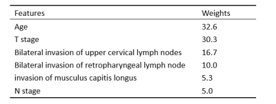 |
11 | A time-related random forest survival model based on MR imaging markers to predict the survival of patients with nasopharyngeal carcinoma
Chao Luo1, Haixia Li2, Kan Deng2, and Haojiang Li1
1Sun Yat-sen University Cancer Center, Guangzhou, China, 2Philips Healthcare, Guangzhou, China
This study aimed to identify magnetic resonance (MR) imaging markers associated with the overall survival (OS) of patients with nasopharyngeal carcinoma (NPC) and establish a random survival forest (RSF) model, which is a time-related machine learning model for survival analysis, to predict their survival.
|
||
3139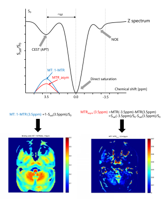 |
12 | Three-dimension Amide Proton Transfer Imaging for Predicting the Treatment Response of Nasopharyngeal Carcinoma
Wenguang Liu1, Yigang Pei1, Simin Xie1, Xiao Wang1, Yu Bai1, Gaofeng Zhou1, Weiyin Vivian Liu2, and Wenzheng Li1
1Department of Radiology, XiangYa Hospital, Central South University, Changsha, China, 2GE Healthcare, Beijing, China
To predict the treatment response for NPC patients is very important. This study aimed to explore the diagnostic performance of 3D APT imaging on predicting the treatment response of NPC patients. A total of 40 patients and 14 healthy volunteers were identified in this study. Result show that the APT and MT values have good and excellent intra- and inter-observer agreement. It is significant difference for MT values between residual and non-residual group. The result suggests that the pre-treatment MT values can be used to predict treatment response of NPC with chemoradiotherapy, and better than the APT and ADC value.
|
||
3140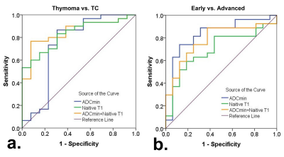 |
13 | Predicting the risk grades of thymic epithelial tumors using magnetic resonance T1 mapping and diffusion weighted imaging: a pilot study
Chenxi Liu1, Shengzhong Wang2, Xiaocheng Wei3, Gang Xiao1, YUchuan Hu1, and Guangbin Cui1
1Tangdu Hospital, Air Force Medical University, Xi'an, China, 2Shaanxi University of Traditional Chinese Medicine, Xi'an, China, 3GE Healthcare, Beijing, China
The pathological staging system of Thymic epithelial tumors (TETs) which called Masaoka staging, is an important prognostic factor, and its impact on survival is much greater than that of WHO classification. However, as the Masaoka stage can only be established postoperatively, preoperative imaging has important implications concerning treatment strategy. This study analyzes the differences of T1 values and ADC values in different regions, different types and stages of TETs tumors, and establishes a reliable noninvasive imaging technology to distinguish the risk classification and stage of tumors.
|
||
 Back to Meeting Home
 Back to the Program-at-a-Glance
The International Society for Magnetic Resonance in Medicine is accredited by the Accreditation Council for Continuing Medical Education to provide continuing medical education for physicians.
 Back to Meeting Home
 Back to the Program-at-a-Glance
 View the Presentation