Online Gather.town Pitches
Clinical Brain Imaging, Including COVID-19
Joint Annual Meeting ISMRM-ESMRMB & ISMRT 31st Annual Meeting • 07-12 May 2022 • London, UK

Online Gather.town Pitches
Clinical Brain Imaging, Including COVID-19
| Booth # | ||||
|---|---|---|---|---|
4954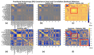 |
1 | Structural covariance of diffusion metrics in mild COVID19: Spatially coherent effect on fractional anisotropy but not free water.
Nick Teller1, Jordan A. Chad1,2, Eugenie Roudaia1, Ali Hashemi3, Haddas Grosbein1, Asaf Gilboa1,2, Maged Goubran2,4, Ivy Cheng2,4, Sandra E. Black2,4, Robert Fowler2,4, Chris Heyn2,4, Fuqiang Gao4, Mario Masellis2,4, Jennifer
Rabin2,4, Xiang Ji4, Aravinthan Jegatheesan2,4, Benjamin Lam2,4, Allison B. Sekuler1,2,3, Bradley J. MacIntosh2,4, Simon J. Graham2,4, and J. Jean Chen1,2
1Rotman Research Institute, North York, ON, Canada, 2University of Toronto, Toronto, ON, Canada, 3Psychology, Neuroscience & Behaviour, McMaster University, Hamilton, ON, Canada, 4Sunnybrook Research Institute, Toronto, ON, Canada
The impact of COVID19 on the brain’s microstructural integrity remains unclear. In this study, we examine self-isolated COVID19 patients and controls using diffusion-tensor and free-water imaging, based on single- and multi-shell acquisitions, respectively. We identify several differences in spatial covariance among patients in fractional anisotropy (in cingulate-frontal and temporal-parietal regions), but not free water fraction. Our results indicate COVID19’s implications in long-term, measurable brain deficits.
|
||
4955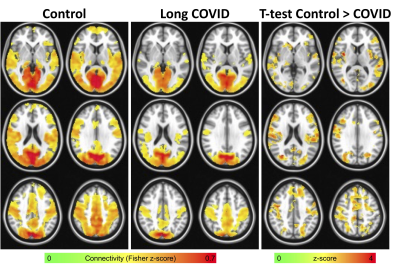 |
2 | Lower functional connectivity in areas of reduced cerebrovascular reactivity in elderly patients with long COVID
Alexander D. Cohen1, Laura Umfleet2, Malgorzata Franczak2, Sara Swanson2, Jessica Pommy2, Mohit Agarwal2, Baolian Yang3, Suchandrima Banerjee3, and Yang Wang1
1Radiology, Medical College of Wisconsin, Milwaukee, WI, United States, 2Medical College of Wisconsin, Milwaukee, WI, United States, 3GE Healthcare, Waukesha, WI, United States
Patients suffering from long COVID report cognitive symptoms months after disease onset, which may be related to neurovascular changes. Here, we evaluated functional connectivity in long COVID elderly patients and control subjects using a region of lower cerebrovascular reactivity (CVR) as the seed. We found significantly lower connectivity in long COVID patients that was widespread potentially linking alterations in CVR to functional connectivity and reduced cognitive ability in long COVID patients.
|
||
4956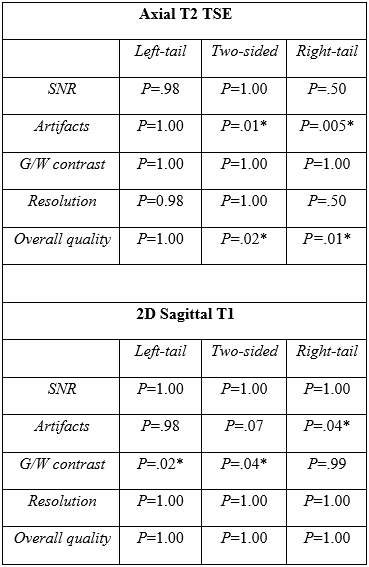 |
3 | COVID-19 as an Opportunity for Quality Improvement in MR Neuro Imaging.
Emanuele Camerucci1, Jose Thulasee1, Matthew Bernstein1, Steven Messina1, Peter Kollasch1, and John Huston1
1Mayo Clinic, Rochester, MN, United States
We took advantage of the COVID-19-related decrease in clinical volume to conduct a quality improvement project; we established a panel of 24 experts for reviewing currently used brain MRI sequences and proposing ways to improve image quality and/or decrease acquisition time. The proposed sequences were integrated in existing protocols and compared with the standard ones. We were able to improve 14 of the most used brain MRI sequences with an equal or improved image quality and reduced acquisition time. Hence, we were able to reduce the acquisition time of our Brain protocol and Epilepsy protocol by 29.2% and 40%, respectively.
|
||
4957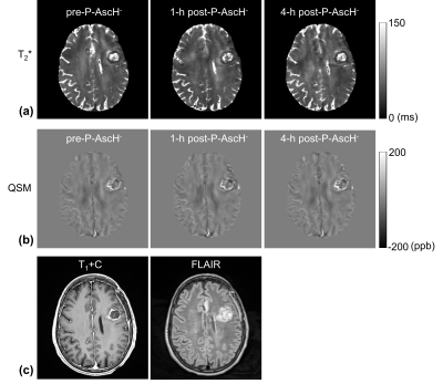 |
4 | Assessing redox changes induced by treatment with pharmacological ascorbate in glioblastoma using T2* and QSM
Chu-Yu Lee1, Michael S Petronek2, Cameron M Cushing3, Garry R Buettner2, Joel J St-Aubin2, Varun Monga4, John M Buatti2, Joseph J Cullen5, Douglas R Spitz2, Bryan G Allen2, and Vincent A Magnotta1
1Department of Radiology, The University of Iowa, Iowa City, IA, United States, 2Department of Radiation Oncology, Free Radical and Radiation Biology Program, The University of Iowa, Iowa City, IA, United States, 3Skope Magnetic Resonance Technologies AG, Zurich, Switzerland, 4Department of Internal Medicine, The University of Iowa, Iowa City, IA, United States, 5Department of Surgery, The University of Iowa, Iowa City, IA, United States
Pharmacological ascorbate (P-AscH-), as an adjuvant to standard treatment for glioblastoma, may selectively enhance cancer cell killing. The mechanisms of P-AscH- have been associated with redox changes in the labile iron pool. This study applies T2* and QSM to assess the redox changes in 40 glioblastoma patients receiving standard treatment and adjuvant P-AscH-. The results demonstrate an increase in T2* values and a decrease in QSM values within contrast-enhancing lesions 1 hour after P-AscH- infusion. These changes in T2* and QSM may result from the redox changes by P-AscH-, supporting the potential for imaging assessment of response to P-AscH-.
|
||
 |
4958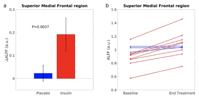 |
5 | Intranasal Insulin Enhances Resting Neurovascular Fluctuations in Type 2 Diabetes
Zongpai Zhang1, Faizan Khan2, Peter Novak2, Christos Mantzoros3, Long Ngo3, Vera Novak2, and Weiying Dai1
1State University of New York at Binghamton, Binghamton, NY, United States, 2Department of Neurology, Harvard Medical School, Boston, ME, United States, 3Department of Medicine, Harvard Medical School, Boston, ME, United States
We aimed to investigate the effect of intranasal insulin (INI) on cognition, balance, and brain neurovascular activity in type 2 diabetes mellitus (T2DM) participants of the MemAID trial. Cognition, balance, and brain neurovascular fluctuations - amplitude of low-frequency fluctuations (ALFF) were measured in 11 subjects before and after 24-week treatment with INI or placebo. INI treatment significantly increased ALFF in the medial-frontal region. The ALFF increases at the end-of INI treatment were associated with improved balance. These findings suggest that INI may increase brain neurovascular activity in T2DM in the regions related to balance control and sustained attention.
|
|
4959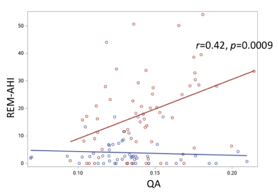 |
6 | Association of White Matter Integrity of Locus Coeruleus Pathways to Hypothalamus and Sleep Apnea in Military Mild Traumatic Brain Injury
Ping-Hong Yeh1, J. Kent Werner, Jr2,3, Chihwa Song1, Rujirutana Srikanchana1, Wei Liu1, Kimbra Kenney1,2, Treven Pickett1,2, Grant Bonavia1,2, Gerard Riedy1,2, and John Ollinger1
1National Intrepid Center of Excellence, Bethesda, MD, United States, 2Uniformed Services University of the Health Sciences, Bethesda, MD, United States, 3Walter Reed National Military Medical Center, Bethesda, MD, United States
Sleep disturbances are common following traumatic brain injury (TBI). The intent of this study is to characterize the role of the noradrenergic locus coeruleus (LC) for sleep maintenance in service members following mild TBI by associating microstructural features, derived from diffusion MRI, of the LC–noradrenergic (NA) system with objective sleep measures. We found that severity of sleep apnea, particularly in rapid eye movement stage during sleep, significantly correlated with microstructural changes in the LC pathways to hypothalamus in mTBI participants. This result suggests sleep apnea following mild TBI may be modulated by sympathetic activity via pathways interconnecting LC and hypothalamus.
|
||
4960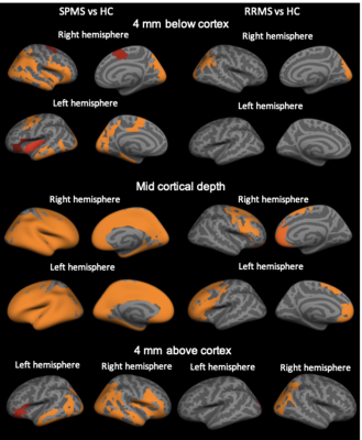 |
7 | Evidence of meningeal inflammation in multiple sclerosis: a combined in vivo 11C-PBR28 MR-PET and post-mortem study Video Permission Withheld
Elena Herranz1,2, Valeria Barletta1,2, Constantina A. Treaba1,2, Ambica Mehendiratta1, Russell Ouellette1,3,4, Eric Klawiter5,6, Jacob Sloane 2,7, Carolina Ionette8, Suma Babu2,5, Marco L. Loggia1,2, Meena M Makary1,2,9, Jacob M Hooker1,2, Ciprian Catana1,2,
Revere Kinkel10, Roberta Magliozzi11,12, and Caterina Mainero1,2
1Radiology, Massachusetts General Hospital, Boston, MA, United States, 2Harvard Medical School, Boston, MA, United States, 3Department of Clinical Neuroscience, Karolinska Institutet, Stockholm, Sweden, 4Department of Neurology, 4. Karolinska University Hospital, Stockholm, Sweden, 5Neurology, Massachusetts General Hospital, Boston, MA, United States, 6Massachusetts General Hospital, Boston, MA, United States, 7Beth Israel Deaconess Medical Center, Boston, MA, United States, 8Department of Neurology, UMass Memorial Medical Center, Worcester, MA, United States, 9Systems and Biomedical Engineering Department, Faculty of Engineering, Cairo University, Giza, Egypt, 10Department of Neuroscience, University of California, San Diego, CA, United States, 11Neurology Section, Department of Neurosciences, Biomedicine and Movement Sciences, University of Verona, Verona, Italy, 12Division of Neuroscience, Department of Brain Sciences, Imperial College London, London, United Kingdom
In multiple sclerosis (MS), neuropathological studies suggest that meningeal inflammation involving T-, B-lymphocytes, plasma cells, macrophages may trigger underlying cortical neuroinflammation and demyelination. Using MR-PET targeting the18kDa mitochondrial translocator protein, which is overexpressed in activated glia/macrophages, we detected in 49 MS patients cortical inflammatory changes along with neuroinflammation in both meninges and juxtacortical white matter. Meningeal inflammation correlated with worse neurological disability and cognitive performance. Histochemistry performed in meningeal tissue from 40 post-mortem progressive MS cases confirmed in vivo findings. This study provides in vivo PET imaging evidence implicating meningeal neuroinflammation in the pathophysiology of MS.
|
||
| 4961 | 8 | Parameters associated with durable clinical success in a multi-center trial using MR guided focused ultrasound treat uterine leiomyomas
Rachel Bitton1, Angela Fast2, Gina Hesley3, Steven Raman4, Alan Matsumoto5, Eric Dolan6, Maureen Kohi7, Thomas Price8, Fiona Fenessey9, and Pejman Ghanouni1
1Radiology, Stanford University, Stanford, CA, United States, 2Allegheny Health Network, Pittsburgh, PA, United States, 3Radiology, Mayo Clinic, Rochester, MN, United States, 4Radiology, UCLA, Los Angeles, CA, United States, 5Radiology and Medical Imaging, University of Virginia, Charlottesville, VA, United States, 6Interventional Radiology, Ohio Health, Columbus, OH, United States, 7Radiology, UCSF, San Francisco, CA, United States, 8Obstetrics and Gynecology, Duke University, Durham, NC, United States, 9Radiology, Brigham and Women's Hospital, Boston, MA, United States
This study presents results of a prospective multicenter clinical trial of 99 women treated with MR guided focused ultrasound (MRgFUS) for symptomatic uterine leiomyomas to assess treatment durability and to evaluate potential demographic, imaging, and technical characteristics associated with lasting clinical outcomes. Following treatment, there was an improvement in symptoms, with a significant decrease in mean symptom severity score at the 6, 12, 24 and 36 month follow up (p<0.001). In a multivariate model, a new parameter of interest, the ratio of non-perfused volume to total fibroid volume (NPV/TFV) was found to be predictive of durable clinical success (p=0.03).
|
||
4962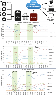 |
9 | Coronavirus Disease 2019 (COVID-19): Impact of First and Second Waves on CT and MRI Patient Volumes at a Large Tertiary Center
Stanley Chu1, Mitchell Collins1, Maurice H Pradella2, Rachel Davids 3, Martin Kramer 3, Mathis Zimmerman3, Sarah Fopma2, Alexander Korutz2, Ryan Avery2, Michael Markl2, Bradley Allen2, Blair Faber2, and James Carr2
1Radiology, Northwestern University, Chicago, IL, United States, 2Northwestern University, Chicago, IL, United States, 3Siemens Medical Solutions, Chicago, IL, United States
The impact of the first wave of COVID-19 (Feb-Apr. 2020) on imaging volumes has been widely explored, but responses to a subsequent wave (Oct-Dec. 2020) remain to be investigated. In this study, a cloud-based analytics tool (CBAT) collected monthly CT/MRI scan volumes at a large tertiary center from Oct 2019 to Aug 2021. The first wave of COVID-19 significantly lowered all forms of CT/MRI imaging. The second COVID-19 wave did not significantly impact CT/MRI scan volumes, which remained at pre-pandemic levels during/afterwards. CBAT has the ability to assess impact/response towards two waves of COVID-19 at a large tertiary center.
|
||
4963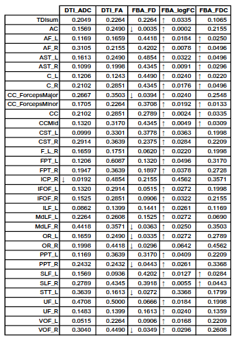 |
10 | Analysis of white matter tissue integrity of COVID-19 recovered patients using diffusion weighted imaging
Alejandro Santos-Díaz1,2, Berenice Sanchez-Alvarado3, Oscar Marrufo Melendez4, Roger Carrillo Mezo4, Antonio Arauz Gongora4, and Monica Rodriguez5
1School of Engineering and Science, Tecnologico de Monterrey, Mexico City, Mexico, 2School of Medicine and Health Sciences, Tecnologico de Monterrey, Monterrey, Mexico, 3School of Engineering and Science, Tecnologico de Monterrey, Monterrey, Mexico, 4Neuroimaging, Instituto Nacional de Neurologia y Neurocirugía, Mexico City, Mexico, 5Instituto Nacional de Enfermedades Respiratorias, Mexico City, Mexico
Evidence supports that the virus SARS-CoV-2 has a potential neuroinvasion causing neurological symptoms, even after recovery. Diffusion Weighted Imaging (DWI) allows us to detect the microstructural changes in white matter tissue integrity. In this work, we studied the neurological manifestations of COVID-19 in apparent diffusion coeficient (ADC), fractional anisotropy (FA), and metrics derived from fixel-based analysis (FBA) such as fiber density (FD), fiber cross-section (FC), and fiber density and cross-section (FDC). We found statistically significant differences in 34/43 fiber bundle regions from the IIT Human Brain Atlas in the five different metrics studied.
|
||
4964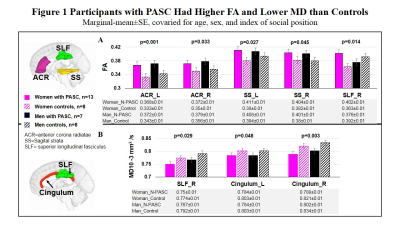 |
11 | High White Matter Fractional Anisotropy and Low Diffusivity in Participants with Post-acute Sequelae SARS-CoV-2 Infection
Huajun Liang1, Thomas Ernst1,2, Kenichi Oishi3, Meghann Ryan1, Eleanor Wilson4, Andrea Levine5, Eric Cunningham1, Shyamasundaran Kottilil4, and Linda Chang1,2,6
1Diagnostic Radiology and Nuclear Medicine, University of Maryland School of Medicine, Baltimore, MD, United States, 2Department of Neurology, Johns Hopkins University School of Medicine, Baltimore, MD, United States, 3Department of Radiology, Johns Hopkins University School of Medicine, Baltimore, MD, United States, 4Institute of Human Virology, Department of Medicine, Division of Infectious Disease, University of Maryland School of Medicine, Baltimore, MD, United States, 5Department of Medicine, Division of Pulmonary & Critical Care Medicine, University of Maryland School of Medicine, Baltimore, MD, United States, 6Department of Neurology, University of Maryland School of Medicine, Baltimore, MD, United States
Diffusion tensor imaging (DTI), a sensitive method to detect inflammatory or cytotoxic tissue changes, has been used to study brain microstructure in patients with COVID-19. At 2-3 months post-acute sequelae SARS-CoV-2 infection (PASC), prior reports found both higher or lower than normal white matter diffusivity in participants who had variable severity of clinical manifestations. We performed DTI and neuropsychiatric assessments in 20 participants with PASC. Individuals with PASC had lower diffusivity 6 months after the diagnosis, which correlated with greater fatigue. These diffusion findings may be due to glial proliferation and immune-response related cytotoxic edema.
|
||
4965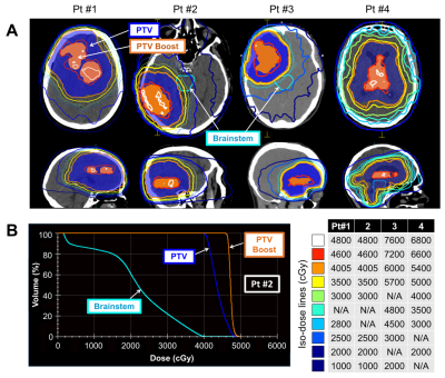 |
12 | Dose-Escalated Radiation Treatment Plans in Glioblastoma Based on Quantitative Magnetization Transfer using a 1.5T MR-Linac
Rachel W. Chan1, Mark Ruschin2, Liam S.P. Lawrence3, James Stewart2, Sten Myrehaug2, Chia-Lin Tseng2, Jay Detsky2, Pejman J. Maralani4, Mary Jane Lim-Fat2, Hany Soliman2, Greg J. Stanisz1,3,5, Arjun Sahgal2, and Angus Z. Lau1,3
1Physical Sciences Platform, Sunnybrook Research Institute, Toronto, ON, Canada, 2Department of Radiation Oncology, Sunnybrook Health Sciences Centre, Toronto, ON, Canada, 3Department of Medical Biophysics, University of Toronto, Toronto, ON, Canada, 4Department of Medical Imaging, Sunnybrook Health Sciences Centre, University of Toronto, Toronto, ON, Canada, 5Department of Neurosurgery and Pediatric Neurosurgery, Medical University of Lublin, Lublin, Poland
Hybrid MR/radiotherapy MR-Linac devices enable dose adaptation based on daily imaging. Saturation transfer MRI, including quantitative magnetization transfer (qMT) and chemical exchange saturation transfer (CEST), have been shown to predict treatment response in glioblastoma. Here, in a proof-of-principle study, we demonstrated the feasibility of retrospectively generating dose-escalated radiation plans based on qMT semi-solid fraction maps acquired on the MR-Linac. Treatment plans with a 115-120% boost dose were successfully generated in all four patients and adhered to clinical organs-at-risk dose constraints.
|
||
4966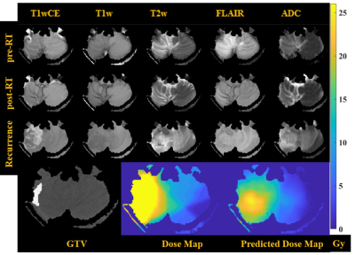 |
13 | Predicting Stereotactic Radiosurgery Dose Maps from Pre-Therapy MR Images using a Deep Neural Network
Shraddha Pandey1,2, Tugce Kutuk3, Matthew N Mills3, Mahmoud Abdalah4, Olya Stringfield4, Kujtim Latifi3, Timothy J Robinson3, Wilfrido Moreno1, Kamran A Ahmed3, and Natarajan Raghunand2,5
1Electrical Engineering, University of South Florida, Tampa, FL, United States, 2Department of Cancer Physiology, H. Lee Moffitt Cancer Center and Research Institute, Tampa, FL, United States, 3Department of Radiation Oncology, H. Lee Moffitt Cancer Center and Research Institute, Tampa, FL, United States, 4Quantitative Imaging Shared Service, H. Lee Moffitt Cancer Center and Research Institute, Tampa, FL, United States, 5Department of Oncologic Sciences, University of South Florida, Tampa, FL, United States
Stereotactic Radiosurgery (SRS) of asymptomatic brain metastases provides lasting tumor control with only minor side effects to healthy brain. An active research area is the development of models to predict tumor response to a given dose of Radiation Treatment (RT) from analysis of pre-RT and post-RT MR images (i.e., the forward problem). Here we propose an approach to train a deep neural net on pre-RT MR images of patients with Breast Cancer Metastases to the Brain (BCMB), for predicting RT dose maps that will yield desired/target tumor voxel intensities on post-RT MR images (i.e., the inverse problem).
|
||
4967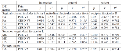 |
14 | Diffusion Tensor Imaging Biomarkers for Chronic Pain Following Mild Traumatic Injury
Ho-Ching Yang1, Kelly Naugle2, Qiuting Wen1, Fletcher A White3,4,5, and Yu-Chien Wu1
1Departments of Radiology and Imaging Sciences, Indiana University School of Medicine, Indianapolis, IN, United States, 2Department of Kinesiology, School of Health and Human Sciences, Indiana University Purdue University Indianapolis, Indianapolis, IN, United States, 3Department of Anesthesia, School of Medicine, Indiana University School of Medicine, Indianapolis, IN, United States, 4Stark Neuroscience Research Institute, Indiana University School of Medicine, Indianapolis, IN, United States, 5Research and Development Services, Richard L. Roudebush VA Medical Center, Indianapolis, IN, United States
To investigate the neuropathogenesis of post-traumatic headache following mild traumatic brain injuries (mTBI), this study applied diffusion tensor imaging and functional magnetic resonance imaging to explore relationships between brain structural and functional changes and sensitization, pain inhibitory capacity, psychological factors and headache pain in mTBI subjects. The results found structural and functional alterations in mTBI. Also, the results showed that DTI metric differences between mTBI subjects and controls can be used to predict pain/psychological measurements, which indicated the relationship between white matter disruption and measurements of endogenous pain modulation and psychological distress in mTBI.
|
||
4968 |
15 | Magnetic Resonance Imaging to Identify Chronicity and Treatability of Migraine and Chronic Headache Disorders
Isaac Vicente Manzanera Esteve1, Adam Evans1, Brian Johnson2, Ryan Robinson3, Saikat Sengupta1, Alonda Pollins1, Wesley Thayer1, and Salam Al Kassis1
1Vanderbilt University Medical Center, Nashville, TN, United States, 2Philips Healthcare, Gainesville, FL, United States, 3Philips Healthcare, Nashville, TN, United States
Migraine Headaches (MH) are the 6th leading cause of years lived with disability worldwide, affect 15% of the US population, and are nearly twice as prevalent in veterans who have been deployed. Our study focuses on pathology of the greater occipital nerve (GON), which occurs with a high prevalence in individuals with a history of trauma (i.e. traffic accidents, explosions, falls). In our pilot MRI scans and operative experience, migraines associated with GON pathology displayed a pathologic thickening of the nerve validated intraoperatively. These results present MRI as a potential biomarker of headache pathologies of MH associated with GON pathology.
|
||
 Back to Meeting Home
 Back to the Program-at-a-Glance
The International Society for Magnetic Resonance in Medicine is accredited by the Accreditation Council for Continuing Medical Education to provide continuing medical education for physicians.
 Back to Meeting Home
 Back to the Program-at-a-Glance
 View the Presentation