Online Gather.town Pitches
RF Coils, Technologies & Sequences II
Joint Annual Meeting ISMRM-ESMRMB & ISMRT 31st Annual Meeting • 07-12 May 2022 • London, UK

Online Gather.town Pitches
RF Coils, Technologies & Sequences II
| Booth # | ||||
|---|---|---|---|---|
3310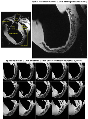 |
1 | A Dedicated Transceiver 8Tx/16Rx pTx Coil Array for Ex-vivo MRI Histology of Post-infarction Myocardium on a Whole-body 7T Scanner
Ibrahim A. Elabyad1, Maxim Terekhov1, and Laura M. Schreiber1
1Chair of Molecular and Cellular Imaging, Comprehensive Heart Failure Center (CHFC), University Hospital Wuerzburg, Wuerzburg, Germany
The purpose of this work was to develop and investigate a dedicated transceiver 16-element antisymmetric loop array for high-resolution imaging of the ex-vivo porcine heart at 7T. The array was interfaced to a 7T scanner in parallel transmit (pTx) mode (8Tx/16Rx). Electromagnetic-field (EMF) simulations were performed with the antisymmetric array loaded with a spherical phantom and a human heart (Billie). The optimization of the B1+-shimming for both sTx and pTx applications was performed using simulated and experimental B1-maps. The array was successfully tested to acquire ultra-high resolution (0.1x0.1x0.8mm voxel) images of the post-infarction scar tissue in the excised pig heart.
|
||
3311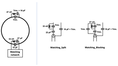 |
2 | A comparison of double-matching networks in a double-tuned coil for multinuclear MRI
Chang-Hoon Choi1, Suk-Min Hong1, Jörg Felder1,2, and N. Jon Shah1,3,4,5
1Institute of Neuroscience and Medicine - 4, Forschungszentrum Juelich, Juelich, Germany, 2RWTH Aachen University, Aachen, Germany, 3Institute of Neuroscience and Medicine - 11, Forschungszentrum Juelich, Juelich, Germany, 4JARA - BRAIN - Translational Medicine, Aachen, Germany, 5Department of Neurology, RWTH Aachen University, Aachen, Germany
X-nuclei MR offers unique information relating to cellular and metabolic processes in tissues. Multi-tuned coils are frequently utilised for X-nuclei measurements, and a number of novel approaches for double tuning coils exist. However, designing a well-performing multi-tuned coil is challenging, and making any improvement in SNR is particularly desirable on the X-nucleus coil due to the intrinsically lower MR sensitivity of X-nuclei. This study focuses on the insertion of double-matching networks in a single-structure, double-tuned 1H/23Na coil and compares their effect on the coil performance. The double-matching networks were built using either split or blocking traps.
|
||
3312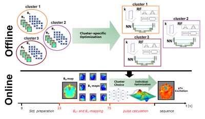 |
3 | Neural Network-supported Fast Online-Customized (FOCUS) parallel transmit (pTx) pulses for slice-selective, large flip angle excitation
Jürgen Herrler1, Patrick Liebig2, Kurt Majewski3, Rene Gumbrecht2, Dieter Ritter2, Christian Richard Meixner4, Andreas Maier5, Arnd Dörfler1, and Armin Michael Nagel4,6
1Department of Neuroradiology, University Hospital Erlangen, Friedrich-Alexander-Universität Erlangen-Nürnberg (FAU), Erlangen, Germany, 2Siemens Healthcare GmbH, Erlangen, Germany, 3Department of Corporate Technology, Siemens, München, Germany, 4Institute of Radiology, University Hospital Erlangen, Friedrich-Alexander-Universität Erlangen-Nürnberg (FAU), Erlangen, Germany, 5Department of Computer Science, Friedrich-Alexander-Universität Erlangen-Nürnberg (FAU), Erlangen, Germany, 6Medical Physics in Radiology, German Cancer Research Center (DKFZ), Heidelberg, Germany
Slice-selective, two-spoke parallel transmit (pTx) pulses for exciting large flip angles can be designed quickly and show robust performance for MRI at 7 Tesla using an 8Tx/32Rx coil. Firstly, clusters of B1+/B0 maps are defined and corresponding pulses are optimized prior to the scan. These serve as initialization for a fast, gradient-descent based online-optimization. For each slice, the best cluster-specific initialization is chosen online via neural networks, which predict their respective spatial distribution of the longitudinal magnetization. This approach outperforms other strategies to initialize the pTx RF pulses such as offline-optimized slab-specific and circularly polarized RF pulses as initializations.
|
||
3313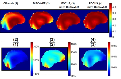 |
4 | Direct Signal Control combined with dynamic, Fast Online Customized (FOCUS) parallel transmit excitation pulses for TSE at 7 Tesla
Jürgen Herrler1, Jonathan Endres1, Raphael Tomi-Tricot2,3,4, Patrick Liebig5, Rene Gumbrecht5, Christian Richard Meixner6, Andreas Maier7, Arnd Dörfler1, Shaihan Malik3,4, and Armin Michael Nagel6,8
1Department of Neuroradiology, University Hospital Erlangen, Friedrich-Alexander-Universität Erlangen-Nürnberg (FAU), Erlangen, Germany, 2MR Research Collaborations, Siemens Healthcare Limited, London, United Kingdom, 3Biomedical Engineering Department, School of Biomedical Engineering and Imaging Sciences, King’s College London, London, United Kingdom, 4Centre for the Developing Brain, School of Biomedical Engineering and Imaging Sciences, King’s College London, London, United Kingdom, 5Siemens Healthcare GmbH, Erlangen, Germany, 6Institute of Radiology, University Hospital Erlangen, Friedrich-Alexander-Universität Erlangen-Nürnberg (FAU), Erlangen, Germany, 7Department of Computer Science, Friedrich-Alexander-Universität Erlangen-Nürnberg (FAU), Erlangen, Germany, 8Medical Physics in Radiology, German Cancer Research Center (DKFZ), Heidelberg, Germany
Direct Signal Control with Variable Excitation and Refocusing (DiSCoVER) was combined with a dynamic, Fast Online-Customized (FOCUS) parallel transmit excitation pulse for a 3D TSE sequence using an 8Tx/32Rx head RF coil at 7 Tesla. DSC could improve the signal, mainly in the cerebellum region compared to CP mode. FOCUS excitation pulses achieve better FA and phase homogeneity than static pTx pulses (standardly used in DiSCoVER). Combining a FOCUS excitation pulse with DiSCoVER-optimized refocusing pulses, led to signal gain across the echoes, which was evaluated in simulations for 132 subjects. Universal and individual DiSCoVER-refocusing pulses showed comparable performance.
|
||
3314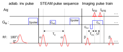 |
5 | iDREAM: T1-bias compensated DREAM B1+ mapping using adiabatic inversion pulses
Kay Nehrke1 and Peter Börnert1
1Philips Research Laboratories, Hamburg, Germany
A refinement of the DREAM B1+ mapping technique is presented, that removes the potential T1-bias from the B1+ maps. It is based on a phase cycling scheme including adiabatic inversion pulses and improves the B1+ mapping accuracy and reliability and extends the dynamic working range of the DREAM approach. The method has been successfully validated on phantoms and in vivo on the brain at 3T.
|
||
3315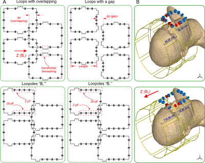 |
6 | Optimization of the Double-Row 16-channel Transmit-only RF Array Coil for Human Brain Imaging at 9.4T.
Anton Nikulin1,2, Klaus Scheffler1,2, and Nikolai Avdievich2
1Department of Biomedical Magnetic Resonance, Eberhard Karls University Tübingen, Tübingen, Germany, 2Magnetic Resonance Center, Max Planck Institute for Biological Cybernetics, Tübingen, Germany
At 9.4T, transmit-only RF array coils for human brain imaging provide mediocre transmit efficiency due to a weak loading factor, which implies low power dissipation in the tissues. In this work we aim to optimize using the full-wave simulations a dual row 16-channel transmit-only array to improve the transmit performance. For that purpose, we simulated eight different array designs containing the loops with overlapping and gaps between the rows and two configurations of loopoles. We have shown that the optimized design with the overlapped loops provides improvement ~15% in Tx-efficiency and 16.5% improvement in SAR-efficiency compared to the reference coil.
|
||
3316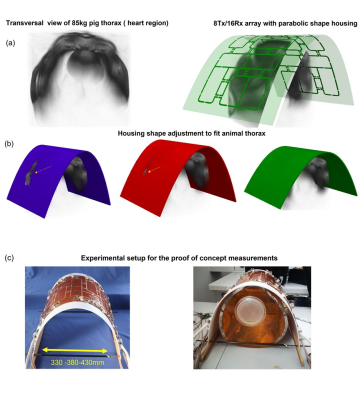 |
7 | Adjustable parabolic shape 8Tx/16Rx array for longitudinal cardiac MRI in large animals at 7T: proof of concept
Maxim Terekhov1, Ibrahim A. Elabyad1, and Laura M. Schreiber1
1Chair of Cellular and Molecular Imaging, Comprehensive Heart Failure Center, University Hospital Würzburg, Wuerzburg, Germany
Cardiovascular MRI at the ultra-high-field is an emerging modality promising a significant increase of the spatial resolution and physical sensitivity of routine cardiac imaging. Pig models play an important role in the establishment and translation of 7T cMRI to humans. The prerequisite for high-quality 7T cMRI data in pigs is dedicated transceiver arrays adapted to the shape of the pig’s thorax. We validated the proof-of-concept of the transceiver pTX cardiac array with adjustable parabolic shape housing for longitudinal studies with pigs. The stability of the transmit and receive characteristics by shape adjustments are tested in-silico and in phantom measurements.
|
||
3317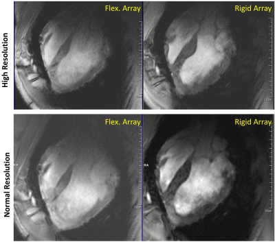 |
8 | A Flexible 16-Element Transceiver 8Tx/16Rx Coil Array for Parallel Transmit Cardiac MRI in Pigs at 7T
Ibrahim A. Elabyad1, Maxim Terekhov1, David Lohr1, Maya Bille1, and Laura M. Schreiber1
1Chair of Molecular and Cellular Imaging, Comprehensive Heart Failure Center (CHFC), University Hospital Wuerzburg, Wuerzburg, Germany
A dedicated flexible 16-element transceiver coil array was developed, simulated, and tested for cardiac MRI (cMRI) in pigs at 7T. The 16-elements of the array are printed on a flexible (Kapton polyimide) substrate to conform to the pig thorax. The performance of the elastic array is evaluated by EM-simulations and MR-measurements in a pig phantom and a 45-kg pig in-vivo. The flexible array supports parallel imaging with acceleration factors up to R=4 with reasonable g-factors (g=1.2) in an ROI at the position heart. The array enables geometrical conformity to pig body weights within a large range of sizes and shapes.
|
||
3318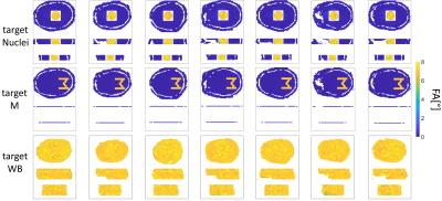 |
9 | Target specific optimization of the transmit k-space trajectory stack-of-spirals and SPINS for pTx pulse design Video Not Available
Ole Geldschläger1, Dario Bosch1,2, and Anke Henning1,3
1High-field Magnetic Resonance, Max-Planck-Institute for biological Cybernetics, Tübingen, Germany, 2Biomedical Magnetic Resonance, University Hospital Tübingen, Tübingen, Germany, 3Advanced Imaging Research Center, University of Texas Southwestern Medical Center, Dallas, TX, United States
Four basis transmit k-space trajectories (a single variable density spiral-in, a two stack of variable density spiral-in, a three stack of variable density spiral-in and a SPINS trajectory) were optimized for pTx radiofrequency pulse design in order to match the excitation target pattern. The parameter to be optimized where the parameter of the analytical equations of the basis trajectories. The procedure was tested on local excitation and whole brain-like excitation target patterns. Optimized trajectories enabled considerably improved radiofrequency pulse performance, compared to radiofrequency pulses based on unsuited trajectories. The optimization code is available online as open source (https://github.com/ole1965/workflow_OTUP.git).
|
||
3319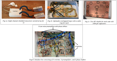 |
10 | An 8-Channel Shielded Transceiver Coil for Carotid Artery imaging at 7T
Pedram Yazdanbakhsh1, Marcus Couch2, Kyle M. Gilbert3, Johnny Derhovagimian1, and Richard Hoge1
1McConnell Brain Imaging Centre, Montreal Neurological Institute, McGill University, Montreal, QC, Canada, 2Siemens Healthcare Limited, Montreal, QC, Canada, 3Centre for Functional and Metabolic Mapping, Western University, London, ON, Canada An 8-channel shielded transmit/receive RF array has been designed and fabricated for imaging the carotid arteries at 7T. The array, similar to that presented in Ref. [1], consists of four overlapping surface loops per side. In addition to the Ref. [1], we have also added two RF shields per side with DC blocking capacitors to mitigate eddy currents. To ensure robust safety of the 7T pTx coil, local SAR matrices, and the commensurate virtual observation points (VOPs), were calculated for online SAR supervision [2,3]. Finally, the phase and amplitude of each channel have been optimized to achieve maximum B1+ homogeneity. |
||
3320 |
11 | Evaluating a Volumetric High-Permittivity (HPM) Helmet for Brain Imaging at 3.0 T
Karthik Lakshmanan1,2, Jezry Walczyk1,2, Sebastian Rupprecht3, Michael T Lanagan3, Qing X Yang3, Ryan Brown1,2,4, and Christopher Collins1,2,4
1Bernard and Irene Schwartz Center for Biomedical Imaging, Department of Radiology, NYU Grossman School of Medicine, Newyork, NY, United States, 2Center for Advanced Imaging Innovation and Research (CAI2R), Department of Radiology, NYU Grossman School of Medicine, New york, NY, United States, 3HyQRS, LLC, State College, PA, United States, 4Vilcek Institute of Graduate Biomedical Science, NYU Grossman School of Medicine, New york, NY, United States A thin (8mm) helmet-shaped shell of high-permittivity material (HPM) is seen to double transmit efficiency and increase SNR by 40% at the center of tissue equivalent head phantom when compared to the body coil alone, with little effect on B1 homogeneity within the brain region. While evaluation of effects on receive arrays is ongoing, from the results shown here it seems an HPM helmet could be used at 3T in place of more electronically cumbersome head transmit coils. |
||
 Back to Meeting Home
 Back to the Program-at-a-Glance
The International Society for Magnetic Resonance in Medicine is accredited by the Accreditation Council for Continuing Medical Education to provide continuing medical education for physicians.
 Back to Meeting Home
 Back to the Program-at-a-Glance
 View the Presentation