Online Gather.town Pitches
Neurological Conditions III
Joint Annual Meeting ISMRM-ESMRMB & ISMRT 31st Annual Meeting • 07-12 May 2022 • London, UK

Online Gather.town Pitches
Neurological Conditions III
| Booth # | ||||
|---|---|---|---|---|
 |
3341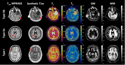 |
1 | Multiparametric Characterization of Focal Cortical Dysplasia using MR Fingerprinting
Zhong Irene Wang1, Joon Yul Choi1, Siyuan Hu2, Yingying Tang1, TingYu Su1,2, Ingmar Blümcke1,3, Stephen Jones4, Ken Sakaie4, Imad Najm1, and Dan Ma2
1Epilepsy Center, Neurological Institute, Cleveland Clinic, Cleveland, OH, United States, 2Biomedical Engineering, Case Western Reserve University, Cleveland, OH, United States, 3Neuropathology, University of Erlangen, Erlangen, Germany, 4Imaging Institute, Cleveland Clinic, Cleveland, OH, United States
Focal cortical dysplasia (FCD) is a common pathology underlying medically intractable focal epilepsies. Conventional MRI can be limited in characterizing subtle FCD, due to the lack of quantitative measurements for tissue properties. Here, we proposed a multiparametric machine learning (ML) approach based on high-resolution 3D MR fingerprinting (MRF) to characterize FCD lesions. The ML model showed robust accuracy of 96%, 89%, and 79% to separate FCD from normal cortex, FCD type I from type II, and FCD type IIa from type IIb, respectively. Our findings suggest the usefulness of the multiparametric MRF ML approach to improve noninvasive epilepsy presurgical evaluation.
|
|
3342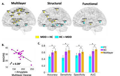 |
2 | Brain Structure-Function Network Hierarchy Abnormalities in Major Depressive Disorder
Yael Jacob1, Laurel Morris1, Sarah Rutter1, Gaurav Verma1, James Murrough1, and Priti Balchandani1
1Icahn School of Medicine at Mount Sinai, New York, NY, United States
Major depressive disorder (MDD) diagnosis, research, and treatment is especially challenging given that current diagnosis entirely depends on clinical symptoms, whereas the underlying brain pathology remains largely unclear. Applying a novel multilayer network analysis, considering communication within functional and structural networks as well as the interactions between them, we found aberrant centrality measures of various brain regions in MDD compared to controls that were not detected using single structural or functional connectomes. In addition, using multilayer network features as predictors in a machine learning algorithm resulted in higher predictive values compared to classification models based on single layer.
|
||
 |
3343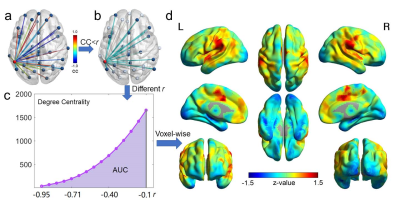 |
3 | Brain Functional Segregation Predicts Symptom Severity and Treatment Response in Psychotic Disorders
Xiaopeng Song1, Tao Song1, Chenyanwen Zhu1, Dost Ongur1, and Fei Du1
1Harvard Medical School, BELMONT, MA, United States
Functional segregation, i.e., anticorrelated neural activities, has not been well-explored in fMRI studies. We introduced the Negative Degree Centrality (NDC) method to quantify functional segregation. We found decreased NDC in psychotic disorder patients compared to controls. Positive, negative, and general psychotic symptoms were associated with impaired NDC in three different brain networks respectively. Using NDC and a machine learning approach, we identified two subgroups of patients with distinct recovery trajectories after one-year treatment. Our findings suggested impaired functional segregation in different brain circuits might be the neurobiological mechanisms associated with various psychotic symptoms and outcome heterogeneity in psychotic disorders.
|
|
3344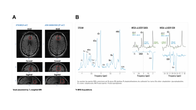 |
4 | Abnormal glutamate metabolism in prefrontal cortex of post-traumatic stress disorder linked to comorbidity with major depression
Kelley M. Swanberg1, Hetty Prinsen2, Christopher L. Averill3,4,5, Leonardo Campos1, Abhinav V. Kurada1, John H. Krystal3,4, Ismene L. Petrakis3,4, Lynnette A. Averill3,4,5, Chadi G. Abdallah4,5, and Christoph Juchem1,2,6,7
1Biomedical Engineering, Columbia University School of Engineering and Applied Science, New York, NY, United States, 2Radiology and Biomedical Imaging, Yale University School of Medicine, New Haven, CT, United States, 3Clinical Neuroscience Division, Department of Veterans Affairs National Center for Posttraumatic Stress Disorder, Veterans Affairs Connecticut Healthcare System, West Haven, CT, United States, 4Psychiatry, Yale University School of Medicine, New Haven, CT, United States, 5Psychiatry and Behavioral Sciences, Baylor College of Medicine, Houston, TX, United States, 6Radiology, Columbia University Medical Center, New York, NY, United States, 7Neurology, Yale University School of Medicine, New Haven, CT, United States
Post-traumatic stress disorder (PTSD) is an anxiety condition evidenced by wide-ranging emotional and cognitive dysfunction. While prefrontal glutamatergic excitotoxicity is thought to contribute to its presentation, no 1H-MRS studies of PTSD have yet been conducted at 7-Tesla field strength facilitating separation of glutamate from metabolic partner glutamine. Here we apply 7-T 1H MRS to investigate medial prefrontal (mPFC) concentrations of glutamate and glutamine with GABA, glutathione, and other metabolites in both PTSD and comorbid major depressive disorder (MDD). We show several PTSD-associated abnormalities in mPFC glutamate metabolism that appear to be driven by MDD status when this comorbidity is considered.
|
||
3345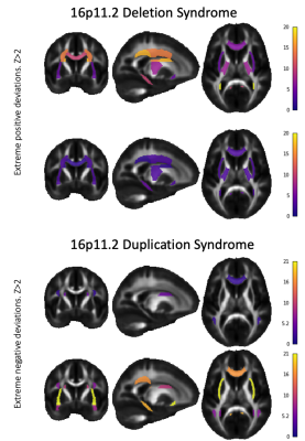 |
5 | Multi-Site Normative Modeling and Hierarchical Bayesian Analysis of DKI metrics in Carriers of 16p11.2 Copy Number Variants
Julio Ernesto Villalon-Reina1, Clara Moreau2, Talia M Nir1, Neda Jahanshad1, Simons Variation in Individuals Project Consortium3, Anne Maillard4, David Romascano4, Bodgan Draganski5, Sarah Lippé6, Carrie E Bearden7, Paul M Thompson1, and Sebastien Jacquemont8
1Mark and Mary Stevens Neuroimaging and Informatics Institute, University of Southern California, Marina del Rey, CA, United States, 2Institut Pasteur, Université de Paris, Paris, France, 3Simons Foundation, New York, NY, United States, 4Service des Troubles du Spectre de l’Autisme et apparentés, Département de psychiatrie, Lausanne University Hospital, Lausanne, Switzerland, 5Laboratoire de Recherche en Neuroimagerie, Université de Lausanne, Lausanne, Switzerland, 6Psychology Department, University of Montreal, Montreal, QC, Canada, 7Departments of Psychiatry and Biobehavioral Sciences and Psychology, Semel Institute for Neuroscience and Human Behavior, University of California, Los Angeles, Los Angeles, CA, United States, 8University of Montreal, Montreal, QC, Canada
We used Hierarchical Bayesian Regression (HBR) to create a normative model of age-effects on Diffusion Kurtosis Imaging (DKI) measures of the white matter. DKI maps from more than 1,300 healthy subjects (3-78 years) from six sites and eight scanners were used to estimate the normative model across the lifespan enabling the multi-site data integration with HBR. 16p11.2 deletions and duplications showed “mirror effects” on diffusion kurtosis metrics.
|
||
3346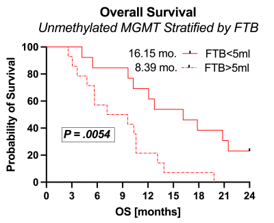 |
6 | Unmethylated MGMT combined with post-CRT perfusion MRI-derived tumor burden informs overall survival in newly diagnosed glioblastoma
Melissa A Prah1, Jennifer M Connelly1, and Kathleen M Schmainda1
1Medical College of Wisconsin, Milwaukee, WI, United States
As treatment response can mimic progressive tumor on standard imaging, clinical outcomes following upfront chemo-radiotherapy in newly diagnosed glioblastoma rely heavily on MGMT promoter methylation status. While MGMT is a reliable marker of outcomes, the purpose of this study was to determine if inclusion of advanced perfusion-MRI derived fractional tumor burden (FTB) volume might better inform survival outcomes. Results revealed that low FTB volume in MGMT unmethylated glioblastoma confers significant survival benefit, approaching that of MGMT methylated glioblastoma. Clinically, combining FTB with MGMT methylation status may identify patients with potentially better outcomes or for whom more aggressive approaches are warranted.
|
||
3347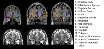 |
7 | Structural abnormalities in olfactory brain regions in early Parkinson’s disease (PD) patients measured by high resolution MRI at 7T
Adrian Grant Paez1,2, Suraj Rajan3,4, Alex Y Pantelyat3, Liana I Rosenthal3, Andreia Faria5, Xinyuan Miao2,6, Ted M Dawson3, Peter C. M. van Zijl1,2, Vidyulata Kamath4, and Jun Hua1,2
1Radiology and Radiological Science, Johns Hopkins University School of Medicine, Baltimore, MD, United States, 2F.M. Kirby Research Center, Kennedy Krieger Institute, Baltimore, MD, United States, 3Neurology, Johns Hopkins University School of Medicine, Baltimore, MD, United States, 4Psychiatry and Behavioral Sciences, Johns Hopkins University School of Medicine, Baltimore, MD, United States, 5Radiology and Radiological Science, Kennedy Krieger Institute, Baltimore, MD, United States, 6Johns Hopkins University School of Medicine, Baltimore, MD, United States
We demonstrate significant structural abnormalities in the OB, primary and secondary olfactory brain regions in early PD patients measured by high resolution MRI at 7T. The MRI measures showed significant correlations with behavioral olfactory deficits in the participants.
|
||
| 3348 | 8 | Metabolic Abnormalities in Bipolar Disorder Observed in the Putamen and Cerebellar Vermis with 7T MRS
Vincent Magnotta1, Jia Xu1, Jess Fiedorowicz2, Aislinn Williams3, Joseph Shaffer4, Gary Christensen5, Jeffrey Long6, Eric Taylor7, Leela Sathyaputri1, Jenny Gringer Richards1, Gail Harmata3, and John Wemmie3
1Radiology, University of Iowa, Iowa City, IA, United States, 2Psychiatry, University of Iowa, Ottawa, ON, Canada, 3Psychiatry, University of Iowa, Iowa City, IA, United States, 4Biosciences, Kansas City University, Kansas City, MO, United States, 5Electrical and Computer Engineering, University of Iowa, Iowa City, IA, United States, 6Paychiatry, University of Iowa, Iowa City, IA, United States, 7University of Iowa, Iowa City, IA, United States
This study used 31P and 1H MRS to study brain metabolic differences in bipolar disorder. Data from 64 participants with BD and 42 controls was acquired from the right putamen and cerebellar vermis were acquired at 7T. The study observed reduced pHi in support of prior work that has proposed mitochondrial dysfunction in BD where there is an impaired ability to utilize pyruvate in oxidative phosphorylation. A shift from oxidative phosphorylation toward glycolytic energy production likely increases lactate acid, CO2 levels, and free protons resulting in tissue acidosis. This shift in energy production may also lead to increased glutathione.
|
||
3349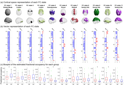 |
9 | Brain functional connectivity in schizophrenia: disrupted network dynamics revealed by fMRI
Miguel Farinha1, Conceição Amado2, and Joana Cabral3,4,5
1Instituto Superior Técnico, University of Lisbon, Lisbon, Portugal, 2Department of Mathematics and CEMAT, Instituto Superior Técnico, University of Lisbon, Lisbon, Portugal, 3Life and Health Sciences Research Institute, School of Medicine, University of Minho, Braga, Portugal, 4Centre for Eudaimonia and Human Flourishing, Linacre College, University of Oxford, Oxford, United Kingdom, 5Center for Music in the Brain, Department of Clinical Medicine, Aarhus University, Aarhus, Denmark
We investigate differences between the dynamic exploration of resting-state functional connectivity (FC) states using fMRI data from 71 schizophrenia (SZ) patients and 74 healthy controls (HCs) by employing the Leading Eigenvector Dynamics Analysis (LEiDA) method to provide potential biomarkers of this disorder. We found a reduced ability of SZ patients to access and remain in a state of global BOLD phase coherence. Functionally meaningful states presented increased occurrence, limiting probability and altered dynamic transitions in SZ patients. These findings expose pronounced differences between SZ patients and HCs - supporting and developing current knowledge regarding disrupted brain dynamics in schizophrenia.
|
||
3350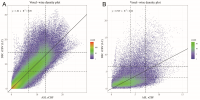 |
10 | Voxel-wise Comparison of Clinical 2D pCASL and DSC Perfusion of Intra-axial Brain Tumors
Johannes Devos1, Ronald Peeters1, and Philippe Demaerel2
1Radiology, UZ Leuven, Leuven, Belgium, 2Neuroradiology, UZ Leuven, Leuven, Belgium
Dynamical susceptibility contrast (DSC) and arterial spin labeling (ASL) are MRI techniques to quantify brain tissue perfusion. A voxel-wise analysis in 23 patients showed a moderate to strong linear relationship between tumoral cerebral blood flow measured by 2D echo planar imaging, pseudo-continuous ASL (pCASL) and leakage corrected, cerebral blood volume measurements by DSC. Additionally, mean and 90th percentile of tumor perfusion of all patients showed a strong linear relationship. In conclusion, 2D EPI pCASL is a viable alternative to DSC for perfusion measurements of brain tumors.
|
||
3351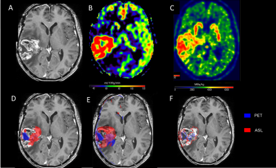 |
11 | Role of PET and ASL simultaneously acquired imaging biomarkers in differentiating progression from radionecrosis in glial brain tumors
Nadya Pyatigorskaya1, Arnaud Pellerin2, Maya Kalife3, Laura Rozenblum-Beddok4, Marc Bertaux2, Marine Soret2, Lydia Yahia-cherif3, and Aurélie Kas2
1CENIR, ICM, Paris, France, 2Pitié-Salpêtrière Hospital, APHP, Sorbonne universite, Paris, France, 3ICM, Paris, France, 4Pitié Salpêtrière, APHP, Paris, France
We aimed at investigating the methods based on coupling of of amino-acid metabolism (18F-DOPA-PET) measurements and cerebral blood flow (CBF) maps to evaluate the diagnostic performance of PET/MRI in glioma imaging. Tumor isocontour maps and T-maps were created using SPM and metabolic/perfusion abnormalities were evaluated with the asymmetry index z-score. SPM map analysis of significant-size clusters and semi-quantitative evaluation were performed and compared to gold standard diagnosis. Both the tumor isocontour maps and T-maps showed the highest specificity for ASL and sensitivity for 18F-DOPA analysis, allowing high diagnostic performance in differentiating between progression and radionecrosis particularly in high-grade treated gliomas.
|
||
3352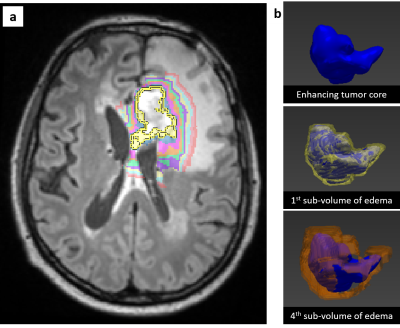 |
12 | Quantitative magnetic resonance imaging combined with three-dimensional image analysis in glioblastoma
Gergely Bertalan1, Julia Onken2, Simone Schwedler3, Bernd Hamm1, Georg Bohner3, and Edzard Wiener3
1Radiology, Charité - Universitätsmedizin Berlin, Berlin, Germany, 2Neurosurgery, Charité - Universitätsmedizin Berlin, Berlin, Germany, 3Neuroradiology, Charité - Universitätsmedizin Berlin, Berlin, Germany
Conventional MRI such as T2 and FLAIR do not show sub-regional intensity changes in peritumoral infiltration zone of glioblastoma and cannot visualize tumor margins. Here, we implemented a new method for sub-volumetric analysis of glioblastoma and investigated T1 and T2 relaxation properties in the peritumoral infiltration zone. The results show a decrease in T1 and T2 relaxation times as a function of distance from the contrast-enhanced T1 solid tumor region to the periphery. Our method may help to better select regions for biopsy, to determine the surgical resection margin and improve radiation therapy planning and post-therapeutic progress controls.
|
||
3353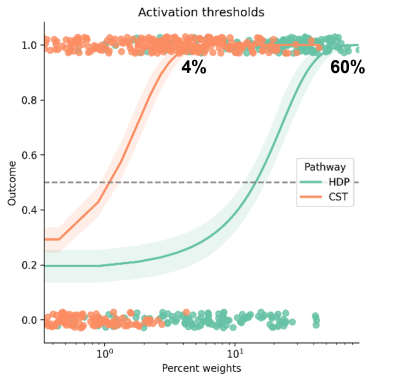 |
13 | Patient-specific hyperdirect pathway activation in DBS for Parkinson's disease
Alba Segura Amil1,2, T.A. Khoa Nguyen1,2, Andreas Nowacki1, and Claudio Pollo1
1Department of Neurosurgery, University Hospital Bern, Bern, Switzerland, 2ARTORG Center for Biomedical Engineering, University of Bern, Bern, Switzerland
Deep brain stimulation is an effective treatment in advanced Parkinson’s disease. The aim of the study was to determine activation thresholds for the hyperdirect pathway and corticospinal tract to control motor symptoms and avoid the appearance of side effects. Patient-specific whole brain tractograpy, DBS leads reconstruction, and generation of volumes of tissue activated were performed in 20 Parkinson patients. The activation threshold for the hyperdirect pathway was 60%, and for the corticospinal was 4%. For newly generated volumes of tissue activated, the average prediction error for the hyperdirect pathway was 1 mA and for the corticospinal tract was 1.5 mA.
|
||
 Back to Meeting Home
 Back to the Program-at-a-Glance
The International Society for Magnetic Resonance in Medicine is accredited by the Accreditation Council for Continuing Medical Education to provide continuing medical education for physicians.
 Back to Meeting Home
 Back to the Program-at-a-Glance
 View the Presentation