Online Gather.town Pitches
Fast, Novel & Robust Acquisitions I
Joint Annual Meeting ISMRM-ESMRMB & ISMRT 31st Annual Meeting • 07-12 May 2022 • London, UK

Online Gather.town Pitches
Fast, Novel & Robust Acquisitions I
| Booth # | ||||
|---|---|---|---|---|
4695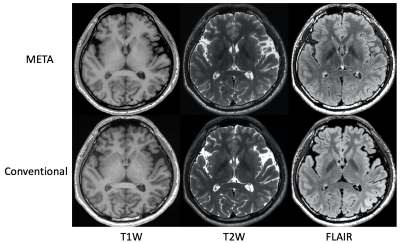 |
1 | Highly Accelerated Volumetric Brain Imaging with META and Deep Learning Reconstruction
Naoyuki Takei1, R Marc Lebel2, Suryanarayanan Kaushik3, Shohei Fujita4,5, Issei Fukunaga4, Shigeki Aoki4, Suchandrima Banerjee6, and Tetsuya Wakayama1
1GE Healthcare, Tokyo, Japan, 2GE Healthcare, Calagary, AB, Canada, 3GE Healthcare, Waukesha, WI, United States, 4Radiology, Juntendo University School of Medicine, Tokyo, Japan, 5Radiology, The University of Tokyo Graduate School of Medicine, Tokyo, Japan, 6GE Healthcare, Menlo Park, CA, United States
We combine advanced acquisition and reconstruction techniques for highly accelerated 3D brain imaging. A hybrid acquisition called META (Mixed Echo Train Acquisition) generates 3D T1W, T2W and FLAIR contrasts simultaneously and with high SNR efficiency. A 3D deep learning reconstruction (AIR Recon DL) reduces image noise, reduces artifacts, and enhances resolution relative to conventional reconstructions, solving the trade-off between SNR, scan time, and resolution. META with the DL Recon achieved 1mm isotropic T1W, T2W and FLAIR images within 4 minutes. These combinations are well suited in high resolution volumetric brain imaging.
|
||
4696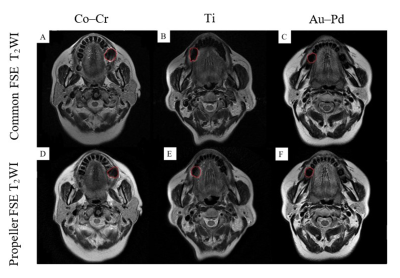 |
2 | Performance of PROPELLER FSE T2WI in reducing metal artifacts for patients with various material porcelain fused to metal
Wenjin Bian1, Jinliang Niu1, Jianting Li1, and Wenqi Wu1
1Department of Imaging, The Second Hospital of Shanxi Medical University, Taiyuan, China The metal artifacts caused by metal copings in porcelain fused to metal (PFM) always impair MRI quality. This study aimed to compare MRI quality between common FSE T2WI with PROPELLER FSE T2WI for patients with various metal copings. Fifty-nine subjects with the most commonly used PFM were recruited and imaged at 1.5T MRI system. By comparing the image quality and artifact areas, we found PROPELLER technique was less sensitive to metal artifacts than common FSE. It can be proposed the PROPELLER FSE T2WI to be used as clinical MRI examination of oral cavity and maxillofacial part for patients with PFM. |
||
4697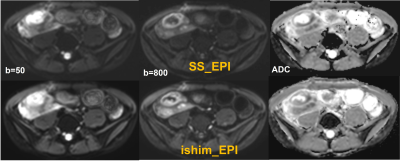 |
3 | Comparison of integrated slice-specific dynamic shimming (ishim) and single shot EPI techniques in patients with Crohn’s disease
Junjiao Hu1, Huiting Zhang2, Hu Guo3, Thomas Benkert4, Shan Jiang1, Weijun Situ1, and Jun Liu1
1Department of Radiology,The Second Xiangya Hospital, Central South University, Changsha, China, 2MR Scientific Marketing, Siemens Healthineers, Wuhan, China, 3MR Application, Siemens Healthineers, Changsha, China, 4MR Application Predevelopment, Siemens Healthcare GmbH, Erlangen, Germany
In this study, image quality and apparent diffusion coefficient (ADC) values of conventional single-shot spin-echo echo-planar imaging (SS-EPI) and prototype integrated slice-specific dynamic shimming EPI (ishim_EPI) diffusion-weighted imaging in patients with Crohn’s disease were compared. Results showed that ishim_EPI had better image quality, including fewer distortion artifacts and clearer edges of the lesions. High correlation and good agreement of ADC value were found between the two techniques. Ishim_EPI DWI technique is recommended to replace conventional SS_EPI for the detection of lesions in patients with Crohn’s disease and further in routine intestinal examination.
|
||
4698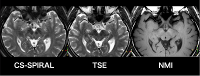 |
4 | Spin-Echo Revisited: Improved Visualization of Accelerated T2W Spin-Echo Spiral Imaging with Compressed Sensing with SENSE (CS-Spiral)
Kosuke Morita1, Masami Yoneyama2, Hiroshi Hamano2, Shogo Fukuda1, Hiroyuki Uetani3, Akira Sasao3, Takeshi Nakaura3, Seitaro Oda3, Masahiro Hatemura1, and Toshinori Hirai3
1Radiology, Kumamoto university, kumamoto-shi, Japan, 2Philips Japan, minato-ku, Japan, 3Diagnostic Radiology, Graduate School of Medical Sciences, Kumamoto university, kumamoto-shi, Japan
CS-Spiral T2WI was good image quality for midbrain. We used 3 sequences; CS-Spiral, conventional-Turbo Spine Echo and neuromelanin image for six healthy volunteers. In this study, we used Compressed Sensing with Sensitivity Encoding (CS-Spiral) technology for SPIRAL acquisition to perform midbrain T2WI for neurodegenerative diseases with clinically usefulness acquisition time. It is expected that parameter settings will provide more clinically useful image quality.
|
||
4699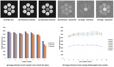 |
5 | Implementation of the QRAPMASTER sequence using a dictionary matching and quantitative evaluation of the magnetization transfer effect
Katsumi Kose1, Ryoichi Kose1, and Yasuhiko Terada2
1MRIsimulations Inc., Tokyo, Japan, 2University of Tsukuba, Tsukuba, Japan
The magnetization transfer effect of the QRAPMASTER sequence was investigated using phantom experiments and Bloch simulations. The phantom consisted of MnCl2 aqueous solution with various proton T1 values and raw chicken breast meat. T1 values of the MnCl2 solution measured using the QRAPMASTER sequence showed excellent linear relation with those measured with the standard method. However, T1 of the chicken breast sample deviated far from the linear regression line. The results suggested that the T1 values of biological samples measured by the QRAPMASTER sequence are underestimated compared to those measured by the standard method.
|
||
4700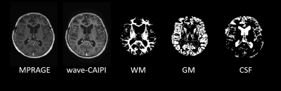 |
6 | Ultra-Fast T1-Weighted Imaging with Wave-CAIPI MPRAGE for Clinical Pediatric Brain MR imaging: A Prospective Quantitative Assessment Study
Yung-Chieh Chen1, Duen-Pang Kuo1,2, and Cheng-Yu Chen1
1Department of Medical Imaging, Taipei Medical University Hospital, Taipei, Taiwan, 2Translational Imaging Research Center, Taipei Medical University Hospital, Taipei, Taiwan
Morphologic neuroimaging in children can be challenging and time-sensitive during routine clinical imaging workflow due to patient discomfort, susceptibility to motion, and frequent need of sedation. Our study quantitatively assessed the imaging performance of ultra-fast T1 3D magnetization-prepared rapid acquisition gradient echo (MPRAGE) using wave-controlled aliasing with parallel imaging (wave-CAIPI) sequence for accelerated pediatric brain MR imaging via whole brain wide analysis under different MR imaging parameter settings with and without contrast administration. Our results show that wave-CAIPI can deliver comparable image quality to standard MPRAGE with significantly reduced scan times with optimized imaging parameters.
|
||
4701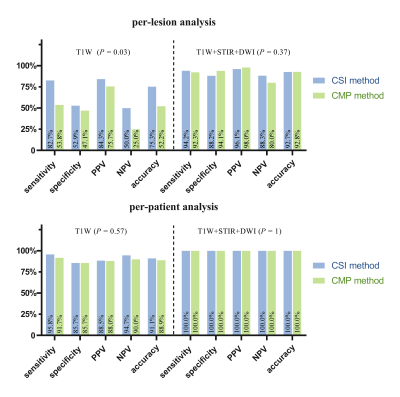 |
7 | Bone metastasis assessment using accelerated whole-body isotropic 3D T1-weighted Dixon acquisition with Compressed SENSE: a feasibility study
Zhenhong Liao1 and Xiaoyong Zhang2
1Department of Radiology, Deyang People's Hospital, Deyang, China, 2Philips Healthcare, Chengdu, China Three-dimensional T1-weighted (T1W) Dixon as morphologic sequence has very high clinical value for the metastatic screening [1]. However, conventional multiplanar (CMP) T1W Dixon is limited by the long acquisition to achieve high resolution whole-body imaging. Therefore, this study aimed to achieve rapid whole-body high-resolution isotropic 3D T1W Dixon examination by Compressed SENSE (CSI T1W Dixon) and to investigate its feasibility in assessing bone metastases by comparing image quality and diagnosis performance with conventional multiplanar (CMP) (coronal, sagittal, axial planes) T1W Dixon scan. The results of this study suggested that high-resolution CSI T1W Dixon has potential to replace CMP T1W Dixon sequence for the assessment of bone metastasis in clinics with higher image quality and diagnostic capacity. |
||
4702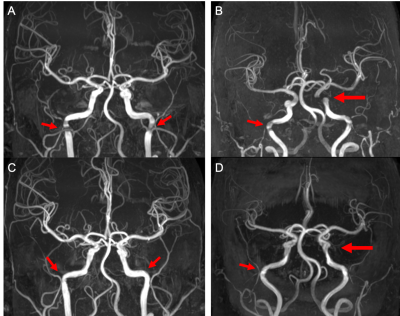 |
8 | Comparison of conventional and spiral TOF-MRA in clinical applications
Taishan Kang1, Liangjie Lin2, Zhigang Wu3, and Jian Wu4
1Magnetic Resonance Center, Zhongshan Hospital Affiliated to Xiamen University, Xiamen, China, 2Philips Healthcare, Beijing, China, 3Philips Healthcare, Shenzhen, China, 4Department of Electronic Science, Xiamen University, Xiamen, China
TOF-MRA is a routine noninvasive method that does not require intravenous contrast agents or exposure to radiation for brain artery assessment. However, artifacts are prone to appear in conventional TOF-MRA under situations of complicated blood vessels or changes in blood flow; and the display of distal arterioles is also not good. The spiral acquisition technique has been previously introduced for scan acceleration of TOF-MRA, while here we compared the performance of conventional and spiral TOF-MRA under similar scan duration. Results indicated that spiral TOF-MRA provide better quality MRA images, and may help improve the clinical diagnosis of vascular problems.
|
||
4703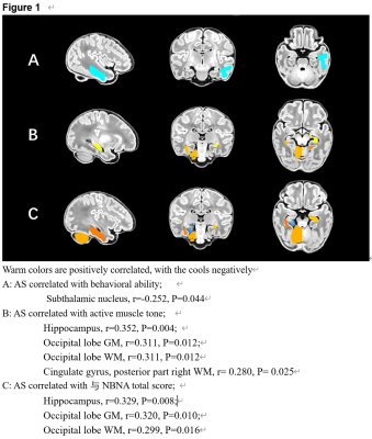 |
9 | Investigation of cerebral asymmetry in full-term newborns and its relationship with neurobehavioural assessment
Pengxuan Bai1, Linlin Zhu1, Yuying Feng1, Na Zhang1, Yao Ge1, Congcong Liu1, Jian Yang1, Yichu He2, Feng Shi2, Xiaocheng Wei3, and Chao Jin1
1The First Affiliated Hospital of Xi’an Jiaotong University, Xi’an, China, 710061, Xi'an, China, 2Department of Research and Development, Shanghai United Imaging Intelligence Co., Ltd., Shanghai, China,200232, Shanghai, China, 3GE Healthcare,Beijing, China,100176, Beijing, China
The specialization of cerebral lateralization and hemispheric function is common in adults. Existing studies have shown that there is already left and right asymmetry in the fetal brain [1]. The study of normal brain structure and development information helps to identify abnormal developmental trajectories. This study takes healthy full-term newborns as the research object, aiming to explore the situation of cerebral asymmetry in the neonatal period and the relationship with neurobehavioral. Meanwhile expand our understanding of normal early brain development, and provide important insights for further revealing the inner connection between brain lateralization and certain diseases in the future.
|
||
4704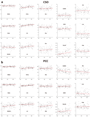 |
10 | Metabolic timecourse of healthy aging
Tao Gong1,2, Steve C.N. Hui3,4, Helge J. Zöllner3,4, Mark Britton5,6,7, Yulu Song3,4, Aaron T. Gudmundson8, Kathleen Hupfeld9, Eric Porges5,6,7, Georg Oeltzschner3,4, Weibo Chen10, Guangbin Wang1,2, and Richard Edden3,4
1Departments of Radiology, Shandong Provincial Hospital Affiliated to Shandong First Medical University, Jinan, Shandong, 250021, China, Jinan, China, 2Departments of Radiology, Shandong Provincial Hospital, Cheeloo College of Medicine, Shandong University, Jinan, Shandong, 250021, China, Jinan, China, 3The Russell H. Morgan Department of Radiology and Radiological Science, Johns Hopkins University School of Medicine, Baltimore, MD, USA, Baltimore, MD, United States, 4F.M. Kirby Research Center for Functional Brain Imaging, Kennedy Krieger Institute, Baltimore, MD, USA, Baltimore, MD, United States, 5Center for Cognitive Aging and Memory, University of Florida, Gainesville, FL, USA, Gainesville, FL, United States, 6McKnight Brain Research Foundation, University of Florida, FL, USA, Gainesville, FL, United States, 7Department of Clinical and Health Psychology, University of Florida, Gainesville, FL, USA, Gainesville, FL, United States, 8Department of Neurobiology and Behavior, University of California, Irvine,USA., Irvine, CA, United States, 9Department of Applied Physiology and Kinesiology, University of Florida, Gainesville, FL, USA, Gainesville, FL, United States, 10Philips Healthcare, Shanghai, China, Shanghai, CA, China
The metabolic timecourse of healthy aging is not well-established, in part due to diversity of quantification methodology. This study recruited a large structured cross-sectional cohort throughout adulthood to investigate metabolic changes as a function of age, and applied consensus-recommended quantification methods. Positive age correlations in tCho, tCr, and mI were observed for CSO and PCC, while none was found for tNAA, Glu or Glx in either region. PCC GABA decreased with age, CSO Scyllo increased, and we observed higher PCC Scyllo for female subjects. Our results establish a metabolic timecourse of healthy aging, as a normative foundation for future work.
|
||
4705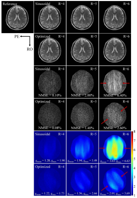 |
11 | Coil Sensitivity Specific Optimization of Wave Encoding Gradient Trajectory for High Acceleration and Low Slew Rate
Shi Su1,2, Zheyuan Yi1,2, Yujiao Zhao1,2, Linfang Xiao1,2, Ziming Huang1,2, Jiahao Hu1,2, Junhao Zhang1,2, Vick Lau1,2, Christopher Man1,2, Alex T.L. Leong1,2, and Ed X. Wu1,2
1Laboratory of Biomedical Imaging and Signal Processing, The University of Hong Kong, Hong Kong, China, 2Department of Electrical and Electronic Engineering, The University of Hong Kong, Hong Kong, China
Wave encoding offers high acceleration in parallel imaging by exploring the coil sensitivity variations in the readout dimension. However, typically preset wave gradients (i.e., sinusoidal trajectory) have never been optimized for specific receiver array coil, thus intrinsically limiting the maximum acceleration factor. We propose to optimize the gradient trajectory in a coil sensitivity specific manner by minimizing the squared L2-norm of off-diagonal elements in encoding correlation matrix. To guarantee an allowed maximum gradient slew rate, a bandlimited constraint is also introduced in optimization. This procedure leads to significant improvements in g-factor map and artifact reduction, especially at very high acceleration.
|
||
4706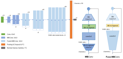 |
12 | MGMT Promoter Methylation Prediction by End-to-end Evidential-Efficient Net
Yingjie Feng1, Junbo Zhao1, Huai Chen2, Xiaoyin Xu3, and Min Zhang1
1Zhejiang Univerisity, Hangzhou, China, 2The First Affiliated Hospital of Guangzhou Medical University, Guangzhou, China, 3Brigham and Women's Hospital,Harvard Medical School, Boston, MA, United States
Malignant brain tumor affects a large number of patients and often have poor prognosis and low response to therapeutics. An indicator of the progress of brain tumor and its response to treatment is the DNA repair protein, O6-methylguanine-DNA methyltransferase (MGMT). As such, accurate assessment of MGMT is of great clinical significance. Biospy is not only invasive but also has the risk of undersampling the tumorous tissue. We presented a novel deep learning model that uses multi-MRI modalities to assess the expression of MGMT in glioblastoma (GBM) patients. Results showed that the model can achieve good performance.
|
||
4707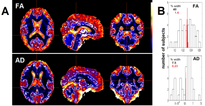 |
13 | Inter-subject variability in global diffusivity metrics degrades cross-sectional statistics
Leighton BARNDEN1, Kiran Thapaliya1, Donald Staines1, Jiasheng SU1, and Sonya Marshall-Gradisnik1
1NCNED, Griffith University, Southport, QLD, Australia
Diffusion metrics from brain Diffusion Tensor Imaging (DTI) characterise axonal structure and include fractional anisotropy (FA), axial diffusivity (AD) and radial diffusivity (RD). Cross-sectional studies of DTI metric maps apply voxel-based statistical analysis of the metric values generated by standard MRTrix and FSL algorithms and assumes that the global values for these metrics are consistent across the populations analysed. This study found that inter-subject global levels vary appreciably for the full range of diffusion metrics. Removal of the variance associated with the global levels markedly improved the statistical inference for differences in a study of ME/CFS patients and healthy controls.
|
||
4708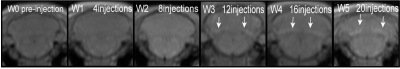 |
14 | Quantitative magnetic resonance imaging in gadolinium deposition in rat brain
xucong wang1, bing chen2, jian li2, zhen hua wang 1, and Xiaocheng Wei3
1ningxia medical university, yinchuan, China, 2Ningxia Medical University General Hospital, yinchuan, China, 3GE Healthcare, Beijing, China To investigate the application of quantitative magnetic resonance imaging in the quantitative measurement of gadolinium deposition in the deep cerebellar nucleus (DCN) of rats.The absolute correlation coefficients between DCN T1 value and DCN gadolinium concentration, and DCN/ cerebrar cortex T1WI signal intensity ratio and DCN gadolinium concentration were 0.925 (P<0.001) and 0.849 (P<0.001).These results suggest that quantitative magnetic resonance imaging has a good expression of T1 relaxation time in the dentate nucleus region of cerebellum, which provides a basis for quantitative magnetic resonance estimation of clinical gadolinium deposition. |
||
4709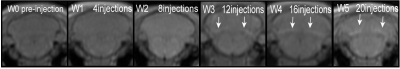 |
15 | Experimental study on intracranial gadolinium deposition in rats after linear conversion to macrocyclic gadolinium contrast agent
xucong wang1, bing chen2, jian li2, zhen hua wang 1, and Xiaocheng Wei3
1ningxia medical university, yinchuan, China, 2Ningxia Medical University General Hospital, yinchuan, China, 3GE Healthcare, Beijing, China To investigate the changes of intracranial gadolinium deposition in rats with linear gadolinium deposition after continuous injection of large ring gadolinium contrast agent. The intracranial gadolinium deposition in these rats increased but was relatively small. The DCN T1 value measured on MRI ensemble sequence scan images was significantly negatively correlated with gadolinium concentration in cerebellar DCN. (ALL P values were less than 0.05). |
||
 Back to Meeting Home
 Back to the Program-at-a-Glance
The International Society for Magnetic Resonance in Medicine is accredited by the Accreditation Council for Continuing Medical Education to provide continuing medical education for physicians.
 Back to Meeting Home
 Back to the Program-at-a-Glance
 View the Presentation